

Osteoclast-Released Wnt-10b Underlies Cinacalcet Related Bone Improvement in Chronic Kidney Disease
- PMID: 31181716
- PMCID: PMC6600662
- DOI: 10.3390/ijms20112800
Osteoclast-Released Wnt-10b Underlies Cinacalcet Related Bone Improvement in Chronic Kidney Disease
Abstract
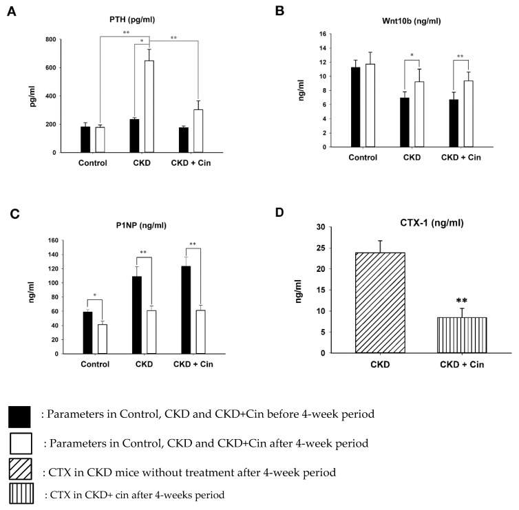
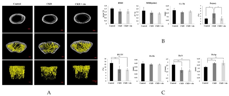
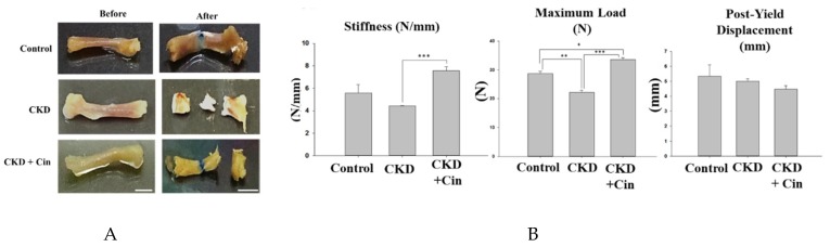
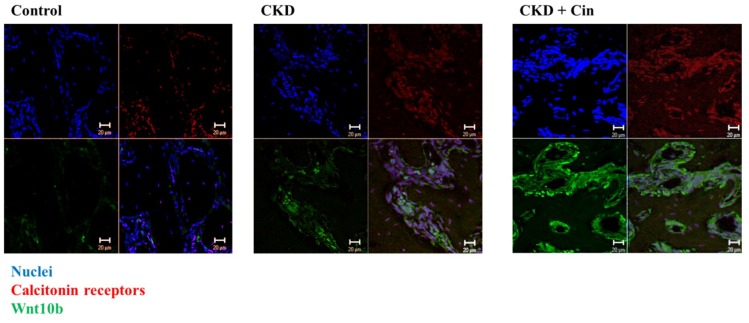
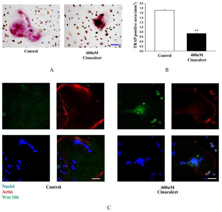
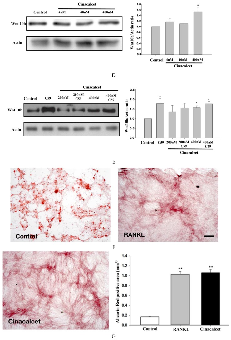
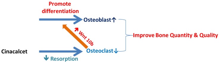
Secondary hyperparathyroidism (SHPT) relates to high turnover bone loss and is responsible for most bone fractures among chronic kidney disease (CKD) patients. Changes in the Wingless/beta-catenin signaling (Wnt/β-catenin) pathway and Wnt inhibitors have been found to play a critical role in CKD related bone loss. A calcimimetic agent, cinacalcet, is widely used for SHPT and found to be similarly effective for parathyroidectomy clinically. A significant decrease in hip fracture rates is noted among US hemodialysis Medicare patients since 2004, which is probably related to the cinacalcet era. In our previous clinical study, it was proven that cinacalcet improved the bone mineral density (BMD) even among severe SHPT patients. In this study, the influence of cinacalcet use on bone mass among CKD mice was determined. Cinacalcet significantly reduced the cortical porosity in femoral bones of treated CKD mice. It also improved the whole-bone structural properties through increased stiffness and maximum load. Cinacalcet increased femoral bone wingless 10b (Wnt10b) expression in CKD mice. In vitro studies revealed that cinacalcet decreased osteoclast bone resorption and increased Wnt 10b release from osteoclasts. Cinacalcet increased bone mineralization when culturing the osteoblasts with cinacalcet treated osteoclast supernatant. In conclusion, cinacalcet increased bone quantity and quality in CKD mice, probably through increased bone mineralization related with osteoclast Wnt 10b secretion.
Keywords: Wnt 10b; chronic kidney disease; cinacalcet; osteoclast; renal osteodystrophy.
Conflict of interest statement
The authors declare no conflict of interest.
Figures
 Decreased;
  Increased.
Similar articles
-
Therapeutic Effect of Calcimimetics on Osteoclast-Osteoblast Crosslink in Chronic Kidney Disease and Mineral Bone Disease.Int J Mol Sci. 2020 Nov 18;21(22):8712. doi: 10.3390/ijms21228712. Int J Mol Sci. 2020. PMID: 33218086 Free PMC article.
-
Association of Anabolic Effect of Calcitriol with Osteoclast-Derived Wnt 10b Secretion.Nutrients. 2018 Aug 25;10(9):1164. doi: 10.3390/nu10091164. Nutrients. 2018. PMID: 30149605 Free PMC article.
-
Treatment Based on Cinacalcet Reduces Oxidative Stress in Hemodialysis Patients with Secondary Hyperparathyroidism.Nephron. 2018;139(4):286-292. doi: 10.1159/000489278. Epub 2018 Jun 7. Nephron. 2018. PMID: 29879701
-
Mineral and bone disorders in conventional hemodialysis: Challenges and solutions.Semin Dial. 2018 Nov;31(6):592-598. doi: 10.1111/sdi.12729. Epub 2018 Jun 13. Semin Dial. 2018. PMID: 29900589 Review.
-
Regulation of bone metabolism by Wnt signals.J Biochem. 2016 Apr;159(4):387-92. doi: 10.1093/jb/mvv124. Epub 2015 Dec 28. J Biochem. 2016. PMID: 26711238 Free PMC article. Review.
Cited by
-
Current perspectives on the multiple roles of osteoclasts: Mechanisms of osteoclast-osteoblast communication and potential clinical implications.Elife. 2024 Apr 9;13:e95083. doi: 10.7554/eLife.95083. Elife. 2024. PMID: 38591777 Free PMC article. Review.
-
Calcitonin Induces Bone Formation by Increasing Expression of Wnt10b in Osteoclasts in Ovariectomy-Induced Osteoporotic Rats.Front Endocrinol (Lausanne). 2020 Sep 8;11:613. doi: 10.3389/fendo.2020.00613. eCollection 2020. Front Endocrinol (Lausanne). 2020. PMID: 33013696 Free PMC article.
-
The role of WNT10B in physiology and disease: A 10-year update.Front Cell Dev Biol. 2023 Feb 6;11:1120365. doi: 10.3389/fcell.2023.1120365. eCollection 2023. Front Cell Dev Biol. 2023. PMID: 36814601 Free PMC article. Review.
-
p38 Signaling Mediates Naringin-Induced Osteogenic Differentiation of Porcine Metanephric Mesenchymal Cells.Chin J Integr Med. 2024 Sep;30(9):818-825. doi: 10.1007/s11655-024-3761-1. Epub 2024 Jun 8. Chin J Integr Med. 2024. PMID: 38850479
-
Mouse Models of Mineral Bone Disorders Associated with Chronic Kidney Disease.Int J Mol Sci. 2023 Mar 10;24(6):5325. doi: 10.3390/ijms24065325. Int J Mol Sci. 2023. PMID: 36982400 Free PMC article. Review.
References
-
- Di Benedetto A., Marcelli D., D’Andrea A., Cice G., D’Isa S., Cappabianca F., Pacchiano G., D’Amato R., Oggero A.R., Bonanno D., et al. Risk factors and underlying cardiovascular diseases in incident esrd patients. J. Nephrol. 2005;18:592–598. - PubMed
-
- Yajima A., Tsuchiya K., Yokota H., Nitta K. [bone disease in the field of ckd-mbd] Clin. Calcium. 2016;26:875–880. - PubMed
MeSH terms
Substances
LinkOut - more resources
Full Text Sources
Medical
