Peficitinib Inhibits the Chemotactic Activity of Monocytes via Proinflammatory Cytokine Production in Rheumatoid Arthritis Fibroblast-Like Synoviocytes
- PMID: 31181818
- PMCID: PMC6627593
- DOI: 10.3390/cells8060561
Peficitinib Inhibits the Chemotactic Activity of Monocytes via Proinflammatory Cytokine Production in Rheumatoid Arthritis Fibroblast-Like Synoviocytes
Abstract
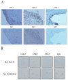
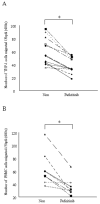
Background: This study was performed to examine the effects of the Janus kinase (JAK) inhibitor peficitinib on fibroblast-like synoviocytes (FLS) obtained from patients with rheumatoid arthritis (RA). Methods: To examine the expression of JAK1, JAK2, and JAK3 in RA synovial tissue (ST) and FLS, immunohistochemistry was performed. We investigated the effects of peficitinib on interleukin 6 and IL-6 receptor responses in RA FLS. Phosphorylation of STAT was determined by western blot. To examine the functional analysis of peficitinib, we performed a proliferation and chemotaxis assays with FLS using THP-1 and peripheral blood mononuclear cells (PBMC). The inflammatory mediator expression of FLS was estimated by enzyme-linked immunosorbent assay. Results: JAK1, JAK2, and JAK3 were expressed in RA STs and FLS. Phosphorylation of STAT1, STAT3, and STAT5 in RA FLS was suppressed by peficitinib in a concentration-dependent manner. Peficitinib-treated RA FLS-conditioned medium reduced THP-1 and PBMC migration (p < 0.05) and proliferation of RA FLS (p < 0.05). Peficitinib suppressed the secretion of MCP-1/CCL2 in the RA FLS supernatant (p < 0.05). Conclusion: Peficitinib suppressed the JAK-STAT pathway in RA FLS and also suppressed monocyte chemotaxis and proliferation of FLS through inhibition of inflammatory cytokines.
Keywords: fibroblast-like synoviocytes; monocyte chemotaxis; peficitinib; rheumatoid arthritis.
Conflict of interest statement
We declare that we have no potential conflict of interest to disclose.
Figures


Similar articles
-
Role of JAK-STAT signaling in the pathogenic behavior of fibroblast-like synoviocytes in rheumatoid arthritis: Effect of the novel JAK inhibitor peficitinib.Eur J Pharmacol. 2020 Sep 5;882:173238. doi: 10.1016/j.ejphar.2020.173238. Epub 2020 Jun 16. Eur J Pharmacol. 2020. PMID: 32561292
-
JAK inhibitors inhibit angiogenesis by reducing VEGF production from rheumatoid arthritis-derived fibroblast-like synoviocytes.Clin Rheumatol. 2024 Nov;43(11):3525-3536. doi: 10.1007/s10067-024-07142-9. Epub 2024 Sep 20. Clin Rheumatol. 2024. PMID: 39302595
-
Targeting Activated Synovial Fibroblasts in Rheumatoid Arthritis by Peficitinib.Front Immunol. 2019 Mar 26;10:541. doi: 10.3389/fimmu.2019.00541. eCollection 2019. Front Immunol. 2019. PMID: 30984167 Free PMC article.
-
JAK3-selective inhibitor peficitinib for the treatment of rheumatoid arthritis.Expert Rev Clin Pharmacol. 2019 Jun;12(6):547-554. doi: 10.1080/17512433.2019.1615443. Epub 2019 May 13. Expert Rev Clin Pharmacol. 2019. PMID: 31059310 Review.
-
Peficitinib: First Global Approval.Drugs. 2019 Jun;79(8):887-891. doi: 10.1007/s40265-019-01131-y. Drugs. 2019. PMID: 31093950 Review.
Cited by
-
Biologic Drugs for Rheumatoid Arthritis in the Context of Biosimilars, Genetics, Epigenetics and COVID-19 Treatment.Cells. 2021 Feb 4;10(2):323. doi: 10.3390/cells10020323. Cells. 2021. PMID: 33557301 Free PMC article. Review.
-
JAK Inhibitors in Rheumatoid Arthritis: Immunomodulatory Properties and Clinical Efficacy.Int J Mol Sci. 2024 Jul 30;25(15):8327. doi: 10.3390/ijms25158327. Int J Mol Sci. 2024. PMID: 39125897 Free PMC article. Review.
-
Chrysoeriol suppresses hyperproliferation of rheumatoid arthritis fibroblast-like synoviocytes and inhibits JAK2/STAT3 signaling.BMC Complement Med Ther. 2022 Mar 16;22(1):73. doi: 10.1186/s12906-022-03553-w. BMC Complement Med Ther. 2022. PMID: 35296317 Free PMC article.
-
Baricitinib therapy response in rheumatoid arthritis patients associates to STAT1 phosphorylation in monocytes.Front Immunol. 2022 Jul 25;13:932240. doi: 10.3389/fimmu.2022.932240. eCollection 2022. Front Immunol. 2022. PMID: 35958600 Free PMC article.
-
Role and mechanism of fibroblast-activated protein-α expression on the surface of fibroblast-like synoviocytes in rheumatoid arthritis.Front Immunol. 2023 Mar 17;14:1135384. doi: 10.3389/fimmu.2023.1135384. eCollection 2023. Front Immunol. 2023. PMID: 37006278 Free PMC article. Review.
References
MeSH terms
Substances
LinkOut - more resources
Full Text Sources
Medical
Research Materials
Miscellaneous

