. 2019 Jun 10;17(1):60.
doi: 10.1186/s12964-019-0376-9.

Elevated Kallikrein-binding protein in diabetes impairs wound healing through inducing macrophage M1 polarization

Affiliations

Elevated Kallikrein-binding protein in diabetes impairs wound healing through inducing macrophage M1 polarization

Juan Feng et al. Cell Commun Signal. .

Abstract

Background: The accumulation of M1-polarized macrophages and excessive inflammation are important in the pathogenesis of diabetic foot ulcer (DFU). However, the underlying mechanism of DFU pathogenesis and the crucial regulators of DFU are less well known. Our previous study reported that kallikrein-binding protein (KBP), an angiogenesis inhibitor, was significantly upregulated in diabetic patients compared to its levels in controls. The effects of KBP on monocyte chemotaxis and macrophage M1 polarization were elucidated in this study.

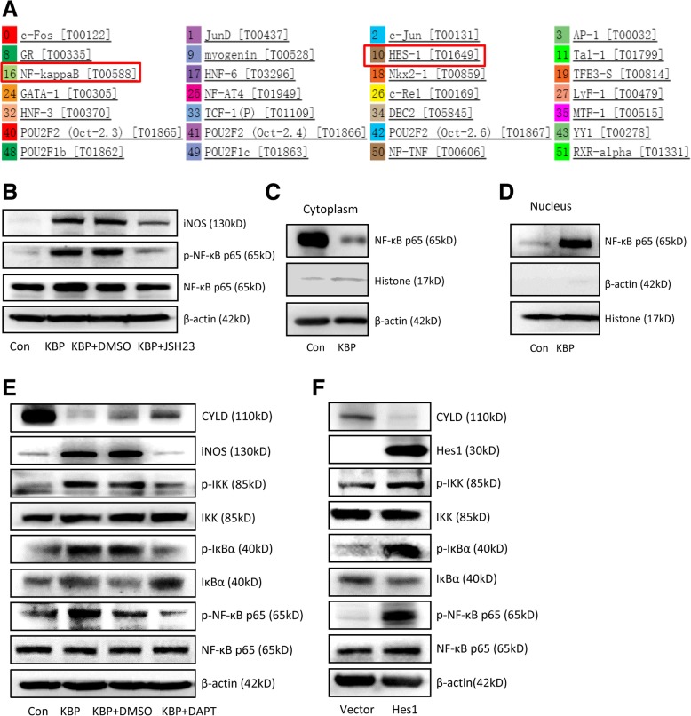
Methods: Plasma KBP levels and monocyte counts were assessed by ELISA and flow cytometry. Wound closure rates in different groups were monitored daily. The phenotype and recruitment of macrophages were measured by real-time PCR, western blot and immunofluorescence assays. The expression of Notch and NF-κB signalling pathway members was determined by the methods mentioned above. ChIP and dual-luciferase reporter gene assays were employed to explore the binding and transcriptional regulation of Hes1 and iNOS.

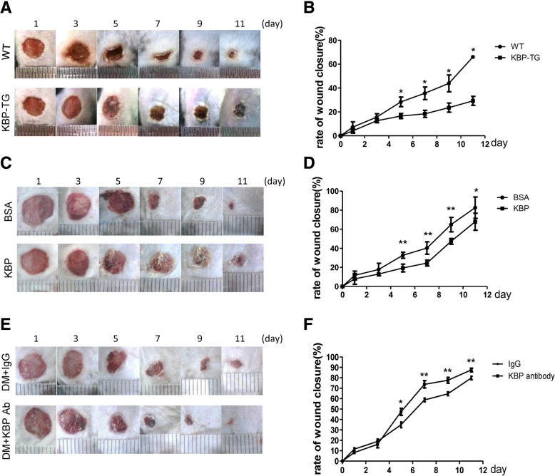
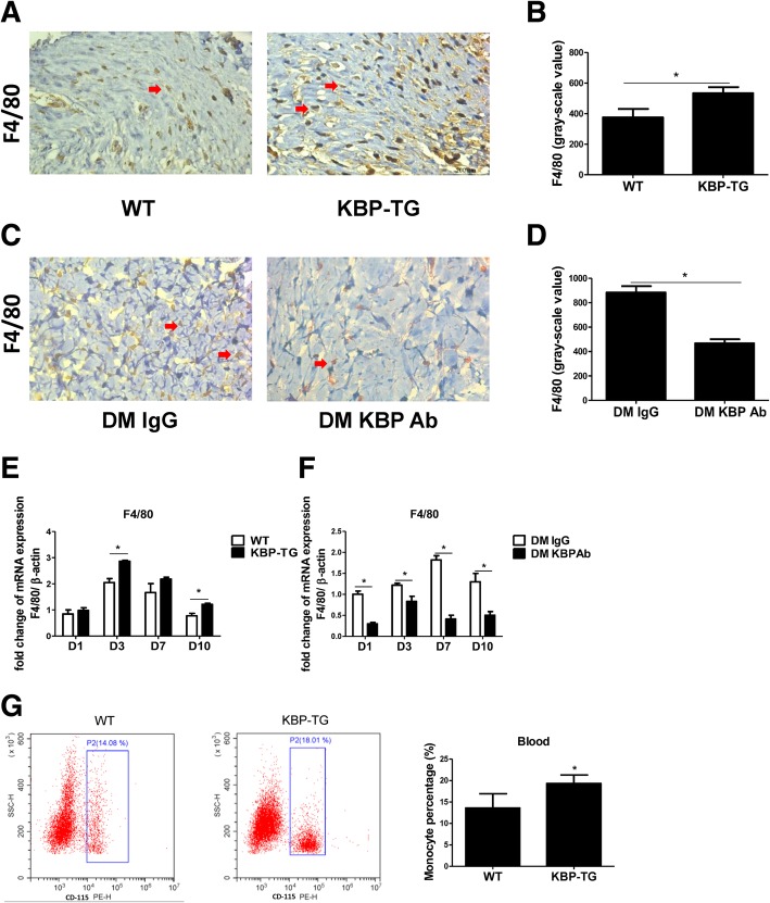
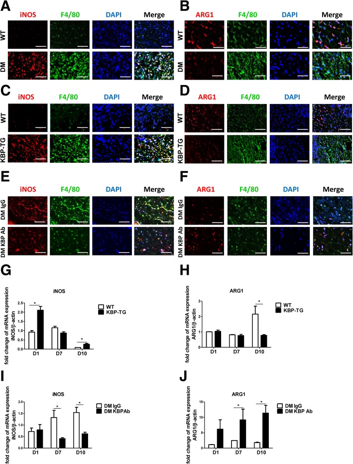
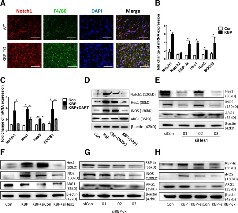
Results: We found that plasma KBP levels and circulating monocytes were elevated in diabetic patients compared to those in nondiabetic controls, and both were higher in diabetic patients with DFU than in diabetic patients without DFU. KBP delayed wound healing in normal mice; correspondingly, KBP-neutralizing antibody ameliorated delayed wound healing in diabetic mice. Circulating monocytes and macrophage infiltration in the wound were upregulated in KBP-TG mice compared to those in control mice. KBP promoted the recruitment and M1 polarization of macrophages. Mechanistically, KBP upregulated iNOS by activating the Notch1/RBP-Jκ/Hes1 signalling pathway. Hes1 downregulated CYLD, a negative regulator of NF-κB signalling, and then activated the IKK/IκBα/NF-κB signalling pathway.

Conclusions: Our findings demonstrate that KBP is the key regulator of excessive inflammation in DFUs and provide a novel target for DFU therapy.

Keywords: Diabetic wound healing; Kallikrein-binding protein; Monocyte-macrophages; Notch/NF-κB signalling.


Conflict of interest statement

The authors declare that they have no competing interests.

Figures

Fig. 1
Fig. 1
Clinical data and the role of KBP in wound healing. a The plasma level of KBP in NDM, DM, DM w/o DFU and DM w/ DFU patients. b Monocyte counts in the blood of NDM, DM, DM w/o DFU and DM w/ DFU patients. The collected data refer to the clinical data detected by a blood cell analyser. c The correlation of KBP and monocyte counts in the patients in all groups. NDM, n = 61; DM, n = 69; DM w/o DFU, n = 44; DM w/ DFU, n = 25
Fig. 2
Fig. 2
The role of KBP in wound healing. a, b Representative images showing wound healing and the wound closure rates in KBP-TG and WT mice. c, d Representative images showing wound healing and the wound closure rates in KBP-treated and BSA-treated mice. e, f Representative images showing wound healing and the wound closure rates in KBP antibody-treated type 2 diabetic mice and IgG-treated type 2 diabetic mice. Data are presented as the mean ± SD. n = 5; * p < 0.05, **p < 0.01
Fig. 3
Fig. 3
KBP increases monocyte counts in the blood and macrophage accumulation in wounds. a, b Representative immunohistochemical results and greyscale analysis of F4/80 (a macrophage marker) in the wounds of WT/KBP-TG mice at D10. c, d Representative immunohistochemical results and greyscale analysis of F4/80 in the wounds of diabetic mice treated with IgG/KBP antibody at D10. e The mRNA expression of F4/80 in the wounds of WT/KBP-TG mice at different time points. f The mRNA expression of F4/80 in the wounds of diabetic mice treated with IgG/KBP antibody at different time points. g Representative FACS results and the quantification of CD115+ monocytes in the peripheral blood of WT/KBP-TG mice. Data are presented as the mean ± SD. n = 3; * p < 0.05
Fig. 4
Fig. 4
KBP promotes the M1 polarization of macrophages in vivo. a, b Representative images showing immunofluorescent staining of wound tissue sections of WT and DM mice at D10. c, d Representative images showing immunofluorescent staining of wound tissue sections of WT and KBP-TG mice at D10. e, f Representative images showing immunofluorescent staining of wound tissue sections of DM mice treated with IgG or KBP antibody at D10. (A-F) Scale bar = 50 μm. F4/80: macrophage marker, iNOS: M1 marker, ARG1: M2 marker. g, h The mRNA expression of iNOS and ARG1 in the wounds of WT and KBP-TG mice at different time points. i, j The expression of iNOS and ARG1 in the wounds of diabetic mice treated with IgG or KBP antibody. Data are presented as the mean ± SD. n = 3; * p < 0.05
Fig. 5
Fig. 5
KBP stimulates M1 polarization in vitro. The mRNA expression levels of different markers/cytokines of M1 or M2 macrophages were measured, and the M1/M2 ratio was calculated. a The M1/M2 ratio in THP-1 cells. b The M1/M2 ratios of different groups in RAW264.7 cells. c The M1/M2 ratios of different groups in BMDMs. d The levels of type-1 immune cytokines (TNFα and IL-6) in the supernatants of different groups of RAW264.7 cells. e-g Western blot to detect iNOS and ARG1 in THP-1 cells, RAW264.7 cells and BMDMs together with a greyscale histogram. Data are presented as the mean ± SD. n = 3; * p < 0.05
Fig. 6
Fig. 6
KBP promotes the M1 polarization of macrophages via activating the Notch signalling pathway. a The expression of Notch1 in the wounds of WT and KBP-TG mice. Scale bar = 50 μm. b The mRNA expression of Notch1, Notch2 and other transcription factors (RBP-Jκ) or target genes associated with Notch signalling in RAW264.7 cells. c DAPT inhibits the effect of KBP on the activation of Notch signalling in RAW264.7 cells. d Western blot to detect Notch1, Hes1, iNOS and ARG1 following the treatment of RAW264.7 cells with KBP and DAPT. e, g The expression of iNOS and ARG1 in RAW264.7 cells following treatment with siHes1 and siRBP-Jκ. (F, H) The expression of iNOS and ARG1 in RAW264.7 cells following treatment with KBP and siHes1 or siRBP-Jκ. Data are presented as the mean ± SD. n = 3; * p < 0.05. Three independent experiments were employed
Fig. 7
Fig. 7
KBP stimulates the M1 polarization of macrophages via cross-activation of the Notch and NF-κB signalling pathways. a Bioinformatics prediction via the PROMO website of the possible transcription factors that bind to the iNOS promoter region. b The expression of iNOS and molecules in the NF-κB signalling pathway in RAW264.7 cells treated with KBP with or without JSH23. c The expression of NF-κB p65 in the cytoplasm of RAW264.7 cells. d The expression of NF-κB p65 in the nuclei of RAW264.7 cells. e The expression of CYLD, iNOS and molecules in the NF-κB signalling pathway in RAW264.7 cells treated with KBP with or without DAPT. f The expression of CYLD, iNOS and molecules in the NF-κB signalling pathway in RAW264.7 cells transfected with Hes1 or control vector. Three independent experiments were employed
Fig. 8
Fig. 8
KBP increases the production of M-CSF and MCP-1, which employs the differentiation and migration of monocytes and macrophages. a The plasma level of M-CSF in WT and KBP-TG mice. b The level of M-CSF in the cell supernatant of RAW 264.7 cells. c The mRNA expression of M-CSF in RAW264.7 cells treated with KBP versus that in the Con group. d KBP stimulates the migration of macrophages. Representative images of the Transwell migration assay and a statistical histogram. e The mRNA expression of MCP-1 in the wounds of WT and KBP-TG mice at different time points. f The mRNA expression of MCP-1 in the wounds of DM mice treated with IgG and KBP antibody at different time points. g The plasma level of MCP-1 in WT and KBP-TG mice. h The level of MCP-1 in the cell supernatant of RAW264.7 cells treated with KBP versus that in the Con group. The supernatant of RAW264.7 cells treated with KBP versus that of the Con group. I. The mRNA expression of MCP-1 in RAW264.7 cells treated with KBP versus that in the Con group. Data are presented as the mean ± SD. n = 3; * p < 0.05

References

    1. Boulton AJ, Vileikyte L, Ragnarson-Tennvall G, Apelqvist J. The global burden of diabetic foot disease. Lancet. 2005;366:1719–1724. doi: 10.1016/S0140-6736(05)67698-2. - DOI - PubMed
    1. White R, McIntosh C. A review of the literature on topical therapies for diabetic foot ulcers. Part 2: advanced treatments. J Wound Care. 2009;18:335–341. doi: 10.12968/jowc.2009.18.8.43633. - DOI - PubMed
    1. Singer AJ, Clark RA. Cutaneous wound healing. N Engl J Med. 1999;341:738–746. doi: 10.1056/NEJM199909023411006. - DOI - PubMed
    1. Qi W, Yang C, Dai Z, Che D, Feng J, Mao Y, Cheng R, Wang Z, He X, Zhou T, et al. High levels of pigment epithelium-derived factor in diabetes impair wound healing through suppression of Wnt signaling. Diabetes. 2015;64:1407–1419. doi: 10.2337/db14-1111. - DOI - PubMed
    1. Bennett CL, Djulbegovic B, Norris LB, Armitage JO. Colony-stimulating factors for febrile neutropenia during cancer therapy. N Engl J Med. 2013;368:1131–1139. doi: 10.1056/NEJMct1210890. - DOI - PMC - PubMed

Publication types

MeSH terms