. 2019 Jun 10;38(1):245.
doi: 10.1186/s13046-019-1256-2.

AKR1C1 controls cisplatin-resistance in head and neck squamous cell carcinoma through cross-talk with the STAT1/3 signaling pathway

Affiliations

AKR1C1 controls cisplatin-resistance in head and neck squamous cell carcinoma through cross-talk with the STAT1/3 signaling pathway

Wei-Min Chang et al. J Exp Clin Cancer Res. .

Abstract

Background: Cisplatin is the first-line chemotherapy used against most upper aerodigestive tract carcinomas. In head and neck cancer, sensitivity to cisplatin remains the key issue in treatment response and outcome. Genetic heterogeneity and aberrant gene expression may be the intrinsic factors that cause primary cisplatin-resistance.

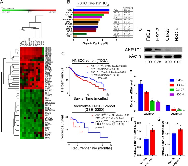
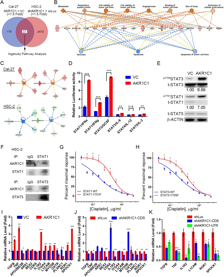
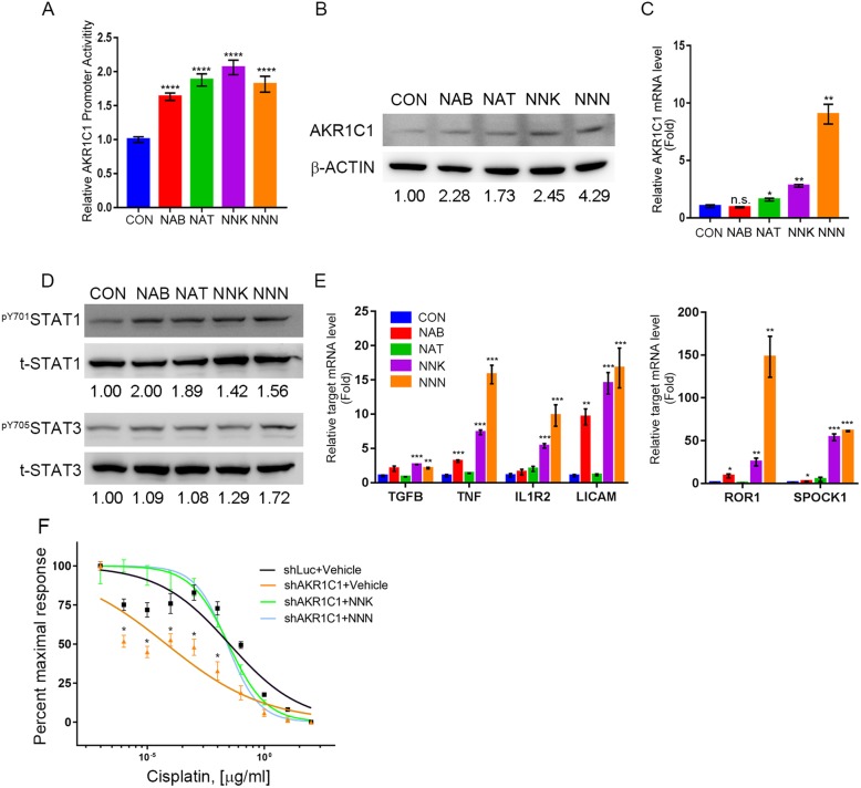
Methods: Combination of the HNSCC gene expression data and the cisplatin sensitivity results from public database. We found that aldo-keto reductase family 1 member C1 (AKR1C1) may be associated with cisplatin sensitivity in HNSCC treatment of naïve cells. We examined the AKR1C1 expression and its correlation with cisplatin IC50 and prognosis in patients. The in vitro and in vivo AKR1C1 functions in cisplatin-resistance through overexpression or knockdown assays, respectively. cDNA microarrays were used to identify the upstream regulators that modulate AKR1C1-induced signaling in HNSCC. Finally, we used the cigarette metabolites to promote AKR1C1 expression and ruxolitinib to overcome AKR1C1-induced cisplatin-resistance.

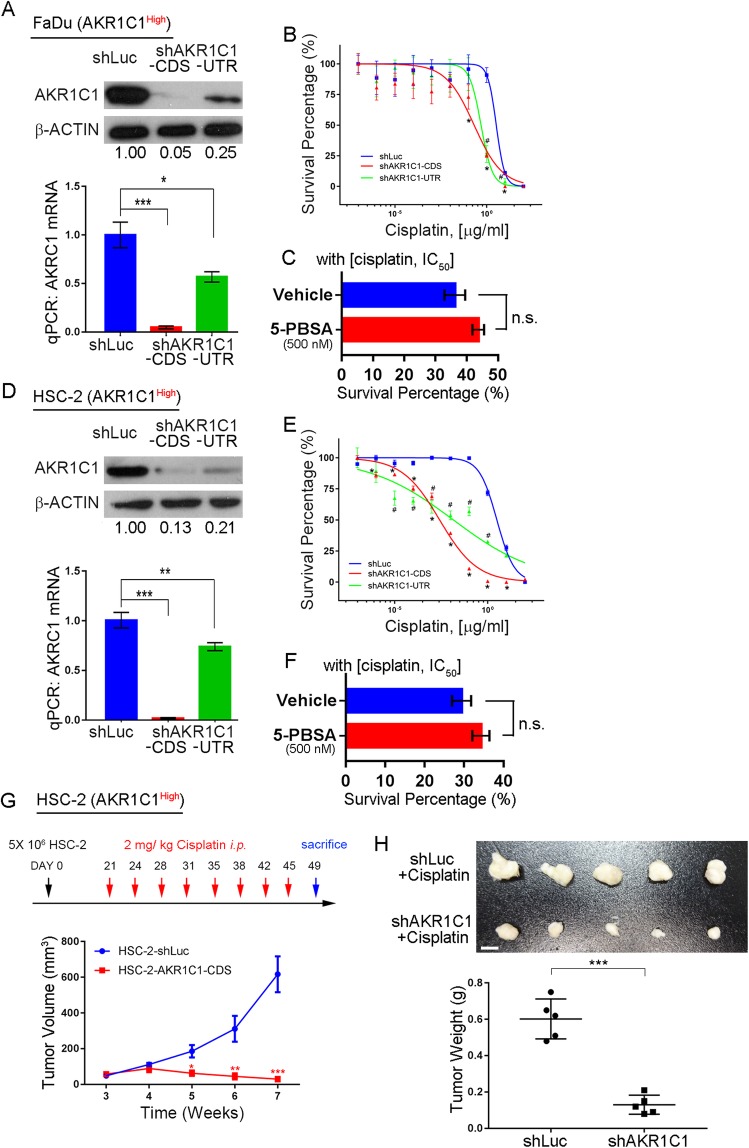
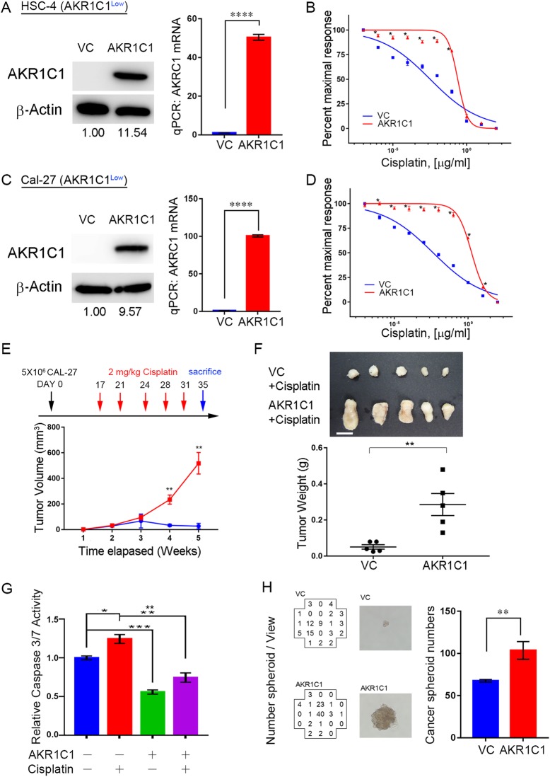
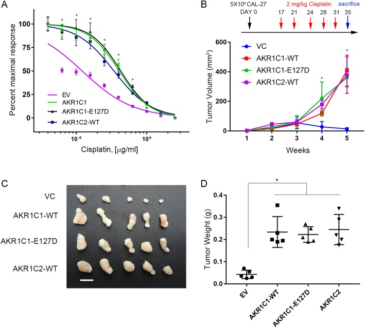
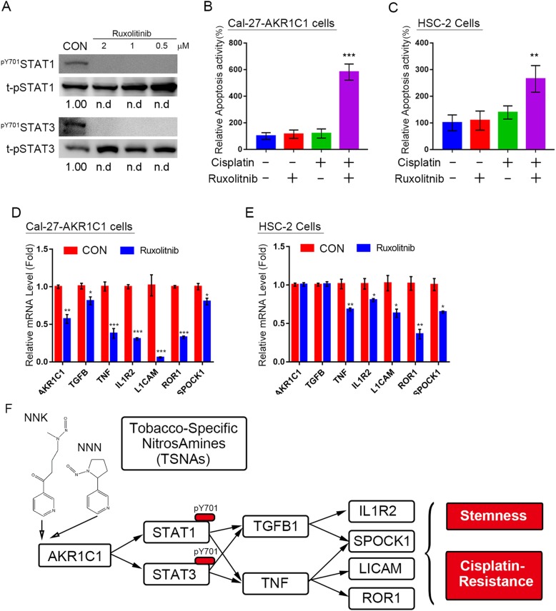
Results: AKR1C1 positively correlates to cisplatin-resistance in HNSCC cells. AKR1C1 is a poor prognostic factor for recurrence and death of HNSCC patients. Silencing of AKR1C1 not only reduced in vitro IC50 but also increased in vivo cisplatin responses and vise versa in overexpression cells. Cigarette metabolites also promote AKR1C1 expression. Transcriptome analyses revealed that STAT1 and STAT3 activation enable AKR1C1-induced cisplatin-resistance and can be overcome by ruxolitinib treatment.

Conclusions: AKR1C1 is a crucial regulator for cisplatin-resistance in HNSCC and also poor prognostic marker for patients. Targeting the AKR1C1-STAT axis may provide a new therapeutic strategy to treat patients who are refractory to cisplatin treatment.

Keywords: AKR1C1; Cisplatin-resistance; HNSCC; Ruxolitinib; STATs.


Conflict of interest statement

The authors declare that they have no competing interests.

Figures

Fig. 1
Fig. 1
AKR1C1 expression correlates to cisplatin response and functions as a poor prognostic marker in HNSCC. a Hierarchical clustering of differentially expressed genes between cisplatin IC50 from GDSC and gene expression profiles from CCLE. Note that AKR1C1 and C2 are highly correlated with cisplatin IC50. b HNSCC cisplatin IC50 value from the GDSC database. c AKR1C1 expression was correlated with a poor survival rate in HNSCC patients in the TCGA (Upper panel, n = 519, HR = 1.84, p = 0.035) and a short recurrence-free time from the GSE10300 HNSCC cohort (Bottom panel, n = 44, HR = 1.76, p = 0.041) from the SurvExpress database. d and e. Expressions of AKR1C1 were assessed by immunoblotting (d) and RT-Q-PCR (e) in the indicated cell lines. f and g. The AKR1C1 (f) andAKR1C2 (g) mRNA expression level in Cal-27 cells with or without cisplatin treatment. The statistical significance was analyzed by Student’s t-test. **p < 0.01, ***p < 0.001
Fig. 2
Fig. 2
Silencing of AKR1C1 can increase the cisplatin response activity in HNSCC cells through enzyme-independent functioning. a to f The in vitro cell viability assay after combining cisplatin and shAKR1C1 lentiviral particles or enzymatic AKR1C1 inhibitor, 5-PBSA, in highly AKR1C1 expressed cells. a and d AKR1C1 protein (upper) and mRNA (bottom) expression after knockdown of AKR1C1. b and e Dose-response curve after knockdown of AKR1C1. c and f The cell viability assay under cisplatin IC50 and with or without AKR1C1 inhibitor, 5-PBSA (500 nM). g and h The HSC-2 in vivo cisplatin response assay in which cells were infected with or without AKR1C1-CDS knockdown clones. g The cisplatin regimen (upper) and in vivo tumor burden (bottom, n = 5). The cisplatin was given 2 mg/kg through intraperitoneal injection (i.p.).h The tumor image and tumor weights from (g) and the scale bar indicates 0.5 cm length. The statistical significance was analyzed by Student’s t-test. **p < 0.01, ***p < 0.001
Fig. 3
Fig. 3
Ectopic AKR1C1 can promote cisplatin-resistance, anti-apoptosis response and cancer stemness in HNSCC cells. a to d The in vitro cell viability assay after combining cisplatin and ectopic AKR1C1 lentiviral particles. a and c AKR1C1 protein (left) and mRNA (right) expression after enforced expression of AKR1C1. b and d Dose-response curve after enforced expression of AKR1C1. e and f The Cal-27 in vivo cisplatin response assay in which cells were infected with or without AKR1C1 overexpression clones. e The cisplatin regimen (upper) and in vivo tumor burden (bottom, n = 5). The cisplatin was given 2 mg /kg through intraperitoneal injection (i.p.). f The tumor image and tumor weights from (e) and the scale bar indicates 1 cm length. g The cisplatin-induced caspase 3/7 activity assay with or without AKR1C1 expression. h The cancer spheroid formation assays with or without AKR1C1 expression in Cal-27 cells. The left panels indicate spheroid numbers which were calculated by ImageXpress XLS High-content system and the middle panels indicate the representative spheroid image in high magnification. The statistical significance was analyzed by Student’s t-test. **p < 0.01, ***p < 0.001
Fig. 4
Fig. 4
AKR1C1 promotes cisplatin-resistance enzymatic-independent manner. a Cisplatin dose-response curve in wild type AKR1C1, AKR1C2, and domain-negative AKR1C1-E127D Cal-27 cells. b to d. b The cisplatin regimen (upper) and in vivo tumor burden (bottom, n = 5). The cisplatin was given 2 mg /kg through intraperitoneal injection (i.p.). c and d. The tumor image and tumor weights (d) from (c) and the scale bar indicates 1 cm length
Fig. 5
Fig. 5
AKR1C1 controls anti-cell death pathways and inflammatory gene networks in HNSCC cells. a The flowchart of identifying the AKR1C1 downstream genes with 1.5-fold change cutoff compared to control vectors and their possible regulators in HNSCC cells. b The disease and biological function results from the IPA database c AKR1C1 regulates inflammation proteins, such as TNF-α and TGF-β networks, by IPA in HSC-2 (d) and Cal-27 (e) cells. D. STAT family reporter activity assays. The reporter activity was normalized by the expression level of pGL4-miniP reporters in the stable cell lines. e Phosphorylation level of pY705STAT3 and pY701STAT1 in AKR1C1-expressing Cal-27 cells. f Immunoprecipitation STAT1 and STAT3 in HSC-2 cells. g and h Cisplatin dose-response curve in Cal-27 expression with wild type STAT1 or constitutive activation form STAT1-Y701F (g) or STAT3 or STAT3-Y705F. I to k. The real-time PCR validation of microarray candidates in AKR1C1 overexpression (I, Cal-27) and knockdown (J, HSC-2; K, FaDu) cells. The gene expressions were normalized with endogenous GAPDH expression. The statistical significance was analyzed by Student’s t-test. *p < 0.05 **p < 0.01, ***p < 0.001
Fig. 6
Fig. 6
Cigarette metabolites control AKR1C1 expression. a to c AKR1C1 promoter activity assay (a), protein (b) and mRNA level (c) in Cal-27 cells exposed to tobacco-specific nitrosamines (TSNA), such as NAB, NAT, NNK and NNN and vehicle at concentration (10 μM) for 24 h. d The phosphorylation status of pY701STAT1 and Py705STAT3 in Cal-27 cells exposed to TSNA. e The AKR1C1 induced STAT1 and 3 downstream gene expression in Cal-27 exposed to TSNA. The gene expressions were normalized by endogenous GAPDH expression. f Cisplatin dose-response curve after knockdown of AKR1C1 and co-treat with NNK or NNN. The statistical significance was analyzed by Student’s t-test. *p < 0.05 **p < 0.01, ***p < 0.001
Fig. 7
Fig. 7
The JAK inhibitor ruxolitinib inhibits AKR1C1-induced cisplatin-resistance and JAK-STAT signaling pathway activation. a immunoblotting of phosphorylation pY701STAT1 and pY705STAT3 status under ruxolitinib-treated Cal-27 AKR1C1 cells (2–0.5 μM). b and c The caspase 3/7 activity assay in Cal-27-AKR1C1 (b) and HSC-2 (c) cells. The cisplatin concentration is 5 μM in Cal-27 cells and 10 μM in HSC-2 cells. The ruxolitinib is treated 0.5 μM in both cells. d and e The real-time PCR results of STAT3 downstream gene expression in Cal-27-AKR1C1 (d) and HSC-2 (e) cells. f The hypothetical model of the AKR1C1 contribution to cisplatin-resistance through STAT1 and 3 activation in HNSCC. The gene expressions were normalized with endogenous GAPDH expression. The statistical significance was analyzed by Student’s t-test. n.d: non-detected, *p < 0.05 **p < 0.01, ***p < 0.001

References

    1. Shen DW, Pouliot LM, Hall MD, Gottesman MM. Cisplatin resistance: a cellular self-defense mechanism resulting from multiple epigenetic and genetic changes. Pharmacol Rev. 2012;64:706–721. doi: 10.1124/pr.111.005637. - DOI - PMC - PubMed
    1. Michalke B. Platinum speciation used for elucidating activation or inhibition of Pt-containing anti-cancer drugs. J Trace Elem Med Biol. 2010;24:69–77. doi: 10.1016/j.jtemb.2010.01.006. - DOI - PubMed
    1. Dasari S, Tchounwou PB. Cisplatin in cancer therapy: molecular mechanisms of action. Eur J Pharmacol. 2014;740:364–378. doi: 10.1016/j.ejphar.2014.07.025. - DOI - PMC - PubMed
    1. Urba SG, Moon J, Giri PG, Adelstein DJ, Hanna E, Yoo GH, Leblanc M, Ensley JF, Schuller DE. Organ preservation for advanced resectable cancer of the base of tongue and hypopharynx: a southwest oncology group trial. J Clin Oncol. 2005;23:88–95. doi: 10.1200/JCO.2005.04.017. - DOI - PubMed
    1. Galluzzi L, Senovilla L, Vitale I, Michels J, Martins I, Kepp O, Castedo M, Kroemer G. Molecular mechanisms of cisplatin resistance. Oncogene. 2012;31:1869–1883. doi: 10.1038/onc.2011.384. - DOI - PubMed

MeSH terms