. 2019 Jul 15;203(2):557-568.
doi: 10.4049/jimmunol.1900192. Epub 2019 Jun 10.

Early Notch Signals Induce a Pathogenic Molecular Signature during Priming of Alloantigen-Specific Conventional CD4+ T Cells in Graft-versus-Host Disease

Affiliations

Early Notch Signals Induce a Pathogenic Molecular Signature during Priming of Alloantigen-Specific Conventional CD4+ T Cells in Graft-versus-Host Disease

Jooho Chung et al. J Immunol. .

Abstract

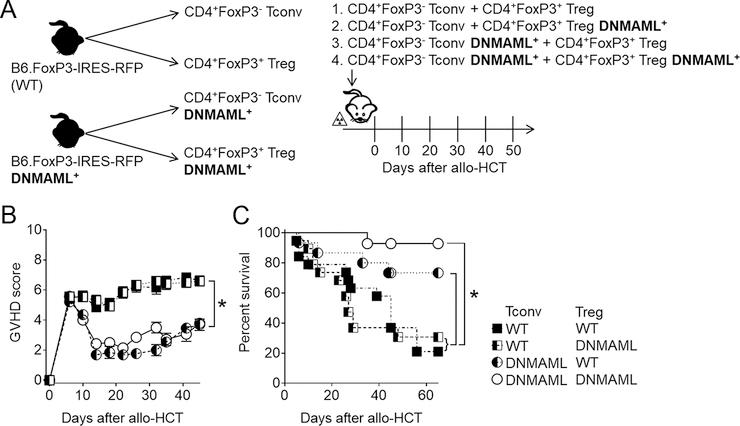
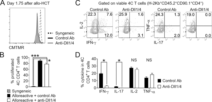
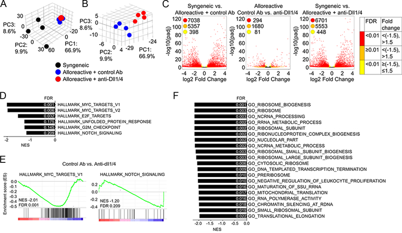
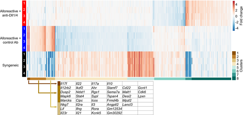
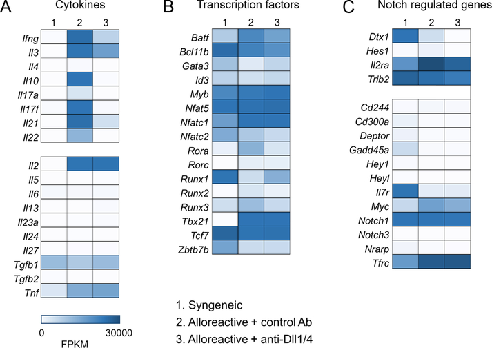
Graft-versus-host disease (GVHD) is the most serious complication of allogeneic hematopoietic cell transplantation. Notch signals delivered during the first 48 h after transplantation drive proinflammatory cytokine production in conventional T cells (Tconv) and inhibit the expansion of regulatory T cells (Tregs). Short-term Notch inhibition induces long-term GVHD protection. However, it remains unknown whether Notch blockade blunts GVHD through its effects on Tconv, Tregs, or both and what early Notch-regulated molecular events occur in alloantigen-specific T cells. To address these questions, we engineered T cell grafts to achieve selective Notch blockade in Tconv versus Tregs and evaluated their capacity to trigger GVHD in mice. Notch blockade in Tconv was essential for GVHD protection as GVHD severity was similar in the recipients of wild-type Tconv combined with Notch-deprived versus wild-type Tregs. To identify the impact of Notch signaling on the earliest steps of T cell activation in vivo, we established a new acute GVHD model mediated by clonal alloantigen-specific 4C CD4+ Tconv. Notch-deprived 4C T cells had preserved early steps of activation, IL-2 production, proliferation, and Th cell polarization. In contrast, Notch inhibition dampened IFN-γ and IL-17 production, diminished mTORC1 and ERK1/2 activation, and impaired transcription of a subset of Myc-regulated genes. The distinct Notch-regulated signature had minimal overlap with known Notch targets in T cell leukemia and developing T cells, highlighting the specific impact of Notch signaling in mature T cells. Our findings uncover a unique molecular program associated with the pathogenic effects of Notch in T cells at the earliest stages of GVHD.


Figures

Figure 1.
Figure 1.. Conventional T cell-intrinsic Notch signaling is essential for GVHD induction after MHC-mismatched allogeneic bone marrow transplantation.
(A) Experimental design to assess the importance of conventional T cells (Tconv) and regulatory T cells (Tregs) in mediating the protective effects of Notch inhibition after allogeneic bone marrow transplantation. Tconv and Tregs were sort purified from CD4+-enriched cells from B6 FoxP3-IRES-mRFP or B6 FoxP3-IRES-mRFP;Tgcd4-cre;ROSADNMAML/+ (FIR-DNMAML) mice, and mixed at a ratio of 8:1 Tconv:Tregs (by analogy to the physiological ratio found in WT donors; 500,000:62,500) to generate four different donor cell groups. (B) Clinical GVHD score and (C) overall survival of lethally irradiated (8 Gy) BALB/c mice transplanted with the four experimental inocula described in (A). *p < 0.05 (Student’s t-test, A; Log-rank test, B). Data are representative of 2 experiments and at least 15 mice per group.
Figure 2.
Figure 2.. Alloantigen-driven 4C T cells induce lethal GVHD after MHC-mismatched allogeneic bone marrow transplantation.
(A) Survival of lethally irradiated BALB/c mice transplanted with 5×10⁶ TCD BM only, or TCD BM supplemented with 5×10⁶ polyclonal B6 splenocytes, 105 4C T cells, 2×10⁴ 4C T cells, or 2×10³ 4C T cells. (B-E) Cellular hallmarks of the alloantigen-specific response in the 4C→BALB/c model of allo-HCT. Lethally irradiated recipients (B6.SJL – syngeneic; BALB/c-CD45.1 – allogeneic) received TCD BM and 5×10⁶ 4C T cells. 1.5 days after allo-HCT, secondary lymphoid organs were processed and lymphocytes isolated for analysis. (B-C) Forward scatter (FSC-A) and side scatter (SSC-A) showed dramatic increase in T cell size and granularity, consistent with acute activation. (D-E) The alloantigen-specific response led to robust 4C T cell activation. Histogram plots and cumulative data showed increased expression of the activation markers CD25, CD44, and CD69, and decrease in Vβ13 TCR chain surface density. *p < 0.05 (Log-rank test, A; Student’s t-test, C and E). Data are representative of at least 2 experiments and at least 10 mice per group.
Figure 3.
Figure 3.. Notch blockade mitigates acute GVHD driven by 4C conventional CD4+ T cells.
Lethally irradiated BALB/c mice were transplanted with 5×10⁶ TCD BM only, or TCD BM supplemented with 2×10³ 4C T cells. 4C T cell recipients were treated with isotype control (blue circles) or anti-Dll1 plus anti-Dll4 (red circles) antibodies (5 mg/kg i.p. on day 0). (A) Percentage of surviving transplanted animals plotted as function of time. *p < 0.05, Log-rank test. (B-E) Lethally irradiated BALB/c mice transplanted with 4C T cells and treated with isotype control or anti-Dll¼ antibodies were sacrificed 5 days after allo-HCT and spleens retrieved for immunophenotypic analysis. (B-C) Notch blockade did not impair 4C Tconv proliferation, as assessed through dilution of CFSE. (D-E) Anti-Dll¼ treatment led to impaired cytokine production in 4C Tconv. Representative flow cytometry plots (D) and cumulative data (E) showing the percentage of donor-derived 4C T cells expressing individual intracellular cytokines after anti-CD3/CD28 restimulation. *p < 0.05, Student’s t-test.
Figure 4.
Figure 4.. Impact of systemic Notch blockade on key cellular events in T cells during their in vivo priming by alloantigens.
(A-F) Lethally irradiated BALB/c mice were transplanted with TCD BM supplemented with 5×10⁶ 4C T cells and treated with isotype control or anti-Dll¼ antibodies on day 0. Animals were sacrificed 24 hours after allo-HCT, and secondary lymphoid organs retrieved for analysis. (A-B) Representative histogram plots (A) and cumulative data of mean fluorescence intensity (B) demonstrate that Notch blockade did not impact overall TCR signaling strength, as seen with indistinguishable Nur77 levels in 4C T cells isolated from mice treated with isotype control or anti-Dll¼ mAb. (C-D) Notch blockade impaired mTORC1 and Ras/MAPK signaling in alloreactive 4C T cells. Histogram plots (C) and cumulative data of mean fluorescence intensity (D) showing the abundance of phosphorylated S6 (S235/S236 and S240/S244 residues) and ERK½ in alloreactive 4C T cells. *p < 0.05, Student’s t-test. Data are representative of at least 3 experiments. (E-F) Notch blockade had limited impact on surface markers of alloreactive 4C T cell activation during in vivo antigen-mediated priming. Histogram plots (E) and cumulative data of mean fluorescence intensity (F) showing flow cytometric analysis of early T cell activation markers CD25 (a direct Notch target), CD69, and CD44. *p < 0.05, Student’s t-test. Data are representative of at least 3 experiments and 10 mice per group.
Figure 5.
Figure 5.. Systemic Notch blockade preserves selected early effector 4C T cell functions after allo-HCT.
B6-SJL (syngeneic) or BALB/c (allogeneic) mice were irradiated and transplanted with TCD BM supplemented with 5×10⁶ 4C T cells. BALB/c recipients were treated with isotype control or anti-Dll¼ mAb on day 0. Mice were sacrificed 42 hours after allo-HCT, and secondary lymphoid organs retrieved for analysis. (A-B) CMTMR dilution by donor 4C cells showed robust initial alloreactive T cell proliferation minimally affected by Notch blockade. (A) Representative histogram plots and (B) cumulative data with 4 animals analyzed from one of three experiments and a total of 10 animals per group. (C-D) Intracellular cytokine staining in donor 4C T cells showed no impact of Notch blockade on IL-2 and TNF-α production, but markedly decreased IFN-γ and IL-17. Representative plots (C) and cumulative data (D) from one of two experiments. * p < 0.05 (Student’s t-test, B; ANOVA, D).
Figure 6.
Figure 6.. Notch signaling has a unique but limited transcriptional footprint during priming of alloreactive T cells.
(A) 5×10⁶ CMTMR labeled 4C T cells were transplanted into lethally irradiated BALB/c (allogeneic) or B6-SJL (syngeneic) recipient. Allogeneic recipients received anti-Dll¼ or isotype control antibody on day 0. 42 hours after transplantation 4C T cells were sort-purified for RNA isolation, library generation, and RNA-Seq. (A-B) Principal component analysis of biological samples used in this study. The percentage of variance captured by each of the three principal components demonstrated alloantigen regulation of T cell response (A) and more limited changes of the alloreactive T cell transcriptome as a result of Notch blockade (B). (C) Volcano plots displaying differentially expressed genes in 4C T cells within the three different comparison groups (syngeneic vs. alloreactive + control antibody (Ab); syngeneic vs. alloreactive + anti-Dll¼ Ab; alloreactive control vs. anti-Dll¼ Ab). Red color-coded dots represent genes with significant differential expression at pre-defined 1.5-fold change. (D-F) GSEA analysis of RNA-Seq data, showing individual gene sets and select enrichment plots from Hallmark (D-E) and gene sets from Gene Ontology (F) collections significantly regulated by Notch blockade in 4C T cells.
Figure 7.
Figure 7.. k-means clustering analysis identifies Notch regulation of multiple GVHD-associated genes in alloreactive 4C T cells.
k-means clustering of 294 genes differentially regulated by Notch blockade in alloreactive 4C T cells during T cell priming period. Individual members of clusters 1–3 are highlighted and reveal several well-defined genes with known mechanistic relevance in GVHD (Il17f, I121, Il2ra, Ahr, Icos). Primary data from these clusters and all other clusters is presented in Supplemental Table 2.
Figure 8.
Figure 8.. The transcriptional effects of Notch signaling in alloreactive T cells are distinct from those induced by oncogenic Notch.
Heatmaps showing differential expression of genes encoding cytokines (A), transcription factors important for T cell homeostasis (B), and genes regulated by Notch signaling in T-ALL (C). Notch regulated expression of many, but not all cytokines with a mechanistic role in GVHD pathogenesis (A). Notch did not impact 4C Th polarization and had no or limited impact on master transcription factors of T helper lineages (B). Notch signaling targets in conventional alloreactive T cells minimally overlapped with those observed in Notch-dependent T-ALL (C).

References

    1. Radtke F, Fasnacht N, and Macdonald HR. 2010. Notch signaling in the immune system. Immunity 32: 14–27. - PubMed
    1. Zhang Y, Sandy AR, Wang J, Radojcic V, Shan GT, Tran IT, Friedman A, Kato K, He S, Cui S, Hexner E, Frank DM, Emerson SG, Pear WS, and Maillard I. 2011. Notch signaling is a critical regulator of allogeneic CD4+ T-cell responses mediating graft-versus-host disease. Blood 117: 299–308. - PMC - PubMed
    1. Riella LV, Ueno T, Batal I, De Serres SA, Bassil R, Elyaman W, Yagita H, Medina-Pestana JO, Chandraker A, and Najafian N. 2011. Blockade of Notch ligand delta1 promotes allograft survival by inhibiting alloreactive Th1 cells and cytotoxic T cell generation. J Immunol 187: 4629–4638. - PMC - PubMed
    1. Sandy AR, Chung J, Toubai T, Shan GT, Tran IT, Friedman A, Blackwell TS, Reddy P, King PD, and Maillard I. 2013. T cell-specific notch inhibition blocks graft-versus-host disease by inducing a hyporesponsive program in alloreactive CD4+ and CD8+ T cells. J Immunol 190: 5818–5828. - PMC - PubMed
    1. Mochizuki K, Xie F, He S, Tong Q, Liu Y, Mochizuki I, Guo Y, Kato K, Yagita H, Mineishi S, and Zhang Y. 2013. Delta-like ligand 4 identifies a previously uncharacterized population of inflammatory dendritic cells that plays important roles in eliciting allogeneic T cell responses in mice. J Immunol 190: 3772–3782. - PMC - PubMed

Publication types

MeSH terms