. 2019 Jun 25;116(26):12642-12647.
doi: 10.1073/pnas.1812334116. Epub 2019 Jun 10.

1,4-Benzoquinone antimicrobial agents against Staphylococcus aureus and Mycobacterium tuberculosis derived from scorpion venom

Affiliations

1,4-Benzoquinone antimicrobial agents against Staphylococcus aureus and Mycobacterium tuberculosis derived from scorpion venom

Edson Norberto Carcamo-Noriega et al. Proc Natl Acad Sci U S A. .

Abstract

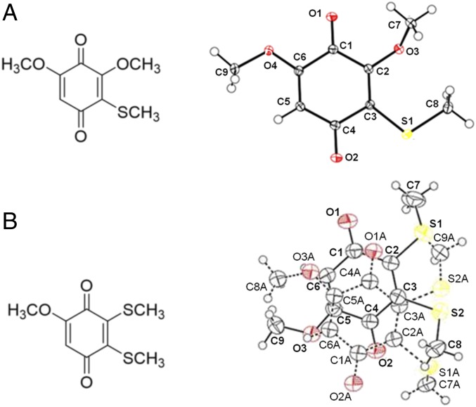
Two 1,4-benzoquinone derivatives, found in the venom of the scorpion Diplocentrus melici following exposure to air, have been isolated, characterized, synthesized, and assessed for antimicrobial activities. Initially a white, viscous liquid, the extracted venom colors within minutes under ambient conditions. From this colored mixture, two compounds, one red, the other blue, were isolated and purified using chromatography. After a variety of NMR and mass spectrometry experiments, the red compound was determined to be 3,5- dimethoxy-2-(methylthio)cyclohexa-2,5-diene-1,4-dione, and the blue compound was determined to be 5-methoxy-2,3- bis(methylthio)cyclohexa-2,5-diene-1,4-dione. Because extremely small amounts of these compounds were isolated from the scorpion venom, we developed laboratory syntheses from commercially available precursors, allowing us to produce sufficient quantities for crystallization and biological assays. The red benzoquinone is effective against Staphylococcus aureus [minimum inhibitory concentration (MIC) = 4 µg/mL], while the blue benzoquinone is active against Mycobacterium tuberculosis (MIC = 4 µg/mL) and even against a multidrug-resistant (MDR) strain with nearly equal effectiveness. The bactericidal effects of both benzoquinones show comparable activity to commercially available antibiotics used against these pathogens and were cytotoxic to neoplastic cell lines, suggesting their potential as lead compounds for the development of novel antimicrobial and anticancer drugs. Importantly, the blue benzoquinone was also effective in vivo with mouse models of MDR tuberculosis infection. After treatment for 2 mo, four mice with late-stage active MDR tuberculosis had a significant decrease in pulmonary bacillary loads and tissue damage. Healthy mice served as negative controls and tolerated treatment well, without adverse side effects.

Keywords: Staphylococcus aureus; antimicrobial activity; benzoquinones; mycobacterium tuberculosis; scorpion venom.


Conflict of interest statement

The authors declare no conflict of interest.

Figures

Fig. 1.
Fig. 1.
(Left) Structures of (A) red and (B) blue compounds extracted from the venom of D. melici. (Right) Corresponding X-ray crystallographic data (Cambridge Crystallographic Data Center No. 0001001197099) of the synthetic molecules (oxygen and sulfur atoms are presented in red and yellow, respectively) (see SI Appendix for details).
Fig. 2.
Fig. 2.
Inhibition of S. aureus. (A) Disk diffusion assay of red and blue benzoquinones showing activity against S. aureus. Ampicillin (5 µg) was used as a positive control. (B) Determination of the MICs of the red and blue benzoquinones against S. aureus. The MICs determined by the broth microdilution assay are 4 µg/mL for the red benzoquinone and 6 µg/mL for the blue benzoquinone. Ampicillin was used as a positive control. Each result is reported as the mean ± SD.
Fig. 3.
Fig. 3.
Inhibitory activity (in vitro) of blue and red benzoquinones against M. tuberculosis (H37Rv and MDR strain). (A) MICs were determined by broth microdilution, and bacterial proliferation was evaluated by a colorimetric assay using Cell Titer 96 Aqueous. For both strains, the MIC of the blue benzoquinone against M. tuberculosis is 4 µg/mL. The red benzoquinone had an MIC of 160 µg/mL, which is considered unsuitable for clinical use. (B) Viability of the bacteria was evaluated by counting the CFUs resulting after treatment at the MIC. Each result is reported as the mean ± SD. (CF) Ultrastructural changes in M. tuberculosis in response to the blue benzoquinone. To examine the cytotoxic effect of benzoquinone against M. tuberculosis, the ultrastructure of bacilli after treatment with the blue benzoquinone was examined. (C) Control untreated bacilli showed a well-defined, homogeneous and slightly electron-lucent cell wall, while the cytoplasm was generally electron-lucent with some lipid medium-sized vacuoles. (D) Incubation with benzoquinone produced substantial abnormalities, such as extensive effacement of cell wall (arrow) and cytoplasmic extraction (asterisk). (E and F) Conglomerates of electron dense reticular filaments located in the central areas of the cytoplasm. (G and H) Similar subcellular changes were induced by isoniazid incubation. (Scale bars: 100 nm.)
Fig. 4.
Fig. 4.
Inhibitory activity (in vivo) of the blue benzoquinone against M. tuberculosis. For this, we used an experimental model of progressive pulmonary tuberculosis consisting of BALB/c mice infected with the MDR CIBIN99 strain. Mice were treated for 2 mo with the blue benzoquinone using a dose of 8 µg administered by intratracheal route every other day. After the 2 mo, the group of mice treated with the blue benzoquinone had a marked improvement in their condition, with (A) a reduction of more than 90% of the lung bacillary load (evaluated by counting the CFUs) compared with the untreated group. (B) A clear reduction in the percentage of lung surface affected by pneumonia (LSAP) was observed in the lungs of the treated group. This difference is confirmed by automated histomorphometry, which shows a 50% reduction of pneumonia posttreatment. (C) Representative micrograph of a lung (hematoxylin-eosin staining) from the untreated group showing extensive areas of pneumonia (asterisk). (D) There is less pneumonia in lungs of the treated group. (E) Representative micrograph of a healthy mouse treated intratracheally for 1 mo with 8 µg of the blue benzoquinone. The pulmonary histology is normal, with the exception of occasional mild inflammatory infiltrates found around venules (arrow). (F) There is no fibrosis in the lungs of these healthy control mice (trichrome Masson staining). (Magnification: C and D, 25×; E and F, 200×.)

References

    1. Epidemiología DGd , “Manual de Procedimientos Estandarizados para la Vigilancia Epidemiológica de la Intoxicación por Picadura de Alacrán” (Secretaría de Salud, Salud SdPyPdl, 2012).
    1. Chippaux J. P., Emerging options for the management of scorpion stings. Drug Des. Devel. Ther. 6, 165–173 (2012). - PMC - PubMed
    1. Espino-Solis G. P., Riaño-Umbarila L., Becerril B., Possani L. D., Antidotes against venomous animals: State of the art and prospectives. J. Proteomics 72, 183–199 (2009). - PubMed
    1. Cahalan M. D., Modification of sodium channel gating in frog myelinated nerve fibres by Centruroides sculpturatus scorpion venom. J. Physiol. 244, 511–534 (1975). - PMC - PubMed
    1. Catterall W. A., Cooperative activation of action potential Na+ ionophore by neurotoxins. Proc. Natl. Acad. Sci. U.S.A. 72, 1782–1786 (1975). - PMC - PubMed

Publication types

MeSH terms