. 2019 Jun 25;116(26):12828-12832.
doi: 10.1073/pnas.1903562116. Epub 2019 Jun 10.

High-resolution cryo-EM structures of outbreak strain human norovirus shells reveal size variations

Affiliations

High-resolution cryo-EM structures of outbreak strain human norovirus shells reveal size variations

James Jung et al. Proc Natl Acad Sci U S A. .

Abstract

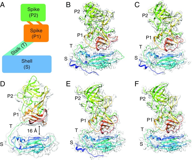
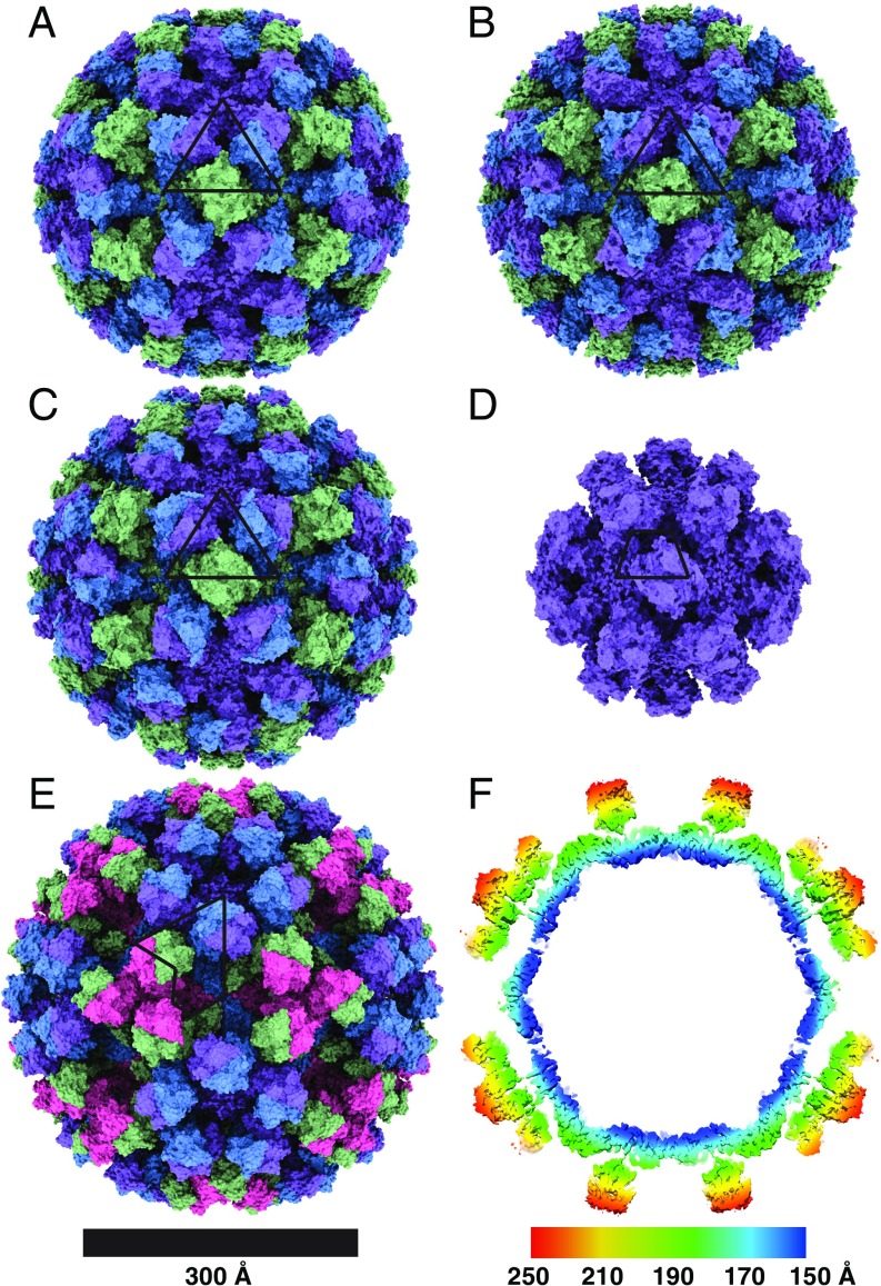
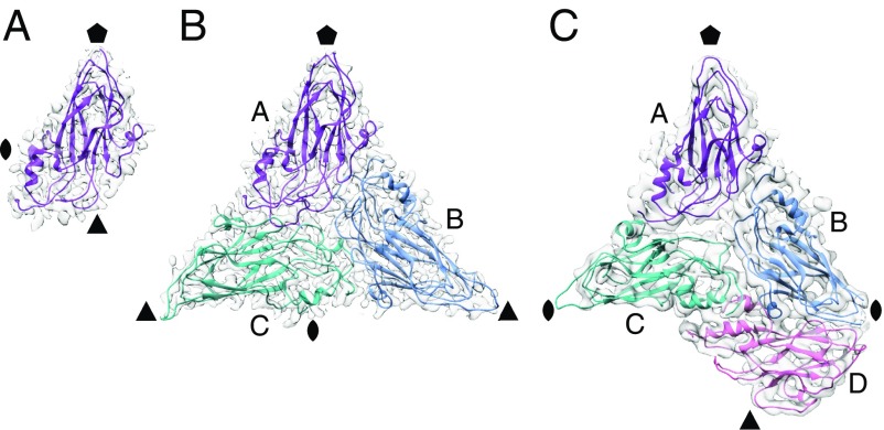
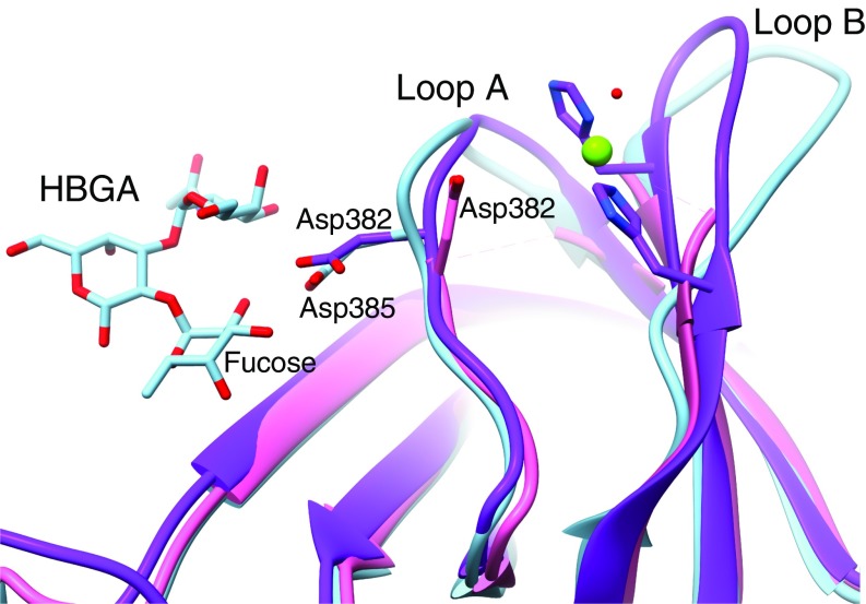
Noroviruses are a leading cause of foodborne illnesses worldwide. Although GII.4 strains have been responsible for most norovirus outbreaks, the assembled virus shell structures have been available in detail for only a single strain (GI.1). We present high-resolution (2.6- to 4.1-Å) cryoelectron microscopy (cryo-EM) structures of GII.4, GII.2, GI.7, and GI.1 human norovirus outbreak strain virus-like particles (VLPs). Although norovirus VLPs have been thought to exist in a single-sized assembly, our structures reveal polymorphism between and within genogroups, with small, medium, and large particle sizes observed. Using asymmetric reconstruction, we were able to resolve a Zn2+ metal ion adjacent to the coreceptor binding site, which affected the structural stability of the shell. Our structures serve as valuable templates for facilitating vaccine formulations.

Keywords: cryo-EM; foodborne illnesses; norovirus.


Conflict of interest statement

The authors declare no conflict of interest.

Figures

Fig. 1.
Fig. 1.
Modular organization of norovirus capsid proteins. Side views of human norovirus major capsid protein A subunits, showing the cryo-EM maps in gray and the fitted atomic models colored in rainbow representation from N to C termini. (A) A schematic diagram showing the modular organization of capsid subunits, consisting of trapezoid-shaped shell, long and flexible stalk, and protruding spike domains. The P domain consists of a P1 subdomain emerging from the S domain and P2, which is an insertion in P1 and positioned at the outermost surface of the virus. (B) GI.1 subunit, showing the P domain placed immediately above and forming contacts with the S domain. (C) GI.7 subunit, with the P domain placed close to the S domain. (D) GII.4 subunit, with the P domain lifted significantly (∼16 Å) above the shell domain through the long and flexible stalk region. (E) GII.2 T = 3 particle subunit, with the P domain making contacts with the S domain. (F) GII.2 T = 1 particle subunit, also with the P domain placed close to the S domain.
Fig. 2.
Fig. 2.
Cryo-EM structures of human norovirus outbreak strain capsids. Shaded depth-cue representations of norovirus VLP structures viewed along the icosahedral twofold axis, colored purple (subunit A), blue (subunit B), green (subunit C), and pink (subunit D). The positions of asymmetric units are identified by black lines. (A) GI.1 Norwalk strain at 2.9-Å resolution in T = 3 icosahedral symmetry with 90 dimeric P-domain spikes assembled from 180 subunits and 410 Å in diameter. (B) GI.7 Houston strain at 2.9-Å resolution in T = 3 symmetry and 420 Å in diameter. (C) GII.2 Snow Mountain virus strain at 3.1-Å resolution in T = 3 symmetry and 430 Å in diameter. The spikes of GII.2 SMV are twisted ∼15° counterclockwise relative to GI.1 and GI.7. (D) GII.2 SMV at 2.7-Å resolution in T = 1 symmetry with 30 spikes and 60 subunits and 310 Å in diameter. The spikes are placed significantly farther apart (∼10–25 Å) in T = 1 symmetry. (E) GII.4 Minerva strain at 4.1-Å resolution in T = 4 symmetry with 120 spikes and 240 subunits and 490 Å in diameter. The spikes of GII.4 are twisted ∼50° clockwise relative to the orientation of GI.1 and GI.7. (F) A central slice view of a GII.4 cryo-EM map colored by radial distance from the center (blue to red), showing a two-layered architecture with a secondary layer of spikes suspended ∼16 Å above the primary layer of the icosahedral shell domain.
Fig. 3.
Fig. 3.
Asymmetric units of GII.2 T = 1, GII.2 T = 3, and GII.4 T = 4 particles. The icosahedral symmetry axes positions are indicated by a pentagon (fivefold), triangle (threefold), and oval (twofold). (A) Shell domain of a single subunit in each ASU of GII.2 T = 1 particles. (B) Shell domains of three subunits in quasi-equivalent positions, A, B, and C, that constitute each ASU of GII.2 T = 3 particles. (C) GII.4 shell domains in the T = 4 particle ASU with four subunits, A, B, C, and D. The C–D dimer takes up a position similar to the C–C dimer in T = 3 particles.
Fig. 4.
Fig. 4.
Histo-blood group antigen binding site in GII.2. The P-domain crystal structure of GII.10 with HBGA bound (PDB ID code 3PA1) was superimposed on the partial P-domain crystal structure of GII.2 (PDB ID code 4RPB) and our GII.2 full-subunit cryo-EM structure without HBGA bound to show the expected positioning of the HBGA binding site of GII.2. In HBGA-binding strains such as GII.10 (light blue), a conserved residue (Asp385) binds the fucose moiety of HBGAs. In the P-domain crystal structure of GII.2 (pink), the side chain of the equivalent residue (Asp382) was turned ∼180° away from the fucose binding site, and the hypervariable loop A bearing Asp382 and loop B were not observed. In our cryo-EM structures (purple), the hypervariable loops A and B are resolved with the Asp382 side chain pointed correctly toward the fucose binding site. A Zn2+ metal ion (green sphere) is bound between loops A and B immediately adjacent to the fucose binding site that may play a role as a coligand that enhances HBGA binding of the GII.2 strain.

References

    1. Centers for Disease Control and Prevention (CDC) , Norovirus worldwide. https://www.cdc.gov/norovirus/trends-outbreaks/worldwide.html. Accessed 22 May 2019.
    1. Hall A. J., et al. ; Division of Viral Diseases, National Center for Immunization and Respiratory Diseases, Centers for Disease Control and Prevention , Updated norovirus outbreak management and disease prevention guidelines. MMWR Recomm. Rep. 60, 1–18 (2011). - PubMed
    1. Kirk M. D., et al. , World Health Organization estimates of the global and regional disease burden of 22 foodborne bacterial, protozoal, and viral diseases, 2010: A data synthesis. PLoS Med. 12, e1001921 (2015). - PMC - PubMed
    1. Teunis P. F. M., et al. , Norwalk virus: How infectious is it? J. Med. Virol. 80, 1468–1476 (2008). - PubMed
    1. de Graaf M., van Beek J., Koopmans M. P. G., Human norovirus transmission and evolution in a changing world. Nat. Rev. Microbiol. 14, 421–433 (2016). - PubMed

Publication types