. 2019 Jun 10;9(1):8420.
doi: 10.1038/s41598-019-44874-0.

Targeting CLDN18.2 by CD3 Bispecific and ADC Modalities for the Treatments of Gastric and Pancreatic Cancer

Affiliations

Targeting CLDN18.2 by CD3 Bispecific and ADC Modalities for the Treatments of Gastric and Pancreatic Cancer

Guoyun Zhu et al. Sci Rep. .

Erratum in

Abstract

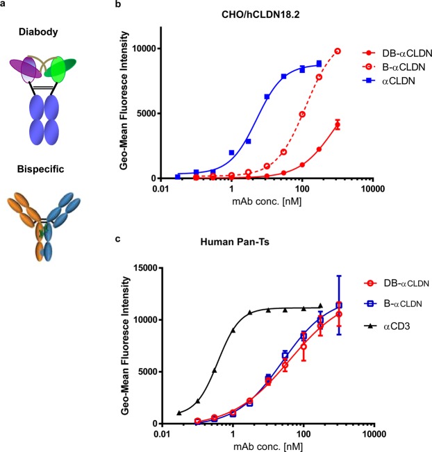
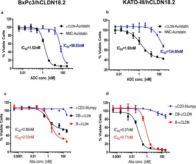
Human CLDN18.2 is highly expressed in a significant proportion of gastric and pancreatic adenocarcinomas, while normal tissue expression is limited to the epithelium of the stomach. The restricted expression makes it a potential drug target for the treatment of gastric and pancreatic adenocarcinoma, as evidenced by efforts to target CLDN18.2 via naked antibody and CAR-T modalities. Herein we describe CLDN18.2-targeting via a CD3-bispecific and an antibody drug conjugate and the characterization of these potential therapeutic molecules in efficacy and preliminary toxicity studies. Anti-hCLDN18.2 ADC, CD3-bispecific and diabody, targeting a protein sequence conserved in rat, mouse and monkey, exhibited in vitro cytotoxicity in BxPC3/hCLDN18.2 (IC50 = 1.52, 2.03, and 0.86 nM) and KATO-III/hCLDN18.2 (IC50 = 1.60, 0.71, and 0.07 nM) respectively and inhibited tumor growth of pancreatic and gastric patient-derived xenograft tumors. In a rat exploratory toxicity study, the ADC was tolerated up to 10 mg/kg. In a preliminary assessment of tolerability, the anti-CLDN18.2 diabody (0.34 mg/kg) did not produce obvious signs of toxicity in the stomach of NSG mice 4 weeks after dosing. Taken together, our data indicate that targeting CLDN18.2 with an ADC or bispecific modality could be a valid therapeutic approach for the treatment of gastric and pancreatic cancer.


Conflict of interest statement

All authors are current or former employees of Pfizer Inc.

Figures

Figure 1
Figure 1
Gastric and pancreatic adenocarcinoma display CLDN18.2 expression (IHC). Tumor tissue samples of primary or metastatic gastric and primary pancreatic adenocarcinoma from tumor microarrays (Tristar or US Biomax) or individual samples were immunostained with an anti-hCLDN18.2 antibody, anti-CLDN-mlgG1, to determine the expression of CLDN18.2. Different levels of CLDN18.2 expression were evaluated by an experienced pathologist using a 4-point scale. H-score: plasma membrane staining intensity + distribution. Sum of staining intensity (0 = negative, 1 = low, 2 = medium, 3 = high) × percentage of cells. All grading is subjective and done by eye by a pathologist. The percentage of CLDN18.2-positive staining with different scores was indicated in the table embedded. (a) Primary gastric adenocarcinoma; (b) primary pancreatic adenocarcinoma; c. primary and metastatic matched gastric adenocarcinoma.
Figure 2
Figure 2
CLDN18.2 is expressed in Lentivirus-transduced tumor cell lines and gastric and pancreatic cancer PDX. Primary BxPC3, KATO-III and hCLDN18.2 engineered BxPC3, KATO-III cells were incubated with 10 μg/mL of anti-hCLDN18.2 mAb. Blue and red peaks represent staining with anti-CLDN18.2 in CLDN18.2 engineered expressing cells and non-engineered expressing cells respectively. (a) Patient-derived tumor sections, Gastric Cancer (CTG-1010, b) and Pancreatic Cancer (Pan-1907, c) were immunostained with an anti-CLDN18.2 antibody. Cytokeratin was stained with mouse anti-human cytokeratin mAb. E-Cadherin was stained with mouse anti-human E-Cadherin.
Figure 3
Figure 3
Anti-hCLDN18.2 or anti-hCD3 antibodies binds CHO/hCLDN18.2 or human Pan-T cells dose-dependently. Schematic of the two anti-CLDN18.2 therapeutic molecule formats: a full length fully human IgG2ΔA D265A bispecific or diabody generated by combining CLDN18.2 and CD3 targeting arms though hinge mutations. (a) CHO engineered expressing hCLDN18.2 and human Pan-T cells were assayed for anti-CLDN18.2-CD3 Bispecific and diabody cell binding. The anti-CLDN18.2 arm of the bispecific and diabody binds in a dose-dependent manner to CHO/CLDN18.2 cell line (b) and the anti-CD3 arm to Pan-T cells (c).
Figure 4
Figure 4
The anti-CLDN18.2 ADC and anti-CLDN18.2 CD3 Bispecific inhibit growth of different hCLDN18.2-tumor cells. The anti-CLDN18.2 ADC generated through site-specific conjugation of cleavable linkers and auristatin drug payloads with a drug-to-antibody ratio (DAR) of 4 dose-dependently inhibited the BxPc3 and KATO-III/hCLDN18.2 cells growth. (a,b) The target cells were incubated with varying concentrations of anti-CLDN18.2 cleavable ADC for 4 days. The cell viability was determined by luminescence by using CellTiter-Glo reagent (Promega). n = 3 technical replicates. NNC-Auristatin: anti-BHV-Auristatin. Bispecific and diabody also effectively eliminates BxPC3 or KATO-III/hCLDN18.2-Luc cells in a dose dependent manner. (c,d) Luciferase-labeled target cells were incubated with varying concentrations of the antibody and fixed ratio of Pan-T vs tumor cells (5:1) for 2 days. After 2 days of incubation, viability of BxPC3 or KATO-III cells were assessed using One-Glo luciferase assay reagent (Promega). The IC50s were then determined by nonlinear regression plot of percent specific cytotoxicity versus Log10 concentration of CLDN18.2 bispecific using GraphPad Prism software, Version 7.04. The results were shown as mean ± standard deviation (SD). αCD3-stumpy: CD3 variable and constant region combined with a truncated antibody consisting of only the hinge/constant region.
Figure 5
Figure 5
The anti-CLDN18.2 ADC and anti-CLDN18.2 CD3 Bispecific Inhibit tumor growths of Pancreatic Pan-1907 and Gastric CTG-1010 PDX. Anti-CLDN18.2 ADC with cleavable linker and DAR4 demonstrated dose-dependent efficacy against an established gastric and pancreatic patient-derived xenograft models after administering a single IV dose of Anti-CLDN18.2 ADC or Negative control (NNC) ADC 50 days (Pancreatic Pan-1907) or 46 days (Gastric CTG1010) after tumor inoculation. (a,b) Anti-CLDN18.2 CD3 bispecific and diabody were also efficacious at all doses tested in an established gastric patient-derived xenograft model. (c) All data shown as mean ± SEM. Tumor growth was monitored by caliper twice-weekly. All studies were performed in immune compromised NSG mice with n = 4–5 mice per group. Statistics represent two-way ANOVA analysis, all groups compared to NNC (for ADC, a,b) or αCD3-Stumpy (for bispecific, c) ****p < 0.0001).
Figure 6
Figure 6
Anti-CLDN18.2 ADC serum concentration-time profiles. Anti-CLDN18.2 ADC serum concentration-time profiles (total mAb and ADC) in rats after intravenous administration of 10 mg/kg on days 1 and 15. Concentration of total mAb and ADC, determined using an ELISA, are shown as solid and dashed lines, respectively.

References

    1. Kumar V, Soni P, Garg M, Kamholz S, Chandra AB. Emerging Therapies in the Management of Advanced-Stage Gastric Cancer. Front Pharmacol. 2018;9:404. doi: 10.3389/fphar.2018.00404. - DOI - PMC - PubMed
    1. Sitarz R, et al. Gastric cancer: epidemiology, prevention, classification, and treatment. Cancer Manag Res. 2018;10:239–248. doi: 10.2147/CMAR.S149619. - DOI - PMC - PubMed
    1. Bray Freddie, Ferlay Jacques, Soerjomataram Isabelle, Siegel Rebecca L., Torre Lindsey A., Jemal Ahmedin. Global cancer statistics 2018: GLOBOCAN estimates of incidence and mortality worldwide for 36 cancers in 185 countries. CA: A Cancer Journal for Clinicians. 2018;68(6):394–424. - PubMed
    1. Pancreatic cancer statistics, https://www.wcrf.org/dietandcancer/cancer-trends/pancreatic-cancer-stati....
    1. Saad AM, Turk T, Al-Husseini MJ, Abdel-Rahman O. Trends in pancreatic adenocarcinoma incidence and mortality in the United States in the last four decades; a SEER-based study. BMC Cancer. 2018;18:688. doi: 10.1186/s12885-018-4610-4. - DOI - PMC - PubMed

Publication types