

Inflammatory and Senescent Phenotype of Pancreatic Stellate Cells Induced by Sqstm1 Downregulation Facilitates Pancreatic Cancer Progression
- PMID: 31182922
- PMCID: PMC6535784
- DOI: 10.7150/ijbs.27825
Inflammatory and Senescent Phenotype of Pancreatic Stellate Cells Induced by Sqstm1 Downregulation Facilitates Pancreatic Cancer Progression
Abstract
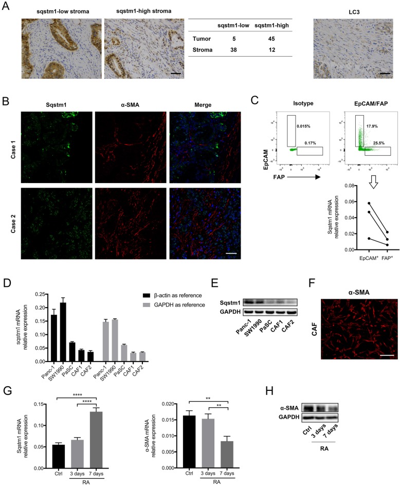
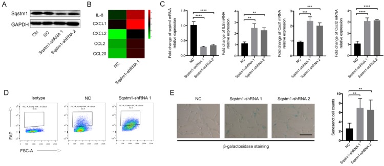
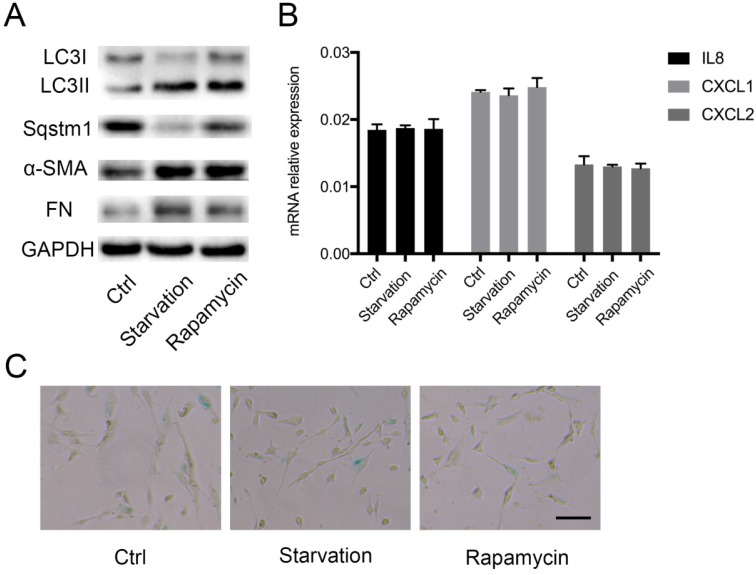
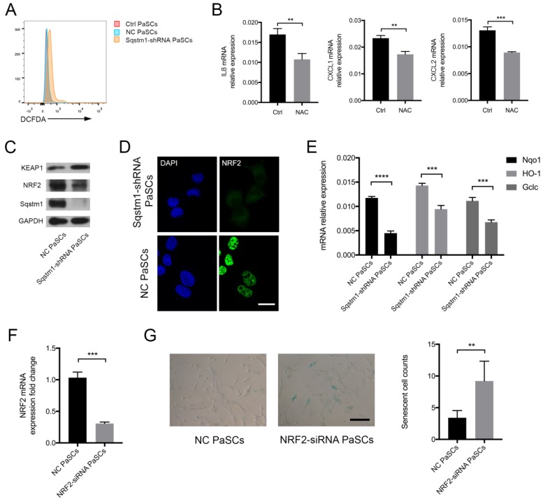
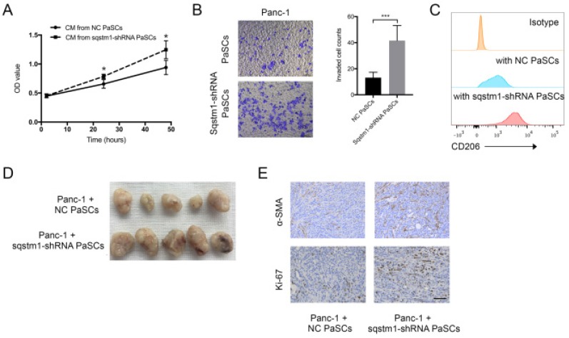
Pancreatic ductal adenocarcinoma (PDAC) has unique microenvironment with extensive infiltration of fibroblasts, which are mainly derived from the resident pancreatic stellate cells (PaSCs). As activated PaSCs constitute a major contributor to pancreatic cancer progression, the mechanisms underlying their activation have been being intensively studied. Previous studies showed that Sequestosome-1 (sqstm1) can modulate the functional status of fibroblasts in cancer. Here, we further delineated the role of sqstm1 in PaSCs. The analysis of PDAC patient samples revealed reduction of sqstm1 expression in activated PaSCs in both mRNA and protein level. Downregulated sqstm1 via shRNA in PaSCs led to an inflammatory and senescent phenotype with increased IL8, CXCL1, and CXCL2 expression. Further analysis demonstrated that increased intracellular reactive oxygen species level contributed to the senescence in sqstm1-downregulated PaSCs. This was mediated via impaired NRF2 activity since reduced sqstm1 resulted in accumulation of KEAP1. Meanwhile, we found that sqstm1 degradation caused by enhanced autophagy was not associated with transformation of senescent phenotype. At last, the data revealed that sqstm1-downregulated PaSCs promoted pancreatic tumor cell growth, invasion, and macrophage phenotype transformation. Collectively, the current study indicated that sqstm1 controlled transformation of senescent phenotype of PaSCs, which in turn is pro-tumorigenic.
Keywords: pancreatic adenocarcinoma; pancreatic stellate cells; reactive oxygen species; senescence; sqstm1.
Conflict of interest statement
Competing Interests: The authors have declared that no competing interest exists.
Figures
Similar articles
-
Regulation of pancreatic stellate cell activation by Notch3.BMC Cancer. 2018 Jan 5;18(1):36. doi: 10.1186/s12885-017-3957-2. BMC Cancer. 2018. PMID: 29304760 Free PMC article.
-
Galectin-1 secreted by activated stellate cells in pancreatic ductal adenocarcinoma stroma promotes proliferation and invasion of pancreatic cancer cells: an in vitro study on the microenvironment of pancreatic ductal adenocarcinoma.Pancreas. 2011 Aug;40(6):832-9. doi: 10.1097/MPA.0b013e318217945e. Pancreas. 2011. PMID: 21747316
-
Galectin-3 Mediates Tumor Cell-Stroma Interactions by Activating Pancreatic Stellate Cells to Produce Cytokines via Integrin Signaling.Gastroenterology. 2018 Apr;154(5):1524-1537.e6. doi: 10.1053/j.gastro.2017.12.014. Epub 2017 Dec 21. Gastroenterology. 2018. PMID: 29274868
-
Persistent activation of pancreatic stellate cells creates a microenvironment favorable for the malignant behavior of pancreatic ductal adenocarcinoma.Int J Cancer. 2013 Mar 1;132(5):993-1003. doi: 10.1002/ijc.27715. Epub 2012 Oct 5. Int J Cancer. 2013. PMID: 22777597 Review.
-
The critical roles of activated stellate cells-mediated paracrine signaling, metabolism and onco-immunology in pancreatic ductal adenocarcinoma.Mol Cancer. 2018 Feb 19;17(1):62. doi: 10.1186/s12943-018-0815-z. Mol Cancer. 2018. PMID: 29458370 Free PMC article. Review.
Cited by
-
Targeting senescence-like fibroblasts radiosensitizes non-small cell lung cancer and reduces radiation-induced pulmonary fibrosis.JCI Insight. 2021 Dec 8;6(23):e146334. doi: 10.1172/jci.insight.146334. JCI Insight. 2021. PMID: 34877934 Free PMC article.
-
The Role of Chemokines in Orchestrating the Immune Response to Pancreatic Ductal Adenocarcinoma.Cancers (Basel). 2024 Jan 28;16(3):559. doi: 10.3390/cancers16030559. Cancers (Basel). 2024. PMID: 38339310 Free PMC article. Review.
-
Senescence-Associated miRNAs and Their Role in Pancreatic Cancer.Pathol Oncol Res. 2022 Apr 29;28:1610156. doi: 10.3389/pore.2022.1610156. eCollection 2022. Pathol Oncol Res. 2022. PMID: 35570840 Free PMC article. Review.
-
Pancreatic Cancer and Cellular Senescence: Tumor Microenvironment under the Spotlight.Int J Mol Sci. 2021 Dec 27;23(1):254. doi: 10.3390/ijms23010254. Int J Mol Sci. 2021. PMID: 35008679 Free PMC article. Review.
-
Senescence program and its reprogramming in pancreatic premalignancy.Cell Death Dis. 2023 Aug 17;14(8):528. doi: 10.1038/s41419-023-06040-3. Cell Death Dis. 2023. PMID: 37591827 Free PMC article. Review.
References
-
- Rahib L, Smith BD, Aizenberg R, Rosenzweig AB, Fleshman JM, Matrisian LM. Projecting cancer incidence and deaths to 2030: the unexpected burden of thyroid, liver, and pancreas cancers in the United States. Cancer research. 2014;74:2913–21. - PubMed
-
- Conroy T, Desseigne F, Ychou M, Bouche O, Guimbaud R, Becouarn Y. et al. FOLFIRINOX versus gemcitabine for metastatic pancreatic cancer. The New England journal of medicine. 2011;364:1817–25. - PubMed
-
- Chiaravalli M, Reni M, O'Reilly EM. Pancreatic ductal adenocarcinoma: State-of-the-art 2017 and new therapeutic strategies. Cancer treatment reviews. 2017;60:32–43. - PubMed
-
- Neesse A, Algul H, Tuveson DA, Gress TM. Stromal biology and therapy in pancreatic cancer: a changing paradigm. Gut. 2015;64:1476–84. - PubMed
Publication types
MeSH terms
Substances
LinkOut - more resources
Full Text Sources
Medical
Research Materials
