. 2019 Apr 28:2019:8173016.
doi: 10.1155/2019/8173016. eCollection 2019.

Modulations of Histone Deacetylase 2 Offer a Protective Effect through the Mitochondrial Apoptosis Pathway in Acute Liver Failure

Affiliations

Modulations of Histone Deacetylase 2 Offer a Protective Effect through the Mitochondrial Apoptosis Pathway in Acute Liver Failure

Yao Wang et al. Oxid Med Cell Longev. .

Abstract

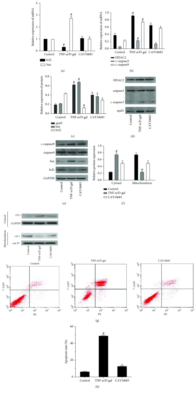
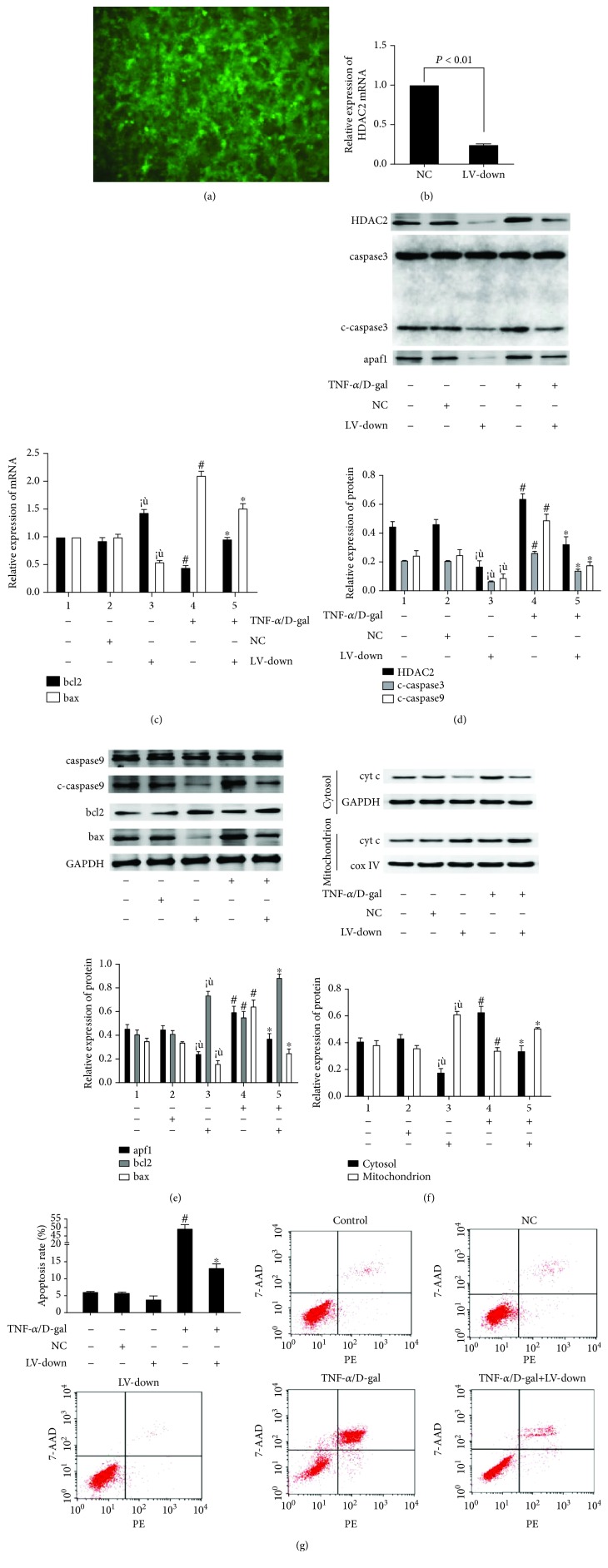
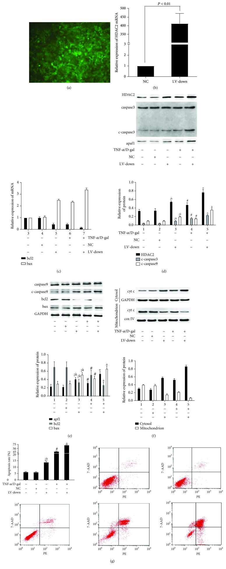
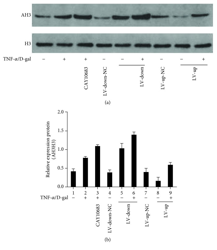
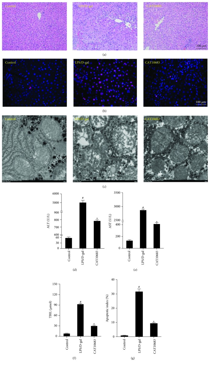
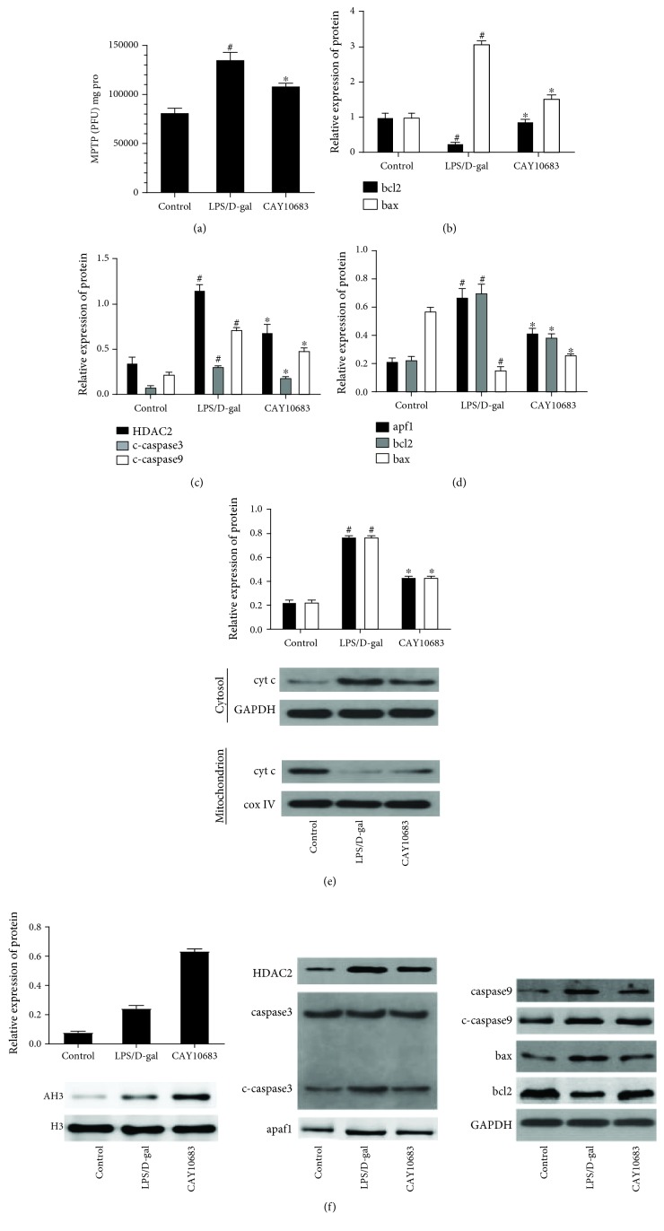
The purpose of this study was to investigate the modulation of histone deacetylase 2 (HDAC2) on mitochondrial apoptosis in acute liver failure (ALF). The cellular model was established with LO2 cells stimulated by tumor necrosis factor alpha (TNF-α)/D-galactosamine (D-gal). Rats were administrated by lipopolysaccharide (LPS)/D-gal as animal model. The cell and animal models were then treated by HDAC2 inhibitor CAY10683. HDAC2 was regulated up or down by lentiviral vector transfection in LO2 cells. The mRNA levels of bcl2 and bax were detected by real-time PCR. The protein levels of HDAC2, bcl2, bax, cytochrome c (cyt c) in mitochondrion and cytosol, apoptosis protease activating factor 1 (apaf1), caspase 3, cleaved-caspase 3, caspase 9, cleaved-caspase 9, acetylated histone H3 (AH3), and histone H3 (H3) were assayed by western blot. Apoptosis was detected by flow cytometry. The serum alanine aminotransferase (ALT), aspartate aminotransferase (AST), and total bilirubin (TBIL) levels were also assayed. The openness degree of the mitochondrial permeability transition pore (MPTP) was detected by ultraviolet spectrophotometry. The apoptosis of hepatocytes in liver tissues was determined by tunnel staining. The liver tissue pathology was detected by hematoxylin eosin (HE) staining. The ultrastructure of liver tissue was observed by electron microscopy. Compared with cell and rat model groups, the bax mRNA level was decreased, and bcl2 mRNA was increased in the CAY10683 treatment group. The protein levels of HDAC2, bax, cyt c in cytosol, apaf1, cleaved-caspase 3, and cleaved-caspase 9 were decreased, and the apoptosis rate was decreased (P < 0.05), whereas the protein level of bcl2 and cyt c in the mitochondrion was elevated (P < 0.05) in the CAY10683 treatment group. In the HDAC2 down- or upregulated LO2 cells, the mitochondrial apoptosis pathway was inhibited or activated, respectively. After being treated with TNF-α/D-gal in HDAC2 down- or upregulated LO2 cells, the mitochondrial apoptosis pathway was further suppressed or activated, respectively. The MPTP value was elevated in CAY10683-treated groups compared with the rat model group (P < 0.05). Liver tissue pathological damage and apoptotic index in the CAY10683-treated group were significantly reduced. In addition, AH3 was elevated in both cell and animal model groups (P < 0.05). Downregulated or overexpressed HDAC2 could accordingly increase or decrease the AH3 level, and TNF-α/D-gal could enhance the acetylation effect. These results suggested that modulations of histone deacetylase 2 offer a protective effect through the mitochondrial apoptosis pathway in acute liver failure.


Figures

Figure 1
Figure 1
CAY10683 inhibited the mitochondrial apoptotic pathway in the TNF-α/D-gal-treated LO2 cells. (a) The mRNA level of bcl2 and bax mRNA levels were detected by RT-PCR. (b-g) The protein expressions of HDAC2, cyt c in mitochondrion, cyt c in cytosol, caspase 3, c-caspase 3, caspase 9, c-caspase 9, apaf1, bcl2, and bax were detected by western blot. (h) The staining with Annexin V-phycoerythrin/7-aminoactinomycin D (Annexin V-PE/7AAD) and flow cytometry were performed to detect the influence LO2 cell apoptosis. #P < 0.05, compared with the control group. P < 0.05, compared with the TNF-α/D-gal-treated model group. c-caspase3: cleaved-caspase 3, c-caspase9: cleaved-caspase 9.
Figure 2
Figure 2
The levels of mitochondrial apoptosis signaling molecules in LO2 cells were decreased after HDAC2 knockdown. (a) The HDAC2 knockdown lentiviral vector was made and transfected into LO2 cells. GFP was observed with a fluorescence microscope after 72 h. (b) At 72 h after transfection, the HDAC2 mRNA level was detected by RT-PCR. (c) The mRNA level of bcl2 and bax mRNA levels were detected by RT-PCR. (d-f) The protein expressions of HDAC2, cyt c in mitochondrion, cyt c in cytosol, caspase 3, c-caspase 3, caspase 9, c-caspase 9, apaf1, bcl2, and bax were detected by western blot. (g) The staining with Annexin V-PE/7AAD and flow cytometry were performed to detect the influence LO2 cell apoptosis. P < 0.05, compared with the control group. #P < 0.05, compared with the control group. P < 0.05, compared with the TNF-α/D-gal-treated model group.
Figure 3
Figure 3
The levels of mitochondrial apoptosis signaling molecules in LO2 cells were increased after HDAC2 overexpression. (a) The HDAC2 overexpression lentiviral vector was made and transfected into LO2 cells. GFP was observed with a fluorescence microscope after 72 h. (b) At 72 h after transfection, the HDAC2 mRNA level was detected by RT-PCR. (c) The mRNA level of bcl2 and bax mRNA levels were detected by RT-PCR. (d-f) The protein expressions of HDAC2, cyt c in mitochondrion, cyt c in cytosol, caspase 3, c-caspase 3, caspase 9, c-caspase 9, apaf1, bcl2, and bax were detected by western blot. (g) The staining with Annexin V-PE/7AAD and flow cytometry were performed to detect the influence LO2 cell apoptosis. P < 0.05, compared with the control group. #P < 0.05, compared with the control group. P < 0.05, compared with the TNF-α/D-gal treated model group.
Figure 4
Figure 4
The level of histone H3 deacetylated by histone deacetylase 2 in different LO2 cell groups. P < 0.05, compared with the NC group. #P < 0.05, compared with the control group. P < 0.05, compared with the TNF-α/D-gal treated model group. NC: normal control.
Figure 5
Figure 5
CAY10683 could alleviate the hepatic pathology and liver function in ALF rats. (a) HE staining was used to detect pathological changes in liver tissue. (b, f) Tunnel staining was used to detect the apoptotic index in liver tissue. (c) The ultrastructure of liver tissue and the mitochondrial membranes were detected by electron microscopy. (d-f) The rat serum ALT, AST, and TBIL levels were detected by the fully automated Aeroset chemistry analyzer. #P < 0.05, compared with the control group. P < 0.05, compared with the LPS/D-gal-treated model group.
Figure 6
Figure 6
CAY10683 alleviates liver damage through regulating the mitochondrial apoptotic pathway in ALF rats. (a) The RFU value was detected by the purified MPTP fluorescence detection kit. (b) The mRNA level of bcl2 and bax mRNA levels were detected by RT-PCR. (c-e) The protein expressions of HDAC2, cyt c in mitochondrion, cyt c in cytosol, caspase 3, c-caspase 3, caspase 9, c-caspase 9, apaf1, bcl2, and bax were detected by western blot. (f) The protein expressions of AH3 and H3 were detected by western blot. #P < 0.05, compared with the control group. P < 0.05, compared with the LPS/D-gal-treated model group.

References

    1. Bernal W., Auzinger G., Dhawan A., Wendon J. Acute liver failure. The Lancet. 2010;376(9736):190–201. doi: 10.1016/S0140-6736(10)60274-7. - DOI - PubMed
    1. Slofstra S. H., Cate H., Spek C. A. Low dose endotoxin priming is accountable for coagulation abnormalities and organ damage observed in the Shwartzman reaction. A comparison between a single-dose endotoxemia model and a double-hit endotoxin-induced Shwartzman reaction. Thrombosis Journal. 2006;4(1):p. 13. doi: 10.1186/1477-9560-4-13. - DOI - PMC - PubMed
    1. Lichtman S. N., Wang J., Zhang C., Lemasters J. J. Endocytosis and Ca2+ are required for endotoxin-stimulated TNF-alpha release by rat Kupffer cells. The American Journal of Physiology. 1996;271(5) Part 1:G920–G928. doi: 10.1152/ajpgi.1996.271.5.G920. - DOI - PubMed
    1. Yoon J. H., Gores G. J. Death receptor-mediated apoptosis and the liver. Journal of Hepatology. 2002;37(3):400–410. doi: 10.1016/S0168-8278(02)00209-X. - DOI - PubMed
    1. Guicciardi M. E., Gores G. J. Apoptosis as a mechanism for liver disease progression. Seminars in Liver Disease. 2010;30(04):402–410. doi: 10.1055/s-0030-1267540. - DOI - PMC - PubMed

MeSH terms