. 2019 Jun 7:9:46.
doi: 10.1186/s13578-019-0308-9. eCollection 2019.

Zika virus antagonizes interferon response in patients and disrupts RIG-I-MAVS interaction through its CARD-TM domains

Affiliations

Zika virus antagonizes interferon response in patients and disrupts RIG-I-MAVS interaction through its CARD-TM domains

Yiwen Hu et al. Cell Biosci. .

Abstract

Background: The emerging threat to global health associated with the Zika virus (ZIKV) epidemics and its link to severe complications highlights a growing need to better understand the pathogenic mechanisms of ZIKV. Accumulating evidence for a critical role of type I interferon (IFN-I) in protecting hosts from ZIKV infection lies in the findings that ZIKV has evolved various strategies to subvert the host defense line by counteracting the early IFN induction or subsequent IFN signaling. Yet, mechanisms underlying the counter-IFN capability of ZIKV and its proteins, which might contribute to the well-recognized broad cellular tropisms and persistence of ZIKV, remain incompletely understood.

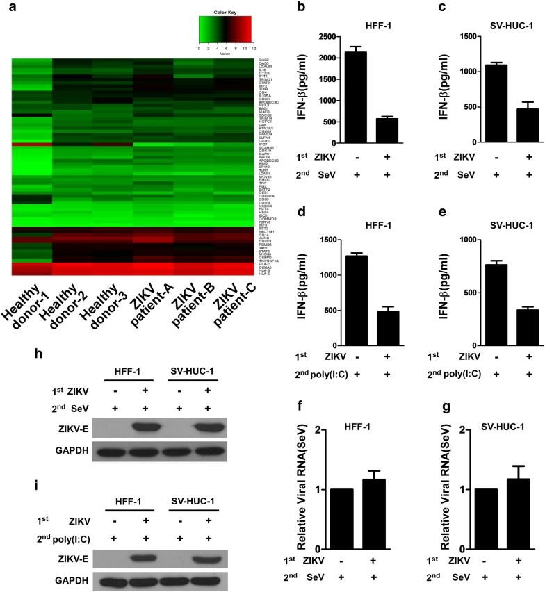
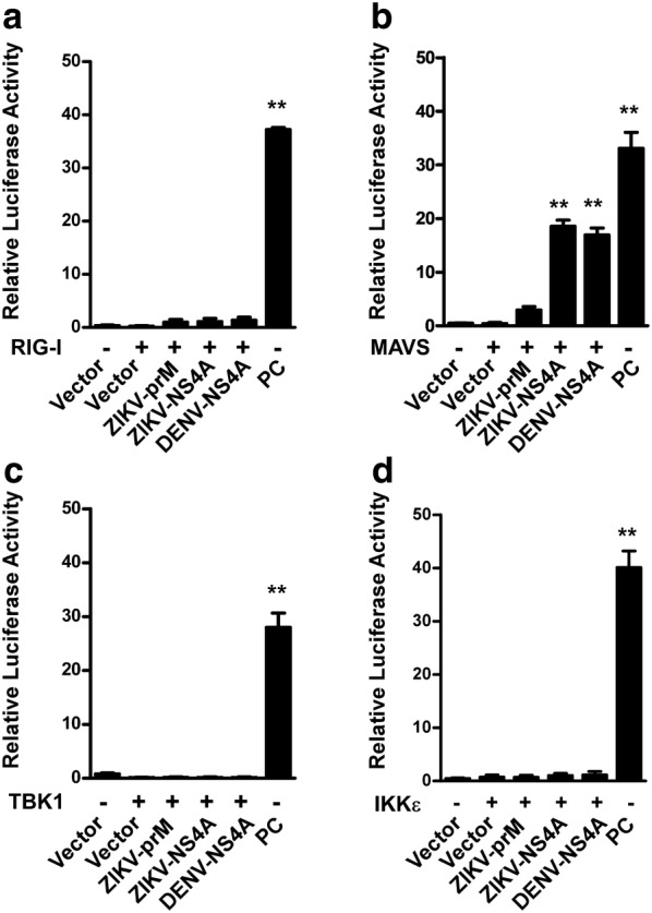
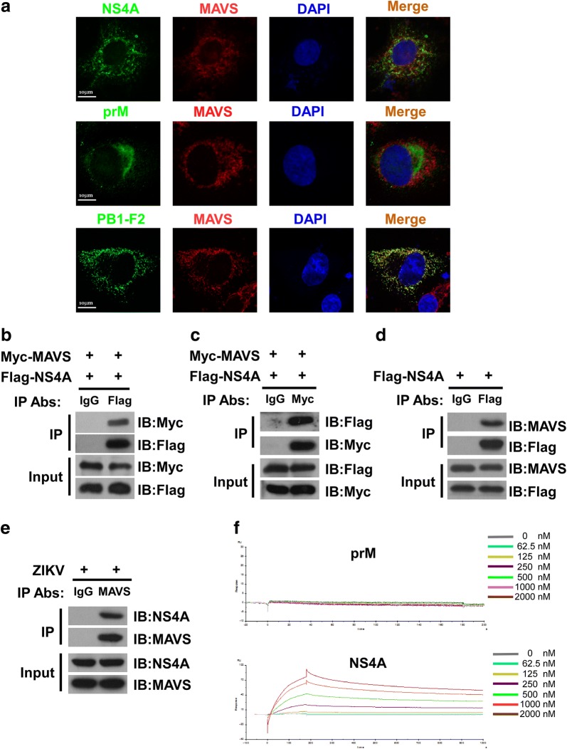
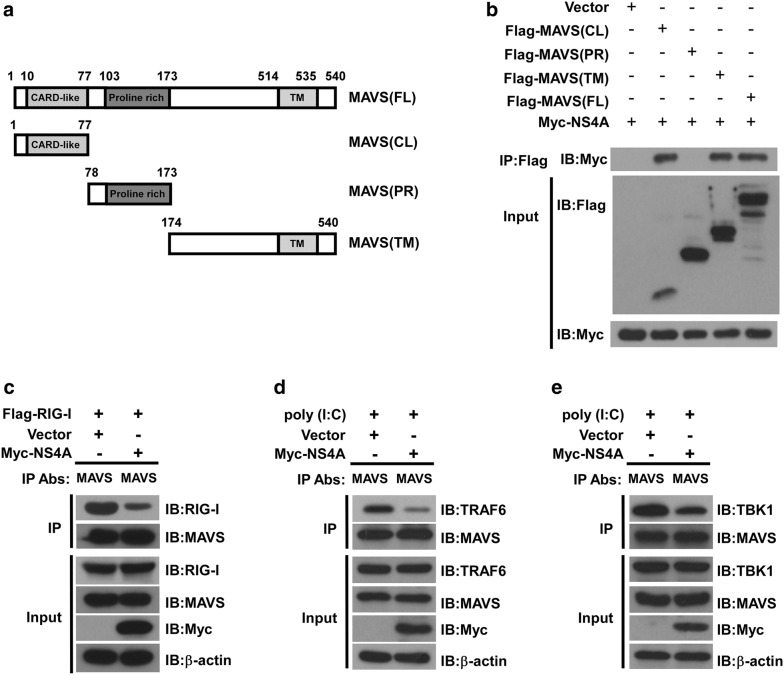
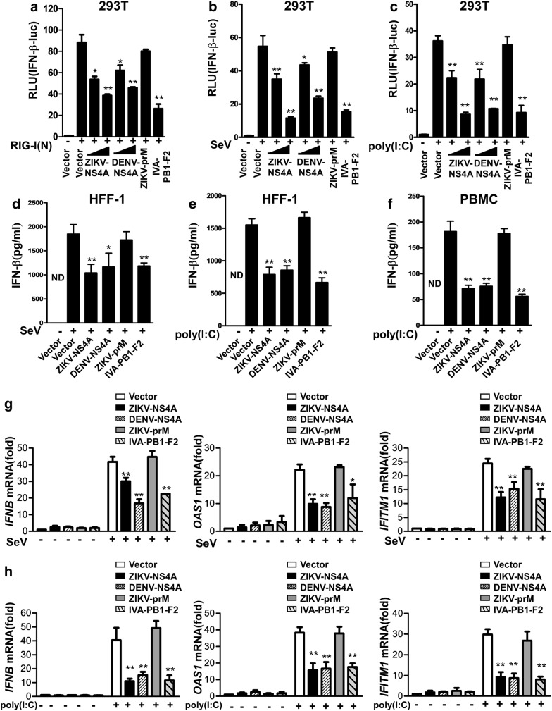
Results: Using RNA sequencing-based transcriptional profiling of whole blood cells isolated from patients acutely infected by ZIKV, we found that transcriptional signature programs of antiviral interferon-stimulated genes and innate immune sensors in ZIKV-infected patients remained inactive as compared to those of healthy donors, suggesting that ZIKV was able to suppress the induction of IFN-I during the natural infection process in humans. Furthermore, by analyzing the molecular interaction in a ZIKV NS4A-overexpression system, or in the context of actual ZIKV infection, we identified that ZIKV NS4A directly bound MAVS and thereby interrupted the RIG-I/MAVS interaction through the CARD-TM domains, leading to attenuated production of IFN-I.

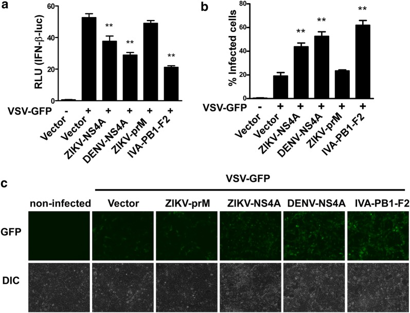
Conclusions: Our findings collectively revealed that ZIKV NS4A targeted MAVS and contributed to ZIKV immune evasion through abrogating MAVS-mediated IFN production. These findings obtained from patient studies have added new knowledge and molecular details to our understanding regarding how ZIKV mediates suppression of the IFN-I system and may provide a new basis for the future development of anti-ZIKV strategies.

Keywords: Interferon; MAVS; Nonstructural protein 4A; RIG-I; Zika virus.


Conflict of interest statement

Competing interestsThe authors declare that they have no competing interests.

Figures

Fig. 1
Fig. 1
ZIKV abrogates the activation of type I IFN signaling. a Heatmap of a pre-defined gene list of type I IFN and ISGs that are differentially expressed (DEGs), generated from comparative RNA-seq data among three ZIKV-infected patients (ZIKV patients ac) and three healthy donors. The expression of read-mapped genes normalized across the entire dataset was analyzed based on the RPKM value, and p-values < 0.05 and log2 fold change ≥ 1.5 relative to the control cohort were set as the threshold to assess the statistical significance of the differential gene expression. be Infection with ZIKV reduces type I IFN production after a secondary infection. HFF-1 and SV-HUC-1 cells were infected with mock or ZIKV at an MOI of 1, and 24 h later, respectively, infected with SeV at 100 HAU/ml (b, c) or transfected with 20 μg/ml of poly(I:C) (d, e). IFN-β protein levels were measured using specific ELISA in the supernatants collected at 36 h after secondary-infection from the two cell lines, and cell lysates were harvested for real time RT-PCR to determine the RNA level of SeV (f, g), and for Western blotting analysis to determine the levels of ZIKV envelope protein (E) and GAPDH protein (h, i). Data are shown as the mean ± SD derived from three repeatedexperiments. *p < 0.05, and **indicates p < 0.01 (Student’s t test)
Fig. 2
Fig. 2
Molecular interaction between ZIKV NS4A protein and the type I IFN induction pathway. GAL4-based mammalian two-hybrid screening assays were performed to identify the molecular targets of ZIKV proteins in RIG-I signaling. 293T cells in 24-well plates were co-transfected with a pGL4.31 vector, a pFN11A (BIND) vector expressing a fusion protein of GAL4-BD and individual ZIKV prM, NS4A or DENV NS4A proteins, and a pFN10A (ACT) vector expressing a RIG-I, b MAVS, c TBK1 or d IKKε. The pFN11A (BIND) vector contained a Renilla luciferase gene that was used as an internal control to normalize DNA transfection efficiency. The pBIND and pACT vectors were used as negative controls, and the pBIND-Id and pACT-MyoD vectors were used as positive controls (PCs) according to the manufacturer’s instructions. At 48 h post-transfection, cell lysates were harvested for the luciferase activity assay. The results are shown as relative luciferase activity after normalization with Renilla luciferase activity. Data are shown as the mean ± SD derived from three repeat experiments. *p < 0.05, and **p < 0.01 (Student’s t test)
Fig. 3
Fig. 3
ZIKV NS4A co-localizes and interacts with MAVS. a HeLa cells transfected with plasmids expressing Flag-tagged NS4A, Flag-tagged ZIKV prM or influenza Flag-tagged PB1-F2 were stained with anti-Flag and anti-MAVS antibodies as well as DAPI. Secondary antibodies conjugated to rhodamine and FITC dye were used to visualize the indicated proteins. Images are representative of three independent experiments. 293T cells co-transfected with plasmids encoding Myc-tagged MAVS and Flag-tagged NS4A were used in a co-IP assay to address whether ZIKV NS4A protein physically interacts with MAVS. Cell lysates were precipitated with an anti-Flag antibody (b), anti-Myc antibody (c), or control mouse IgG, and immunocomplexes were analyzed with the indicated antibodies by western blotting. d 293T cells were transfected with plasmids encoding Flag-tagged NS4A, followed by immunoprecipitation using anti-Flag antibody or control IgG. The immunocomplexes were analyzed with anti-MAVS antibody by Western blotting. e HFF-1 cells were infected with ZIKV at an MOI of 5 followed by immunoprecipitation using anti-MAVS antibody or control mouse IgG. The immunocomplexes that were captured by the protein G Dynabeads were analyzed by Western blotting using anti-NS4A, or anti-MAVS antibodies. f SPR analysis of the interactions between MAVS and NS4A. Direct binding was measured by Biacore assays. MAVS was immobilized on a CM5 chip. The analytes consisted of serial dilutions of NS4A proteins ranging between 0 and 2000 nM. The data shown are representative of three independent experiments with similar results
Fig. 4
Fig. 4
ZIKV NS4A interacts with both the CL and TM domains of MAVS and prevents RIG-I from binding MAVS. a A schematic diagram of the MAVS protein and functional domains: CARD-like domain (aa 10 to 77), proline-rich domain (aa 103 to 173) and transmembrane domain (aa 514 to 535). b Co-IP and western blotting analysis of 293T cells transfected with Myc-tagged NS4A along with vectors expressing the indicated Flag-tagged MAVS truncation forms or full-length MAVS. Empty vector was used as a negative control. c 293T cells were co-transfected with Flag-tagged RIG-I and Myc-tagged NS4A or an empty vector control for 24 h. Whole cell lysates were subjected to immunoprecipitation using an anti-MAVS antibody and analyzed by western blotting for RIG-I (c). 293T cells were transfected with Myc-tagged NS4A or an empty vector control for 24 h later, transfected with 20 μg/ml of poly(I:C). Whole cell lysates were subjected to immunoprecipitation using an anti-MAVS antibody and analyzed by western blotting for TRAF6 (d), or TBK1 (e). The expression of precipitated proteins was determined by western blotting analysis using the indicated antibodies (lower panel). The data shown are representative of three independent experiments with similar results
Fig. 5
Fig. 5
ZIKV NS4A negatively regulates IFN production. a Lysates of 293T cells co-transfected with RIG-I(N), IFN-β reporter, together with ZIKV NS4A (250, or 500 ng), DENV NS4A (250, or 500 ng), ZIKV prM (500 ng) or PB1-F2 (500 ng)were analyzed for luciferase activity. 293T cells were transfected with IFN-β reporter, together with ZIKV NS4A (250, or 500 ng), DENV NS4A (250, or 500 ng), ZIKV prM (500 ng) or PB1-F2 (500 ng), b then infected with SeV at 100 HAU/ml for 24 h, c or transfected with 20 μg/ml of poly(I:C) for 24 h followed by analysis of the cell lysates for luciferase activity. d, e HFF-1 cells or f PBMC were transfected with blank control vector or a vector expressing ZIKV NS4A (500 ng), DENV NS4A (500 ng), ZIKV prM (500 ng) or PB1-F2 (500 ng), respectively, and 24 h later were mock-infected or infected with SeV at 100 HAU/ml or transfected with or without 20 μg/ml of poly(I:C) for 24 h, followed by analysis of the supernatant for IFN-β protein levels. g, h Meanwhile, at 24 h post-infection, total cellular RNA was isolated, and real-time PCR was performed to detect IFNB, OAS1, and IFITM1 mRNA levels in 293T cells. The data are presented as the mean ± SD derived from three repeat experiments. *p < 0.05, and **p < 0.01 (Student’s t-test)
Fig. 6
Fig. 6
ZIKV NS4A blocks VSV-induced IFN production. a 293T cells transfected with IFN-β reporter together with ZIKV-NS4A, DENV-NS4A, ZIKV-prM and IVA-PB1-F2 (500 ng/well) were infected with VSV-GFP for 24 h, and cell lysates were then analyzed for reporter luciferase activity. The data are shown as the mean ± SD derived from three repeat experiments. **p < 0.01 (Student’s t test). b, c 293T cells were transfected with empty vector, and a ZIKV-NS4A-, DENV-NS4A-, ZIKV-prM- or IVA-PB1-F2-expressing plasmid. Twenty-four hours later, the cells were infected with SeV, and the supernatants were UV-treated and used to overlay 293T cells. Twenty-four hours later, the cells were infected with VSV-GFP for 24 h, followed by scoring of the number of GFP-positive cells by flow cytometry (b), and cell images were taken under a fluorescence microscope (c)

Similar articles

Cited by

References

    1. Pierson TC, Diamond MS. The emergence of Zika virus and its new clinical syndromes. Nature. 2018;560(7720):573–581. doi: 10.1038/s41586-018-0446-y. - DOI - PubMed
    1. Martines RB, Bhatnagar J, de Oliveira Ramos AM, Davi HP, Iglezias SD, Kanamura CT, Keating MK, Hale G, Silva-Flannery L, Muehlenbachs A, et al. Pathology of congenital Zika syndrome in Brazil: a case series. Lancet. 2016;388(10047):898–904. doi: 10.1016/S0140-6736(16)30883-2. - DOI - PubMed
    1. http://www.who.int/emergencies/zika-virus/en/.
    1. Li H, Saucedo-Cuevas L, Shresta S, Gleeson JG. The neurobiology of Zika virus. Neuron. 2016;92(5):949–958. doi: 10.1016/j.neuron.2016.11.031. - DOI - PubMed
    1. Yuan L, Huang XY, Liu ZY, Zhang F, Zhu XL, Yu JY, Ji X, Xu YP, Li G, Li C, et al. A single mutation in the prM protein of Zika virus contributes to fetal microcephaly. Science. 2017;358(6365):933–936. doi: 10.1126/science.aam7120. - DOI - PubMed