

Case Reports
doi: 10.1155/2019/9625075.
eCollection 2019.
Pituitary Gland and Neurological Involvement in a Case of Hemophagocytic Syndrome Revealing an Intravascular Large B-Cell Lymphoma
Affiliations
- PMID: 31183225
- PMCID: PMC6512020
- DOI: 10.1155/2019/9625075
Item in Clipboard
Case Reports
Pituitary Gland and Neurological Involvement in a Case of Hemophagocytic Syndrome Revealing an Intravascular Large B-Cell Lymphoma
Case Rep Hematol.
.
Abstract
Intravascular large B-cell lymphoma is a rare entity characterized by the proliferation of neoplastic lymphocytes in the lumen of small blood vessels and high mortality. Diagnosis of intravascular lymphoma is often delayed or established postmortem. Here, we report the case of a 48-year-old woman presenting hemophagocytic syndrome, with pituitary gland and neurological involvement. Diagnosis of intravascular large B-cell lymphoma was made on perisplenic vessels, while liver and bone marrow biopsy was noncontributive. This case demonstrates the importance of thorough histopathologic investigations in the setting of high suspicion.
Figures
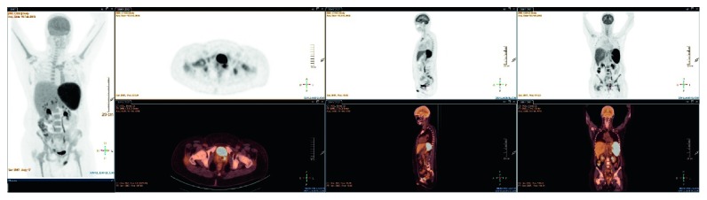
PET-CT hypermetabolic splenomegaly, perihepatic hypermetabolic lymph nodes, and cervical hypermetabolism.
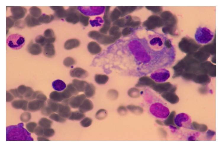
Hemophagocytosis at bone marrow aspiration.
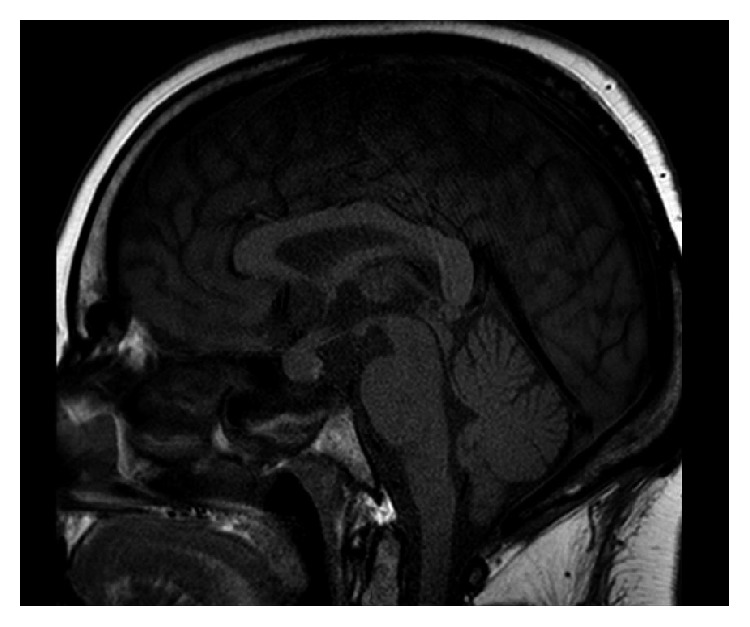
Brain MRI showing enlargement of the pituitary gland and pituitary stalk without evidence of adenoma.
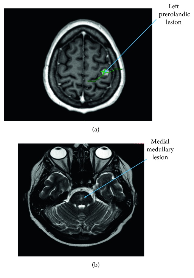
Brain MRI showing left prerolandic lesion and medial medullary hypersignal. (a) Prerolandic lesion with homogeneous corticosubcortical ring enhancement. (b) Medial medullary hypersignal in T2 and flair.
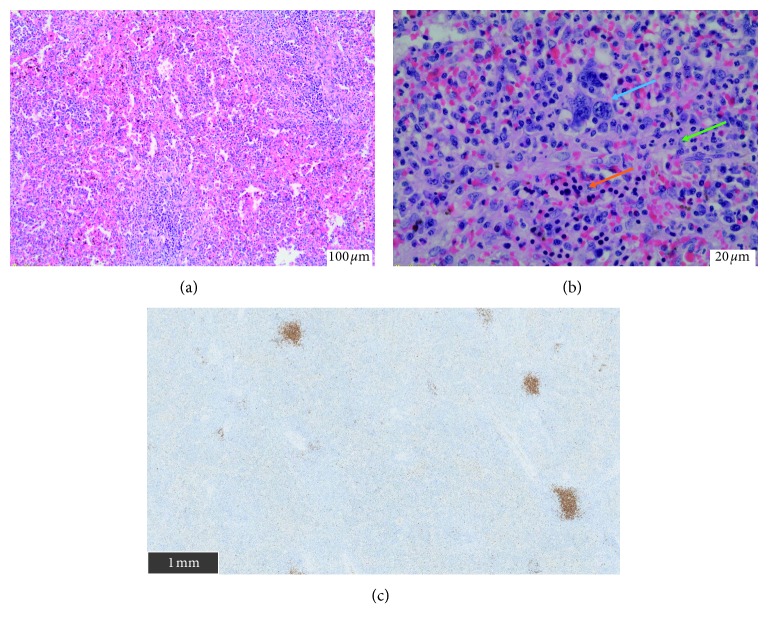
Histopathology of a splenectomy piece. (a) Extramedullary hematopoiesis (H&E, 10x). (b) Extramedullary hematopoiesis (H&E, 40x) with megakaryocytes (blue arrow), erythroblasts (orange arrow), and neutrophils (green arrow). (c) CD20 immunostaining showing no spleen infiltration (scale on image).
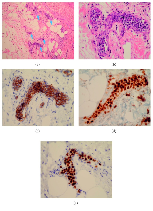
Histopathology of perisplenic tissues. (a) Tumoral intravascular infiltration (arrows) (H&E, 10x). (b) Large hyperchromatic cell inside small vessels around splenic lymph nodes (H&E, 40x). (c) Strong positive CD20 B-cell surface expression. (d) Positive expression of MUM1. (e) Positive expression of PAX5.
Similar articles
-
Intravascular large B-cell lymphoma with hemophagocytic syndrome (Asian variant) in a Caucasian patient.Int J Clin Exp Pathol. 2012;5(5):448-54. Epub 2012 May 23. Int J Clin Exp Pathol. 2012. PMID: 22808298 Free PMC article.
-
[Intravascular large cell lymphoma revealed by diffuse telangiectasia and cauda equina syndrome].Ann Dermatol Venereol. 2002 Mar;129(3):320-4. Ann Dermatol Venereol. 2002. PMID: 11988690 French.
-
A case of intravascular large B-cell lymphoma mimicking erythema nodosum: the importance of multiple skin biopsies.J Cutan Pathol. 2000 Sep;27(8):413-8. doi: 10.1034/j.1600-0560.2000.027008413.x. J Cutan Pathol. 2000. PMID: 10955689
-
Intravascular B-cell lymphoma: report of two cases with different clinical presentation but rapid central nervous system involvement.Leuk Lymphoma. 2003 Aug;44(8):1353-9. doi: 10.1080/1042819031000097393. Leuk Lymphoma. 2003. PMID: 12952229 Review.
-
[An autopsy case of intravascular malignant lymphomatosis with hemophagocytic syndrome, mental confusion and liver dysfunction].Rinsho Ketsueki. 1998 Aug;39(8):586-92. Rinsho Ketsueki. 1998. PMID: 9785977 Review. Japanese.
Cited by
-
Hematologic Malignancies: Two Cases of a Rare Cause of Hypopituitarism.JCEM Case Rep. 2024 Sep 11;2(9):luae147. doi: 10.1210/jcemcr/luae147. eCollection 2024 Sep. JCEM Case Rep. 2024. PMID: 39263278 Free PMC article.
-
ICAM1-Negative Intravascular Large B-Cell Lymphoma of the Pituitary Gland: A Case Report and Literature Review.AACE Clin Case Rep. 2021 Feb 9;7(4):249-255. doi: 10.1016/j.aace.2021.01.011. eCollection 2021 Jul-Aug. AACE Clin Case Rep. 2021. PMID: 34307847 Free PMC article.
-
Intravascular large B-cell lymphoma causing hypopituitarism and respiratory failure due to infiltration into pulmonary capillaries.BMJ Case Rep. 2022 May 24;15(5):e247880. doi: 10.1136/bcr-2021-247880. BMJ Case Rep. 2022. PMID: 35609931 Free PMC article.
-
Hemophagocytic syndrome and neurological involvement in a case of intravascular large B-cell lymphoma.J Int Med Res. 2021 Sep;49(9):3000605211006644. doi: 10.1177/03000605211006644. J Int Med Res. 2021. PMID: 34590922 Free PMC article.
-
Intravascular large B-cell lymphoma affecting multiple cranial nerves: A histopathological study.Neuropathology. 2021 Oct;41(5):396-405. doi: 10.1111/neup.12767. Epub 2021 Sep 19. Neuropathology. 2021. PMID: 34541718 Free PMC article.
References
-
- Fischer M., Iglseder S., Grams A., et al. Intravascular large B-cell lymphoma mimicking central nervous system vasculitis. Human Pathology: Case Reports. 2017;8:3–8. doi: 10.1016/j.ehpc.2016.11.002. - DOI
Publication types
LinkOut - more resources
Full Text Sources
