

Role of computed tomography and magnetic resonance imaging in local complications of acute pancreatitis
- PMID: 31183322
- PMCID: PMC6534767
- DOI: 10.21037/gs.2018.12.07
Role of computed tomography and magnetic resonance imaging in local complications of acute pancreatitis
Abstract
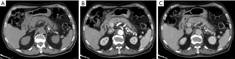
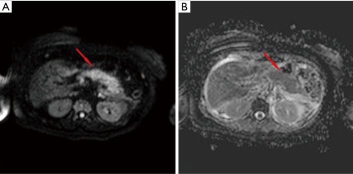
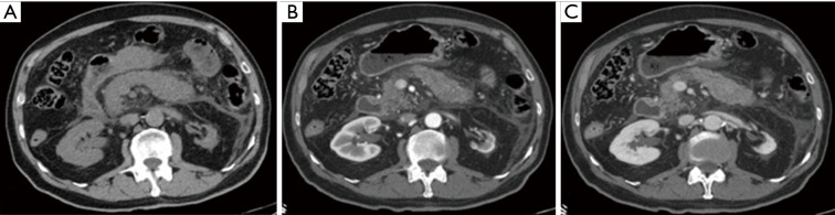
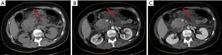
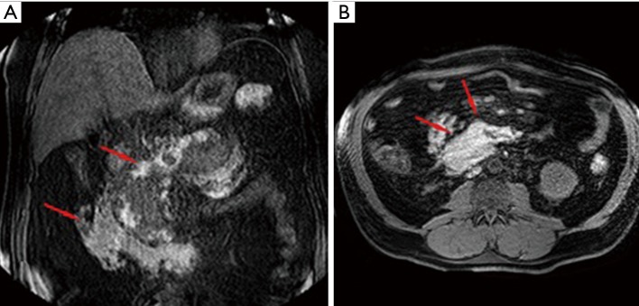
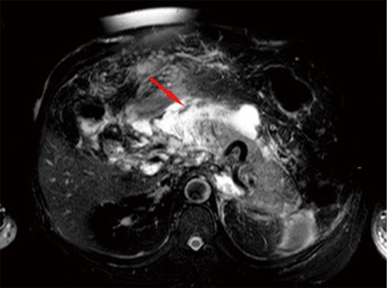
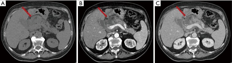
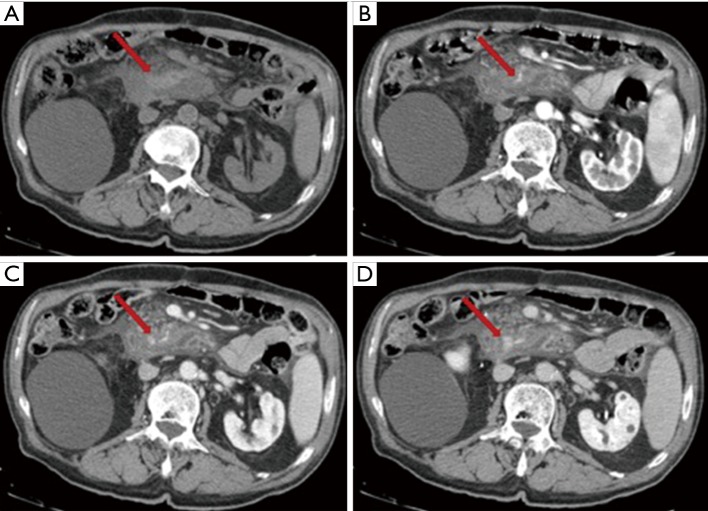
Acute pancreatitis (AP) represents a pancreas inflammation of sudden onset that can present different degrees of severity. AP is a frequent cause of acute abdomen and its complications are still a cause of death. Biliary calculosis and alcohol abuse are the most frequent cause of AP. Computed tomography (CT) and magnetic resonance imaging (MRI) are not necessary for the diagnosis of AP but they are fundamental tools for the identification of the cause, degree severity and AP complications. AP severity assessment is in fact one of the most important issue in disease management. Contrast-enhanced CT is preferred in the emergency setting and is considered the gold standard in patients with AP. MRI is comparable to CT for the diagnosis of AP but requires much more time so it is not usually chosen in the emergency scenario. Complications of AP can be distinguished in localized and generalized. Among the localized complications, we can identify: acute peripancreatic fluid collections (APFC), pseudocysts, acute necrotic collections (ANC), walled off pancreatic necrosis (WOPN), venous thrombosis, pseudoaneurysms and haemorrhage. Multiple organ failure syndrome (MOFS) and sepsis are possible generalized complications of AP. In this review, we focus on CT and MRI findings in local complications of AP and when and how to perform CT and MRI. We paid also attention to recent developments in diagnostic classification of AP complications.
Keywords: Acute pancreatitis (AP); computed tomography (CT); magnetic resonance imaging (MRI).
Conflict of interest statement
Conflicts of Interest: The authors have no conflicts of interest to declare.
Figures
Similar articles
-
Pancreatic fluid collections: What is the ideal imaging technique?World J Gastroenterol. 2015 Dec 28;21(48):13403-10. doi: 10.3748/wjg.v21.i48.13403. World J Gastroenterol. 2015. PMID: 26730150 Free PMC article. Review.
-
Revised Atlanta classification for CT pancreatic and peripancreatic collections in the first month of acute pancreatitis: interobserver agreement.Eur Radiol. 2019 May;29(5):2302-2310. doi: 10.1007/s00330-018-5906-0. Epub 2019 Jan 10. Eur Radiol. 2019. PMID: 30631920
-
The role of nonenhanced magnetic resonance imaging in the early assessment of acute pancreatitis.Am J Gastroenterol. 2007 May;102(5):997-1004. doi: 10.1111/j.1572-0241.2007.01164.x. Epub 2007 Mar 22. Am J Gastroenterol. 2007. PMID: 17378903
-
Updated imaging nomenclature for acute pancreatitis.AJR Am J Roentgenol. 2014 Nov;203(5):W464-9. doi: 10.2214/AJR.13.12222. AJR Am J Roentgenol. 2014. PMID: 25341160 Review.
-
Current concepts for the diagnosis of acute pancreatitis by multiparametric magnetic resonance imaging.Quant Imaging Med Surg. 2019 Dec;9(12):1973-1985. doi: 10.21037/qims.2019.11.10. Quant Imaging Med Surg. 2019. PMID: 31929970 Free PMC article. Review.
Cited by
-
Magnetic resonance severity index assessed by T1-weighted imaging for acute pancreatitis: correlation with clinical outcomes and grading of the revised Atlanta classification-a narrative review.Gland Surg. 2020 Dec;9(6):2312-2320. doi: 10.21037/gs-20-554. Gland Surg. 2020. PMID: 33447582 Free PMC article. Review.
-
Treatment of acute pancreatitis with early pancreatic stenting: a case series of 336 patients.Gland Surg. 2021 Sep;10(9):2780-2789. doi: 10.21037/gs-21-574. Gland Surg. 2021. PMID: 34733727 Free PMC article.
-
Current Progress in the CT- and MRI-Based Detection and Evaluation of Acute Pancreatitis Complications.Med Sci Monit. 2025 Aug 10;31:e948306. doi: 10.12659/MSM.948306. Med Sci Monit. 2025. PMID: 40783779 Free PMC article. Review.
-
Pancreatic neuroendocrine tumours: spectrum of imaging findings.Gland Surg. 2020 Dec;9(6):2215-2224. doi: 10.21037/gs-20-537. Gland Surg. 2020. PMID: 33447574 Free PMC article. Review.
-
Role of MRI and abbreviated MRI in the evaluation and management of acute pancreatitis: a comprehensive review.Abdom Radiol (NY). 2025 Jul 21. doi: 10.1007/s00261-025-05097-2. Online ahead of print. Abdom Radiol (NY). 2025. PMID: 40689954 Review.
References
-
- Ranson JH. Acute pancreatitis: pathogenesis, outcome and treatment. Clin Gastroenterol 1984;13:843-63. - PubMed