

Neuroimaging in emergency: a review of possible role of pineal gland disease
- PMID: 31183323
- PMCID: PMC6534760
- DOI: 10.21037/gs.2019.01.02
Neuroimaging in emergency: a review of possible role of pineal gland disease
Abstract
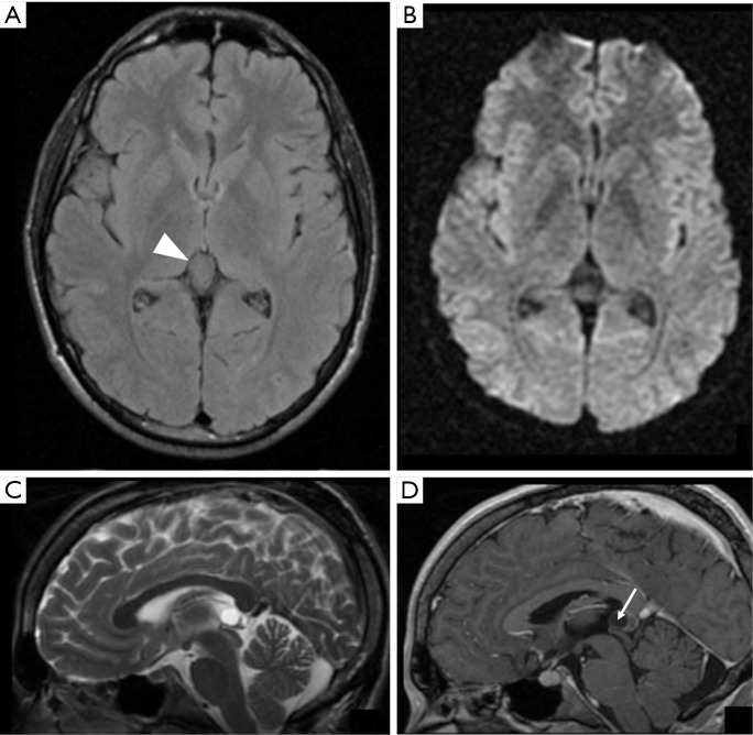
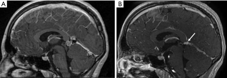
The pineal gland can be involved in a variety of neoplastic and congenital masses and tumors. Pineal gland neoplasms occur more frequently in children, accounting for 3-8% of intracranial tumors in the pediatric population. Pineal cysts are small lesions usually asymptomatic and encountered incidentally. Pathologic processes involving the pineal region produce signs and symptoms related to the mass effect on the adjacent structures and invasion of surrounding structures. These include several acute symptoms, such as increased intracranial pressure syndrome from obstruction of the aqueduct and consequent hydrocephalus, and Parinaud syndrome. Pineal apoplexy is rare and refers to the sudden neurological deterioration following hemorrhage in the pineal gland, most commonly into a pineal cyst. Knowledge of the clinical presentation and imaging features of these lesions is essential to narrow the differential diagnosis, especially when presenting with acute onset.
Keywords: Brain tumors; emergency; magnetic resonance imaging (MRI); pineal gland.
Conflict of interest statement
Conflicts of Interest: The authors have no conflicts of interest to declare.
Figures
References
-
- De Los Reyes EVA, Rivera ID, Santos HM, et al. Mature teratoma of the pineal region in the paediatric age group : A case report and review of the literature. Malays J Pathol 2018;40:175-83. - PubMed
-
- Taraszewska A, Matyja E, Koszewski W, et al. Asymptomatic and symptomatic glial cysts of the pineal gland. Folia Neuropathol 2008;46:186-95. - PubMed
Publication types
LinkOut - more resources
Full Text Sources