

Multi-detector computed tomography in the diagnosis and characterization of adrenal gland traumatic injuries
- PMID: 31183326
- PMCID: PMC6534768
- DOI: 10.21037/gs.2019.01.07
Multi-detector computed tomography in the diagnosis and characterization of adrenal gland traumatic injuries
Abstract
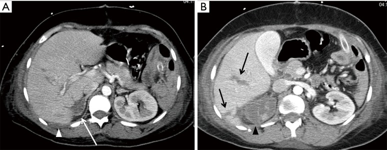
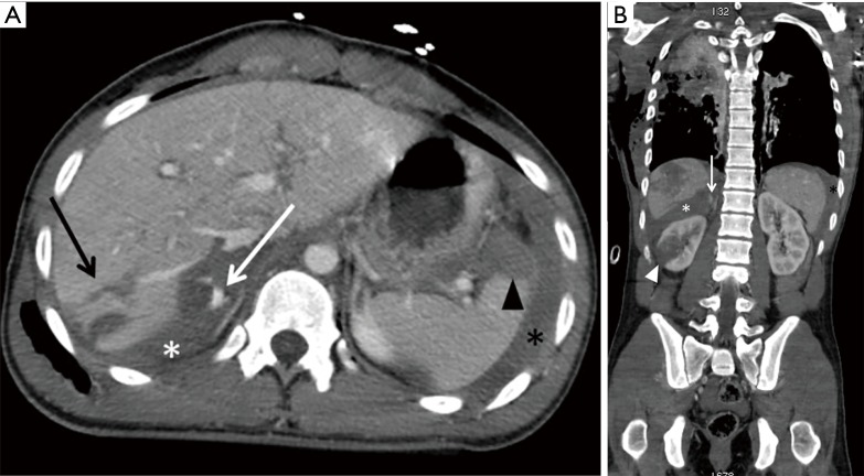
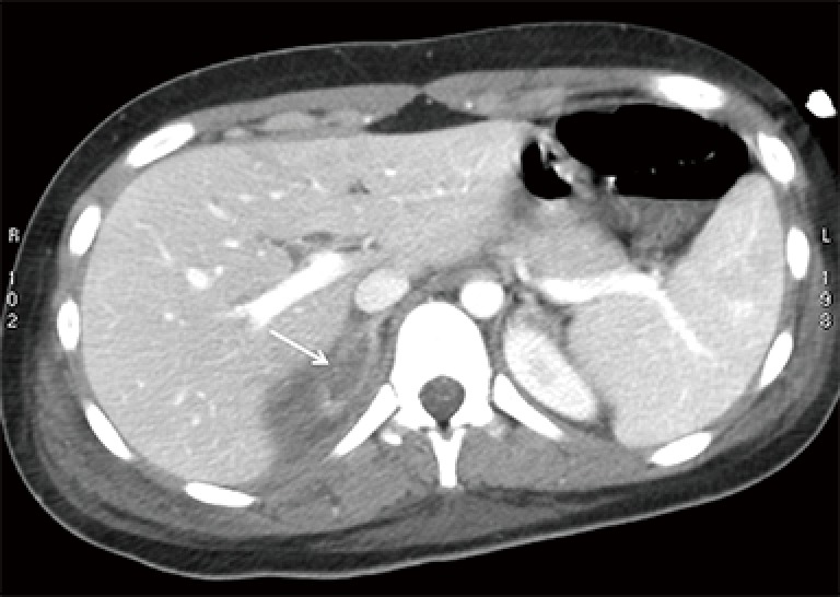
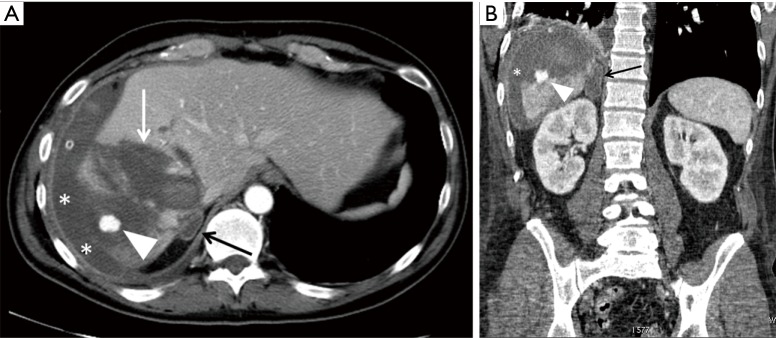
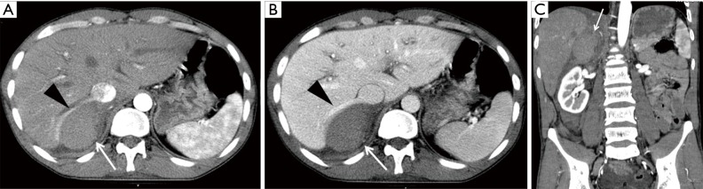
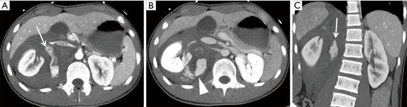
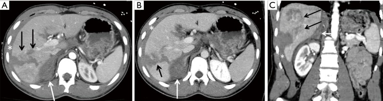
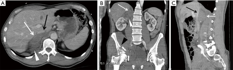
Adrenal gland injuries after a blunt abdominal trauma are rare events and represent important indicators for severe trauma. Multidetector CT evolution with high volumetric resolution and fast acquisition with the use of multiplanar reformatted (MPR) visualization allows for an accurate and fast diagnosis of the adrenal gland for post-traumatic pathologies. While, before its introduction the diagnosis was made mainly postmortem or during surgery. Adrenal injuries are unilateral up to 90% of the cases involving most commonly the right gland; thoracoabdominal organs injuries are often also associated. Bilateral adrenal lesions are asymptomatic, potentially leading to the development of acute adrenal insufficiency. The purpose of the present review was to determine the prevalence, the mechanism of injury and the different CT appearances of adrenal trauma. Prognosis and management of adrenal injury will also be reviewed.
Keywords: Emergency radiology; adrenal glands; adrenal injuries; blunt abdominal trauma; hematoma; hemorrhage; multi-detector computed tomography (MDCT); trauma imaging.
Conflict of interest statement
Conflicts of Interest: The authors have no conflicts of interest to declare.
Figures


Similar articles
-
Role of multidetector row computed tomography in the assessment of adrenal gland injuries.Eur J Radiol. 2006 Sep;59(3):355-8. doi: 10.1016/j.ejrad.2006.04.029. Epub 2006 Jun 19. Eur J Radiol. 2006. PMID: 16784828
-
Traumatic adrenal gland injury: epidemiology and outcomes in a major Australian trauma center.Eur J Trauma Emerg Surg. 2010 Dec;36(6):567-72. doi: 10.1007/s00068-010-0007-z. Epub 2010 Mar 24. Eur J Trauma Emerg Surg. 2010. PMID: 26816312
-
CT manifestations of adrenal trauma: experience with 73 cases.Emerg Radiol. 2007 Mar;13(6):313-8. doi: 10.1007/s10140-006-0563-z. Epub 2007 Jan 25. Emerg Radiol. 2007. PMID: 17252249
-
Isolated unilateral adrenal gland hemorrhage following motor vehicle collision: a case report and review of the literature.J Med Case Rep. 2017 Dec 26;11(1):358. doi: 10.1186/s13256-017-1506-x. J Med Case Rep. 2017. PMID: 29277157 Free PMC article. Review.
-
Blunt adrenal gland trauma in the pediatric population.Asian J Surg. 2011 Jul;34(3):103-10. doi: 10.1016/j.asjsur.2011.08.003. Epub 2011 Oct 22. Asian J Surg. 2011. PMID: 22208684 Review.
Cited by
-
Adrenal gland laceration in adult trauma patients without severe concomitant abdominal organ injuries: Incidence, associated factors, and outcomes from a national trauma database study.Medicine (Baltimore). 2025 Mar 7;104(10):e41756. doi: 10.1097/MD.0000000000041756. Medicine (Baltimore). 2025. PMID: 40068060 Free PMC article.
-
Pitfalls and differential diagnosis on adrenal lesions: current concepts in CT/MR imaging: a narrative review.Gland Surg. 2020 Dec;9(6):2331-2342. doi: 10.21037/gs-20-559. Gland Surg. 2020. PMID: 33447584 Free PMC article. Review.
-
Isolated Adrenal Gland Hemorrhage: A Case of a Car Accident.Bull Emerg Trauma. 2023;11(3):162-165. doi: 10.30476/BEAT.2023.98940.1446. Bull Emerg Trauma. 2023. PMID: 37525649 Free PMC article.
-
Life-Threatening Hypotension in a Brain-Injured, Multi-Trauma Patient With Unilateral Adrenal Gland Damage: How a Single Hydrocortisone Dose Revealed Relative Corticosteroid Insufficiency.Cureus. 2022 Dec 22;14(12):e32843. doi: 10.7759/cureus.32843. eCollection 2022 Dec. Cureus. 2022. PMID: 36694505 Free PMC article.
References
Publication types
LinkOut - more resources
Full Text Sources
Molecular Biology Databases