Editorial
. 2019 Apr;8(2):197-206.
doi: 10.21037/gs.2018.12.08.

Diffusion-weighted MRI: new paradigm for the diagnosis of interstitial oedematous pancreatitis

Affiliations
Editorial

Diffusion-weighted MRI: new paradigm for the diagnosis of interstitial oedematous pancreatitis

Massimo Tonolini et al. Gland Surg. 2019 Apr.
No abstract available


Conflict of interest statement

Conflicts of Interest: The authors have no conflicts of interest to declare.

Figures

Figure 1
Figure 1
Distribution of measured apparent diffusion coefficient (ADC) values (along Y axis, ×10−3 mm2/s) of four pancreatic regions in 16 patients with interstitial oedematous pancreatitis (IEP).
Figure 2
Figure 2
Distribution of ADC values (along Y axis, ×10−3 mm2/s) of four pancreatic regions in 16 patients with IEP, normalized using the ADC value of the iliopsoas muscle in the same patient. ADC, apparent diffusion coefficient; IEP, interstitial oedematous pancreatitis.
Figure 3
Figure 3
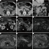
In an 84-year-old male with gallstone-related IEP multidetector CT including precontrast (A), enhanced pancreatic (B) and portal venous (C) CT acquisitions showed near-normal findings concerning pancreatic size, enhancement and surrounding planes. Alternative causes of upper abdominal pain were excluded. Further investigation with MRI confirmed normal size, morphology and signal intensity of the pancreatic head and isthmus (arrowheads) on T2-weighted (D,E with fat suppression) images, and revealed visual hyperintensity on diffusion-weighted imaging (DWI, F,H) and corresponding low signal on apparent diffusion coefficient (ADC) maps (G and I, mean measured value 1.01×10⁻³ mm2/s). The altered DWI decreased along the pancreatic body and normalized at the tail. IEP, interstitial oedematous pancreatitis.
Figure 4
Figure 4
In a 62-year-old male with IEP and history of cholecystectomy, contrast-enhanced CT (A,B) did not reveal any abnormality. At MRI, compared to the normal pancreatic head, the body and tail (arrowheads) showed moderate uniform thickening, increased signal intensity on fat-saturated T2-weighted images (C,D) and hypointensity on T1-weighted images (E,F) reflecting oedema, and strong hyperintensity on DWI (G,H) with corresponding low ADC (I, mean 0.70×10⁻³ mm2/s). IEP, interstitial oedematous pancreatitis; ADC, apparent diffusion coefficient; DWI, diffusion-weighted imaging.
Figure 5
Figure 5
A 44-year-old male with gallstone-related IEP had CT (A,B,C) findings of peripancreatic fat stranding (*), preserved pancreatic size and enhancement. At MRI, fat-suppressed T2-weighted images (D,E) better showed thin fluid effusion along periduodenal planes and retroperitoneal fasciae. Pancreatic diffusion was homogeneously restricted along the entire gland (F,H) with corresponding low ADC signal (G,I). IEP, interstitial oedematous pancreatitis; ADC, apparent diffusion coefficient.
Figure 6
Figure 6
Differential diagnosis with pancreatic carcinoma. In a 58-year-old male with unspecific upper abdominal pain (not included in the personal series) precontrast (A) and enhanced (B) CT images showed fusiform enlargement with smooth margins of the pancreatic tail (arrowheads). At MRI, the lesion (arrowheads) showed moderately increased and inhomogeneous T2-weighted signal (C), restricted diffusion (D), poor inhomogeneous enhancement on pancreatic (E) and portal venous (F) phases of dynamic gadolinium-enhanced acquisition. Additionally, upstream dilatation of the obstructed main pancreatic duct (thin arrows in C,F) reinforced the suspicion of tumour, which was ultimately confirmed at endoscopic ultrasound guided biopsy.

Similar articles

Cited by

References

    1. Roberts SE, Morrison-Rees S, John A, et al. The incidence and aetiology of acute pancreatitis across Europe. Pancreatology 2017;17:155-65. 10.1016/j.pan.2017.01.005 - DOI - PubMed
    1. Gullo L, Migliori M, Olah A, et al. Acute pancreatitis in five European countries: etiology and mortality. Pancreas 2002;24:223-7. 10.1097/00006676-200204000-00003 - DOI - PubMed
    1. Popa CC, Badiu DC, Rusu OC, et al. Mortality prognostic factors in acute pancreatitis. J Med Life 2016;9:413-8. - PMC - PubMed
    1. Pezzilli R, Zerbi A, Di Carlo V, et al. Practical guidelines for acute pancreatitis. Pancreatology 2010;10:523-35. 10.1159/000314602 - DOI - PubMed
    1. Sheu Y, Furlan A, Almusa O, et al. The revised Atlanta classification for acute pancreatitis: a CT imaging guide for radiologists. Emerg Radiol 2012;19:237-43. 10.1007/s10140-011-1001-4 - DOI - PubMed

Publication types