

A Preliminary Study of Neonatal Cranial Venous System by Color Doppler
- PMID: 31183374
- PMCID: PMC6512013
- DOI: 10.1155/2019/7569479
A Preliminary Study of Neonatal Cranial Venous System by Color Doppler
Abstract
Aim: To present anatomic data in the ultrasound planes for the identification of the major veins and the venous sinuses in cerebrum and to establish the sonographic normal reference values for the visualization of vein vessels and vein sinuses and blood flow velocities.
Methods: This study involved 55 healthy full-term neonates for transfontanellar color Doppler sonography. The imaging included both sagittal and coronal planes with LA332E probe, supplemented with PA240 probe as necessary. As low as reasonably achievable (ALARA) principle was obeyed, limiting Doppler exposure time and maximizing signal intensity by increasing gain rather than outputting transducer power settings. The output power was kept at a minimum level consistent with recording an adequate signal. Keeping the newborns in calm state, the total examination time which every neonate required was less than 5 min. All images were stored also in a workstation for further analysis. The description statistics and t-test for statistical analysis were used.
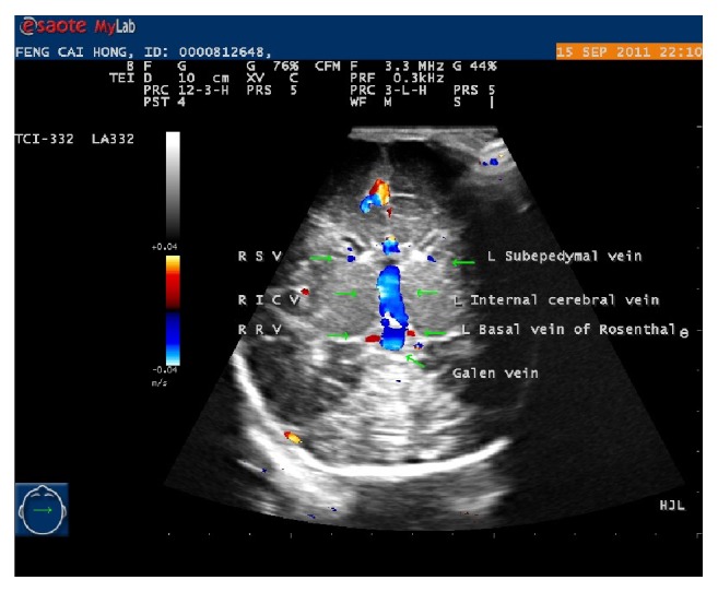
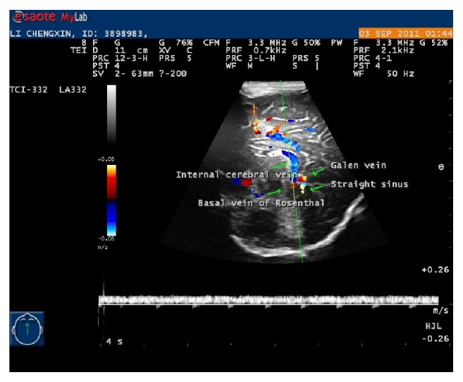
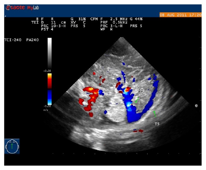
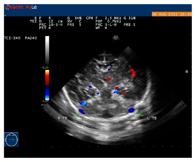
Result: In all studied cases (100% cases), subependymal veins (SV), internal cerebral veins (ICV), Galen vein (GV), straight sinus (SS), superior sagittal sinus (SSS), and transverse sinuses (TS) were visualized. The visualization percentages of inferior sagittal sinus (ISS) or basal veins/Rosenthal veins (BV/RV) were lower than 100%. Based on vessel visualization percentage from high to low, the vessels were ordered as follows: SV, ICV, BV, SS, TS, ISS, and SSS. In SSS and TS, the pulsation percentage was 100%. The descending percentages of vessel pulsation were noted in SS, BV, ICV, and SV. On the basis of the mean of maximum velocities of the vessels from low to high, the vessels were ordered as follows: ISS, BV-L, BV-R, ICV-R, ICV-L, SV-L, SV-R, SSS, TS-L, TS-R, and SS.
Conclusion: The measurements percent of visualization of cerebral deep veins was higher than the percent of cerebral venous sinuses. The pulsation percent of measurement and the velocities of cerebral venous sinuses were absolutely higher than the cerebral deep venous system. The pairs of vascular blood flow velocities were nonsignificantly different from one another.
Figures
Similar articles
-
Transtemporal power- and frequency-based color-coded duplex sonography of cerebral veins and sinuses.AJNR Am J Neuroradiol. 1997 Oct;18(9):1771-81. AJNR Am J Neuroradiol. 1997. PMID: 9367330 Free PMC article.
-
Intracranial venous system in the newborn: evaluation of normal anatomy and flow characteristics with color Doppler US.Radiology. 1992 May;183(2):449-52. doi: 10.1148/radiology.183.2.1561348. Radiology. 1992. PMID: 1561348
-
Transoccipital power-based color-coded duplex sonography of cerebral sinuses and veins.Stroke. 1997 Jul;28(7):1319-23. doi: 10.1161/01.str.28.7.1319. Stroke. 1997. PMID: 9227676
-
Ultrasound of the Fetal Veins Part 3: The Fetal Intracerebral Venous System.Ultraschall Med. 2016 Feb;37(1):6-26. doi: 10.1055/s-0035-1553284. Epub 2015 Jun 26. Ultraschall Med. 2016. PMID: 26114342 Review.
-
Cerebral veins and sinuses.Front Neurol Neurosci. 2006;21:182-193. doi: 10.1159/000092400. Front Neurol Neurosci. 2006. PMID: 17290137 Review.
Cited by
-
Reversal of blood flow in deep cerebral vein in preterm intraventricular hemorrhage: two case reports.BMC Pediatr. 2020 Nov 11;20(1):517. doi: 10.1186/s12887-020-02414-0. BMC Pediatr. 2020. PMID: 33172412 Free PMC article.
-
Paediatric cranial ultrasound: abnormalities of the brain in term neonates and young infants.Insights Imaging. 2025 Jul 22;16(1):159. doi: 10.1186/s13244-025-02031-4. Insights Imaging. 2025. PMID: 40696237 Free PMC article.
-
Use of greyscale and Doppler ultrasound in initial evaluation and follow-up of neurovascular malformations in children.Pediatr Radiol. 2024 Feb;54(2):347-356. doi: 10.1007/s00247-023-05846-9. Epub 2024 Jan 9. Pediatr Radiol. 2024. PMID: 38191809 Review.
-
Doppler Ultrasound Flow Reversal in the Superior Sagittal Sinus to Detect Cerebral Venous Congestion in Vein of Galen Malformation.AJNR Am J Neuroradiol. 2023 Jun;44(6):707-715. doi: 10.3174/ajnr.A7891. Epub 2023 May 25. AJNR Am J Neuroradiol. 2023. PMID: 37230540 Free PMC article.
-
Low frequency cerebral arterial and venous flow oscillations in healthy neonates measured by NeoDoppler.Front Pediatr. 2022 Nov 28;10:929117. doi: 10.3389/fped.2022.929117. eCollection 2022. Front Pediatr. 2022. PMID: 36518773 Free PMC article.
References
-
- Perlman J. M., Rollins N., Burns D., Risser R. Relationship between periventricular intraparenchymal echodensities and germinal matrix-intraventricular hemorrhage in the very low birth weight neonate. Pediatrics. 1993;91(2):474–480. - PubMed
-
- Perlman J. M., Risser R., Broyles R. S. Bilateral cystic periventricular leukomalacia in the premature infant: associated risk factors. Pediatrics. 1996;97:822–827. - PubMed
-
- Vople J. J. Neurology of the Newborn. 3rd. Philadelphia, Pa, USA: WB Saunders Co; 1995.
-
- Philips A. g., Allen W. C., Tito A. M., Wheeler L. R. Intraventricular haemorrhage in preterm infants: declining evidence in the 1980s. Pediatrics. 1989;84:797–801. - PubMed
MeSH terms
LinkOut - more resources
Full Text Sources
Research Materials
