

doi: 10.1007/s40477-019-00388-z.
Epub 2019 Jun 10.
Sonoanatomy of anterior forearm muscles
Affiliations
- PMID: 31183837
- PMCID: PMC6704193
- DOI: 10.1007/s40477-019-00388-z
Item in Clipboard
Sonoanatomy of anterior forearm muscles
J Ultrasound.
2019 Sep.
Abstract
The anterior or volar compartment of the forearm contains eight muscles: five belong to the superficial group (pronator teres, flexor carpi radialis, palmaris longus, flexor digitorum superficialis and flexor carpi ulnaris), and three to the deep group (flexor digitorum profundus, flexor pollicis longus and pronator quadratus). Knowledge of the topographic anatomy is essential for correct performance of ultrasound (US) examinations and correct interpretation of the images provided.
Keywords: Anterior compartment; Flexor muscles; Forearm; Sonoanatomy; Ultrasound.
Conflict of interest statement
The authors have no conflict of interest.
Figures
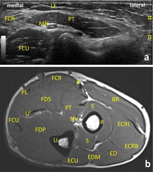
Ultrasound (US) (a) and magnetic resonance imaging (MRI) transversal/axial images of the proximal flexor compartment of the forearm (b). LF lacertus fibrosus, PT pronator teres, FCR flexor carpi radialis, FCU flexor carpi ulnaris, MN median nerve, FDS flexor digitorum superficialis, PL palmaris longus, FDP flexor digitorum profundus, S supinator, BR brachioradialis muscle, ECRL extensor carpi radialis longus, ECRB extensor carpi radialis brevis, ED extensor digitorum, EDM extensor digitorum minimi, ECU extensor carpi ulnaris, R radius, U ulna, NV neurovascular bundle with median nerve, brachial artery, and vein; R’ superior radial nerve, artery, and vein; U’ ulnar nerve, artery, and vein. MRI reprinted from Magnetic Resonance Imaging Clinics of North America, Vol. 19, Vogelius E, Hanna W, Robbin M. Magnetic Resonance Imaging of the Long Bones of the Upper Extremity, pages 567–579. Copyright (2019), with permission from Elsevier

Ultrasound (US) (a) and magnetic resonance imaging (MRI) (b) transversal/axial images of anatomy of the mid-forearm at the level of the deep extensor origins. Flexor digitorum superficialis is the largest of the superficial flexors, and arises by a humero-ulnar and a radial head. Flexor digitorum profundus arises deep to the superficial flexors from the posterior border of the ulna and extends distally almost to pronator quadratus. FDS flexor digitorum superficialis, FPD flexor digitorum profundus, FCR flexor carpis radialis, FCU flexor carpi ulnaris, FDS flexor digitorum superficialis, PL palmaris longus, FDP flexor digitorum profundus, BR brachioradialis muscle, ECRL extensor carpi radialis longus, ECRB extensor carpi radialis brevis, ED extensor digitorum, EDM extensor digitorum minimi, ECU extensor carpi ulnaris, EI extensor indicis, EPL extensor pollicis longus, EPB extensor pollicis brevis, APL abductor pollicis longus, R radius, U ulna, U’ ulnar nerve, artery, and vein; R’ superior radial nerve, artery, and vein; A anterior interosseous nerve, MN median nerve. MRI reprinted from Magnetic Resonance Imaging Clinics of North America, Vol. 19, Vogelius E, Hanna W, Robbin M. Magnetic Resonance Imaging of the Long Bones of the Upper Extremity, pages 567–579. Copyright (2019), with permission from Elsevier
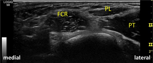
Flexor carpi radialis arises from the common flexor tendon, from the antebrachial fascia and from adjacent intermuscular septa. FCR flexor carpi radialis, PL palmaris longus, PT pronator teres
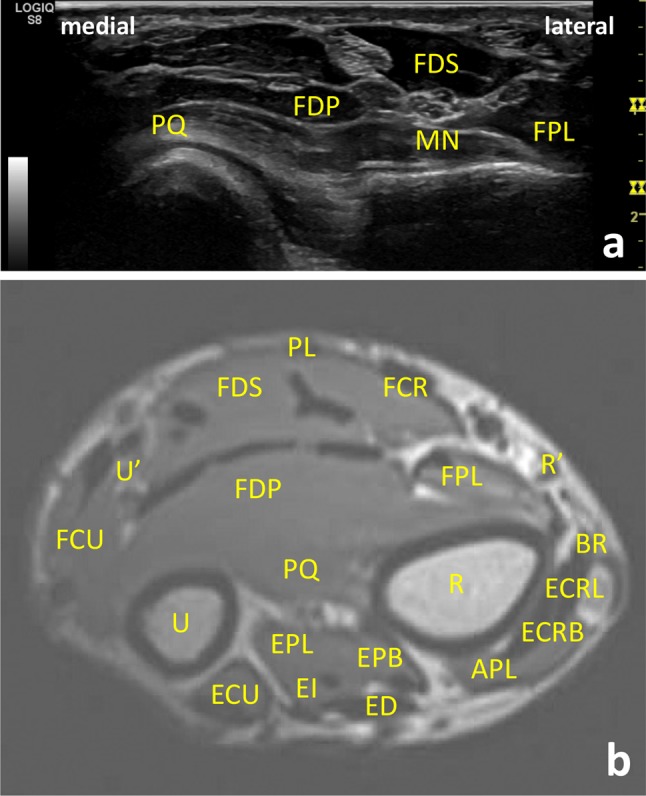
Ultrasound (US) (a) and magnetic resonance imaging (MRI) (b) transversal/axial images of anatomy of the distal forearm at the level of the proximal radial metaphysis. FDP flexor digitorum profundus, FPL flexor pollicis longus, PQ pronator quadratus, FSD flexor digitorum superficialis, FPL flexor pollicis longus, FDS flexor digitorum superficialis, FCR flexor carpis radialis, FCU flexor carpi ulnaris, PL palmaris longus, BR brachioradialis muscle, ECRL extensor carpi radialis longus, ECRB extensor carpi radialis brevis, ED extensor digitorum, ECU extensor carpi ulnaris, EI extensor indicis, EPL extensor pollicis longus, EPB extensor pollicis brevis, APL abductor pollicis longus, R radius, U ulna, U’ ulnar nerve, artery, and vein, R’ superior radial nerve, artery, and vein, MN median nerve. MRI reprinted from Magnetic Resonance Imaging Clinics of North America, Vol. 19, Vogelius E, Hanna W, Robbin M. Magnetic Resonance Imaging of the Long Bones of the Upper Extremity, pages 567–579. Copyright (2019), with permission from Elsevier
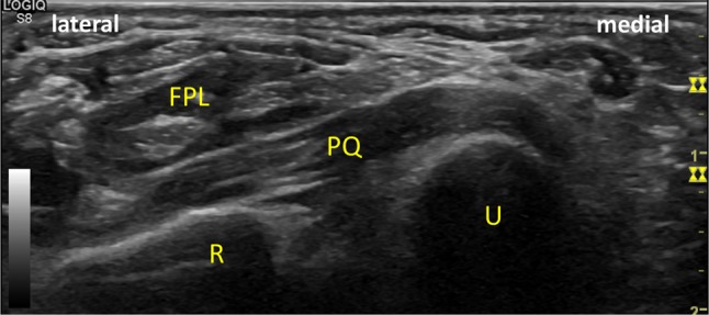
Pronator quadratus extends across the front of the distal parts of the radius and ulna. FPL flexor pollicis longus, PQ pronator quadratus, R radius, U ulna
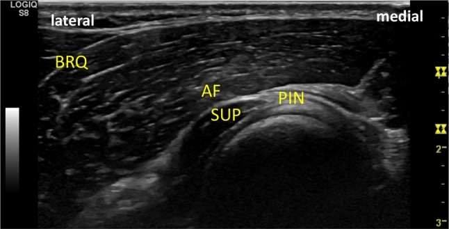
At the radial aspect of the forearm, the extensor carpi radialis brevis and longus and the brachioradialis form the so-called mobile wad. The supinator is the deepest of the lateral muscles. BRQ brachioradialis, SUP supinator
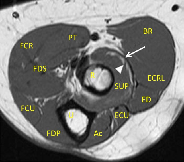
Magnetic resonance imaging (MRI) axial image of anatomy of the proximal forearm. After branching from the main radial nerve at the elbow, the posterior interosseous nerve (PIN) passes beneath the fascia of the supinator muscle (arcade of Frohse). Arrow: arcade of Frohse, arrowhead: PIN, SUP supinator, R radius, U ulna, BR brachioradialis muscle, ECRL extensor carpi radialis longus, ED extensor digitorum, Ac Anconeus muscle, ECU extensor carpi ulnaris, PT pronator teres, FCR flexor carpis radialis, FDS flexor digitorum superficialis, FDP flexor digitorum profundus, FCU flexor carpi ulnaris. MRI—Courtesy Dr. Michael Stadnick, in Stadnick M. Posterior Interosseous Nerve Syndrome. MRI Web Clinic, 2005. Retrieved from https://radsource.us/posterior-interosseous-nerve-syndrome/
References
-
- Lannotti J, Parker R. Netter collection of medical illustrations: part I the upper limb. 2. Amsterdam: Elsevier; 2013. pp. 109–148.
-
- Standing S (2016) Elbow and forearm. In: Gray's anatomy, 41th edn. Elsiever, Amsterdam, pp 837–861
-
- Reavey P, Rafijah G (2017) Anatomy and examination of the hand, wrist, forearm, and elbow. In: Principles of hand surgery and therapy, 3rd edn. Elsevier, Amsterdam, pp e1–e25
-
- Bianchi S, Martinoli C. ultrasound of the musculoskeletal system. New York: Springer; 2007. Forearm; pp. 409–423.
MeSH terms
LinkOut - more resources
Full Text Sources
