Review
. 2019 Nov 1;31(13):954-993.
doi: 10.1089/ars.2019.7818. Epub 2019 Jul 11.

The Role of HMGB1, a Nuclear Damage-Associated Molecular Pattern Molecule, in the Pathogenesis of Lung Diseases

Affiliations
Review

The Role of HMGB1, a Nuclear Damage-Associated Molecular Pattern Molecule, in the Pathogenesis of Lung Diseases

Mao Wang et al. Antioxid Redox Signal. .

Abstract

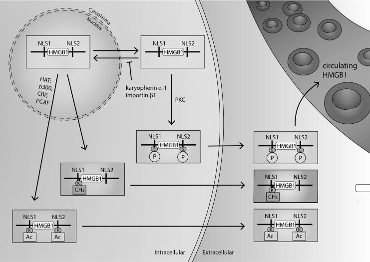
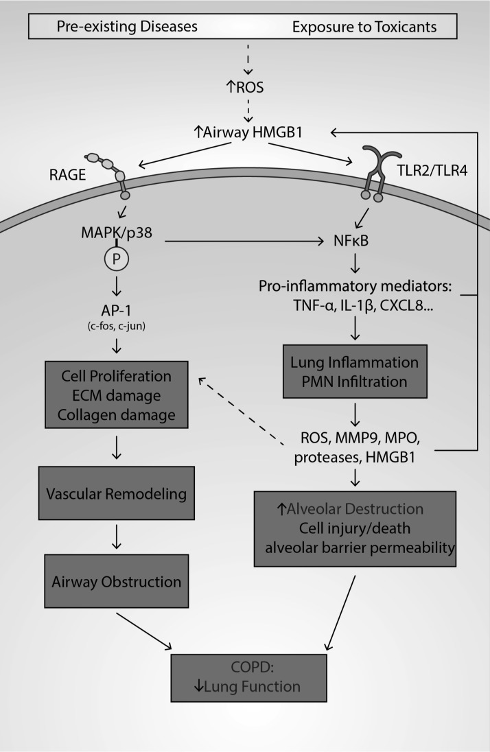
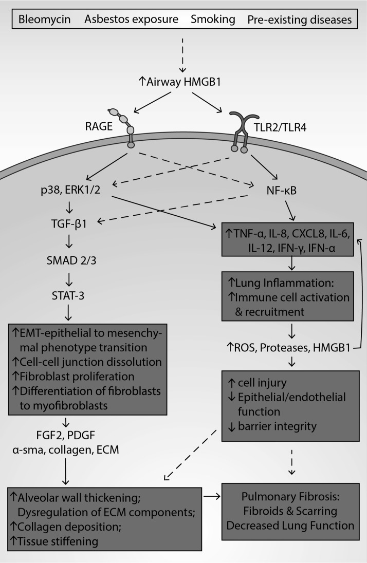
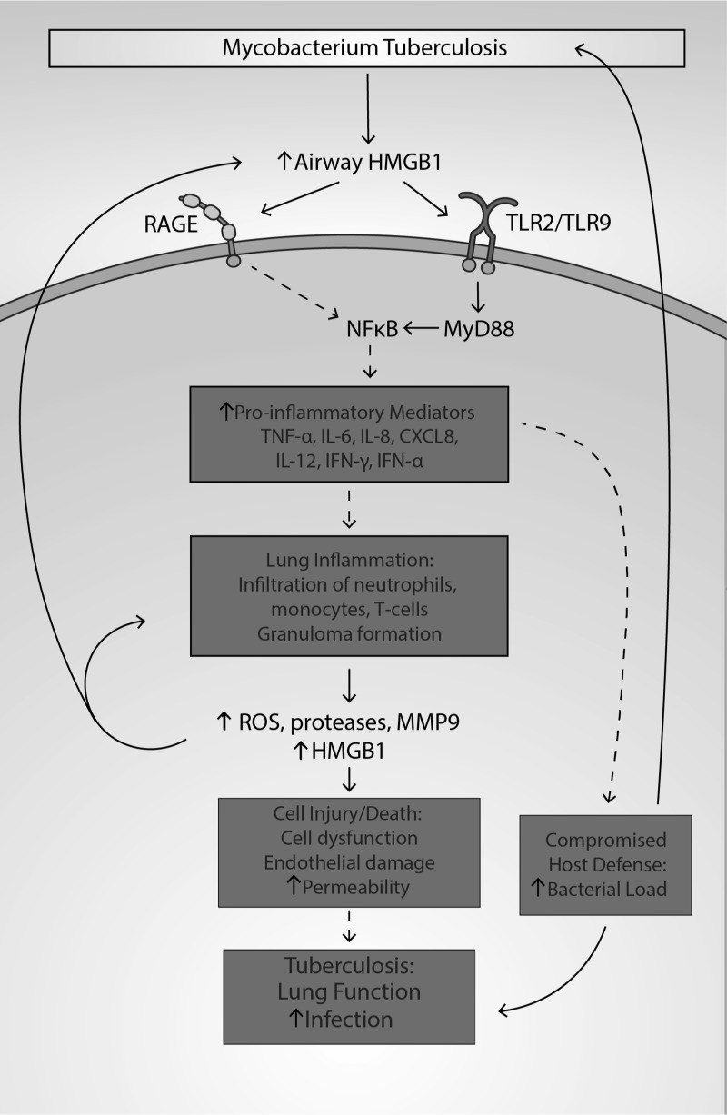
Significance: High-mobility group protein box 1 (HMGB1), a ubiquitous nuclear protein, regulates chromatin structure and modulates the expression of many genes involved in the pathogenesis of lung cancer and many other lung diseases, including those that regulate cell cycle control, cell death, and DNA replication and repair. Extracellular HMGB1, whether passively released or actively secreted, is a danger signal that elicits proinflammatory responses, impairs macrophage phagocytosis and efferocytosis, and alters vascular remodeling. This can result in excessive pulmonary inflammation and compromised host defense against lung infections, causing a deleterious feedback cycle. Recent Advances: HMGB1 has been identified as a biomarker and mediator of the pathogenesis of numerous lung disorders. In addition, post-translational modifications of HMGB1, including acetylation, phosphorylation, and oxidation, have been postulated to affect its localization and physiological and pathophysiological effects, such as the initiation and progression of lung diseases. Critical Issues: The molecular mechanisms underlying how HMGB1 drives the pathogenesis of different lung diseases and novel therapeutic approaches targeting HMGB1 remain to be elucidated. Future Directions: Additional research is needed to identify the roles and functions of modified HMGB1 produced by different post-translational modifications and their significance in the pathogenesis of lung diseases. Such studies will provide information for novel approaches targeting HMGB1 as a treatment for lung diseases.

Keywords: HMGB1; cancer; fibrosis; inflammatory lung injury; pulmonary infection; pulmonary vascular remodeling.


Conflict of interest statement

The authors declare no competing financial interest.

Figures

<b>FIG. 1.</b>
FIG. 1.
The structure and function determining sequence of HMGB1. Human HMGB1 is a protein with 215 amino acids, encoded by the gene located at chromosome 13q12.3. HMGB1 contains two DNA-binding domains: the A Box (amino acids 9–79) and B-Box (amino acids 95–163), and a C-terminal tail (amino acids 186–215), which is involved in promoting the interaction of A and B box with DNA. HMGB1 contains two NLS, which are located at amino acids 28–44 (NLS1) and 179–185 (NLS2), responsible for the nuclear localization of HMGB1 and for regulating HMGB1's translocation between the nucleus and the cytoplasm on post-translational modifications, such as phosphorylation and acetylation. There are three critical cysteines (C23, C45, and C106) subject to redox modifications, which determine whether HMGB1 functions as a cytokine, a chemokine, or an inactive protein. HMGB1 also has a heparin binding site (amino acids 6–12), a TLR4 binding site (amino acids 89–108), and an RAGE binding site (amino acids 150–183). HMGB1, high-mobility group protein box 1; NLS, nuclear localization signals; RAGE, receptor for advanced glycation end products; TLR, toll-like receptor.
<b>FIG. 2.</b>
FIG. 2.
Regulation of HMGB1 localization. HMGB1 is a nuclear nonhistone binding protein that can shuttle between the nucleus and the cytosol through nuclear pores. HMGB1 contains two nuclear localization sequences (NSL1 and NLS2). These NLS are post-translationally modified by hyperacetylating lysine residues within NLS1 and NLS2. Hyperacetylation of NLS by HAT (p300, PCAF, CBP) is required to induce nucleocytoplasmic translocation. Also, the phosphorylation of cytoplasmic HMGB1 by PKC can cause HMGB1 to bind with karyopherin-α-1 and importin-β-1, which can block its nuclear import, keeping it in the cytoplasm. In addition, the methylation of lysine-42 at NLS1 alters HMGB1 conformation, which can result in its decreased ability to bind with DNA and causing HMGB1's passive diffusion to the cytoplasm. Under conditions of oxidative stress, HMGB1 translocates from the nucleus to the cytoplasm through nuclear export factor 1 (CRM1). Cytoplasmic HMGB1 can then be secreted from the cell in secretory vesicle-mediated exocytosis. Alternatively, HMGB1 can be passively secreted from injured or necrotic cells. Whether passively or actively secreted, HMGB1 then can accumulate in extracellular spaces such as the airways and circulating blood. Extracellular HMGB1 also is subjected to redox modifications of three critical cysteine residues (C23, C45, and C106). When HMGB1 fully reduces it can function as a cytokine and when HMGB1 is oxidized it forms a disulfide bond between C23 and C45, which imparts chemokine and cytokine activity. HMGB1 can be terminally oxidized (sulfonic acid) at all three cysteines, which inactivates all activity. CRM1, chromosome region maintenance 1; HAT, histone acetyltransferase; PCAF, p300/CBP-associated factor; PKC, protein kinase C.
<b>FIG. 3.</b>
FIG. 3.
The role of HMGB1 in the pathogenesis of lung diseases. In the studies reviewed in this article, elevated extracellular HMGB1, whether it is in the airways, serum, or other extracellular milieu, can signal as a DAMP through similar pathways that lead to the pathogenesis of different lung diseases. The binding of extracellular HMGB1 to its receptors, such as TLR2, TLR4, TLR9, and RAGE, can initiate innate immune responses by increasing NF-κB and MAPK activities. The activation of NF-κB is responsible for the increased secretion of numerous proinflammatory mediators, including the active release of HMGB1 into the airways. Proinflammatory mediators can induce the infiltration of leukocytes into the lungs and airways, leading to lung inflammation. This further causes an elevation in the levels of proteases and ROS, which feeds back and contributes to increased NF-κB activity. A significant elevation in ROS can damage and cause dysfunction, injury, and death to lung cells, including macrophages and endothelial and epithelial cells. Both ROS and extracellular HMGB1 can directly disrupt epithelial and endothelial intercellular tight junctions, reduce their barrier integrity, and result in increased alveolar permeability. Macrophage injury reduces efferocytosis, which contributes to the higher leukocyte counts, as well as impairs the phagocytosis of invading bacteria, resulting in increased susceptibility to infections. Pulmonary infections and damage to the lung cells further increase the accumulation of extracellular HMGB1, establishing a negative feedback cycle. In addition, the binding of HMGB1 to RAGE receptors increases MAPK activity, altering cell proliferation and inducing vascular remodeling. Overall, HMGB1-induced inflammatory lung cell damage, in combination with vascular remodeling, can significantly compromise lung functions and cause lung diseases. DAMP, damage-associated molecular pattern; MAPK, mitogen-activated protein kinase; NF-κβ, nuclear factor kappa-light-chain-enhancer of activated B cells; ROS, reactive oxygen species.
<b>FIG. 4.</b>
FIG. 4.
The role of HMGB1 in the pathogenesis of asthma. Asthma is pathologically characterized by airway inflammation, vascular remodeling, airway hyperreactivity, and constriction. Extracellular HMGB1 levels have been reported to be increased in asthma patients. The increased levels of airway HMGB1 in asthma can produce inflammation and vascular remodeling. Airway HMGB1 can bind to RAGE, TLR2, and TLR4. The binding of HMGB1 to these receptors can activate NF-κB and MAPK pathways. NF-κB activation stimulates the expression and secretion of proinflammatory mediators such as IL-8, IL-4, IL-5, IFN-γ, IFN-α, IL-13, IL-6, and IL-17, initiating the infiltration of eosinophils, neutrophils, and helper T cells into the airway. The presence of activated neutrophils in the lungs can increase the levels of ROS, proinflammatory cytokines, proteases (e.g., MMP9), and HMGB1, which can produce cell injury, further increasing neutrophil infiltration. HMGB1-mediated NF-κB activation can also promote the secretion of VEGF and TGF-β, which are involved in vascular remodeling. The subsequent vascular remodeling is characterized by loss of epithelial integrity, increased smooth muscle mass, smooth muscle cell proliferation, and subepithelial fibrosis. TGF-β-induced remodeling can result in an increase in lung fibroblast migration, mucus production, and secretion of ECM components as well as levels of MMP9, collagen, and α-sma. The airway injury that occurs as a result of inflammation and vascular remodeling produces airway hyperreactivity and reversible airway obstruction. ECM, extracellular matrix; IFN, interferon; IL, interleukin; MMP9, matrix metalloproteinase 9; sma, smooth muscle actin; TGF-β, transforming growth factor-β; VEGF, vascular endothelial growth factor.
<b>FIG. 5.</b>
FIG. 5.
The role of HMGB1 in the pathogenesis of COPD. COPD, caused by chronic inhalation of environmental pollutants, cigarette smoking, or preexisting diseases, is pathologically characterized by chronic obstruction of airways, alveolar dysfunction, and airway inflammation. Increased levels of ROS in the lungs of subjects with COPD can induce the accumulation of airway HMGB1. Airway HMGB1, via a number of mechanisms, can exacerbate COPD by inducing excessive lung inflammation and vascular remodeling. Lung inflammation is induced by HMGB1 binding to TLR2, TLR4, and RAGE, which increases the likelihood of NF-κB translocating to the nucleus. Subsequently, NF-κB increases the expression and secretion of proinflammatory cytokines such as IL-1β, IL-8, TNF-β, IFN-γ, and MCP-1, which induce leukocyte infiltration and produce inflammation. Increased MPO activity, MMP9, and ROS levels from the neutrophils/monocytes can cause damage to the tight junctions of endothelial/epithelial cells, causing a loss of barrier integrity. Furthermore, the increased levels of elastase, proteases, MPO, and ROS can damage epithelial/endothelial cells, causing increased vascular and alveolar permeability, subsequently contributing to further HMGB1 accumulation in the airways and exacerbating lung inflammation. The injury and death of endothelial and epithelial cells can further result in alveolar destruction and a decline in lung function. HMGB1 also signals through RAGE to activate MAPK pathways. MAPK can activate the transcription factor AP-1, promoting proliferation of pulmonary endothelial and smooth muscle cells. Neutrophil elastase and MMP9 contribute to ECM and elastic fiber damage. Overall, these aforementioned changes promote vascular remodeling, which contributes to airway obstruction. AP-1, activator protein 1; COPD, chronic obstructive pulmonary disease; MCP-1, monocyte chemoattractant protein-1; MPO, myeloperoxidase; TNF, tumor necrosis factor.
<b>FIG. 6.</b>
FIG. 6.
The role of HMGB1 in the pathogenesis of ALI and ARDS. ALI and ARDS are caused by various conditions and characterized by excessive lung inflammation, increased alveolar permeability, and impairment of respiratory function. Increased levels of airway HMGB1 in patients and animals with ALI and ARDS induce the accumulation of airway HMGB1. The deleterious effects of airway HMGB1 are mediated by excessive lung inflammation and alveolar destruction. Airway HMGB1 induces lung inflammation by binding to TLR2, TLR4, and RAGE, which increases the likelihood of NF-κB translocating to the nucleus, where it increases the transcription of genes that code for proinflammatory cytokines such as IL-1β, TNF-α, IL-4, and IL-6, causing leukocyte infiltration and inflammation. Leukocytes increase the levels of MPO activity, ROS production, and proteases, thereby damaging endothelial/epithelial cells and their tight junctions, causing the dysfunction of endothelial/epithelial and other lung cells, which compromises alveolar barrier and lung function. In addition, the activation of NF-κB is associated with HMGB1 hyperacetylation, which leads to its subsequent translocation to the cytoplasm and its active release into the airways. The increased production of ROS, such as NO, can inhibit SIRT-1 (a deacetylase enzyme) and can further stimulate HMGB1 release. Airway HMGB1 can also signal through RAGE-activated MAPK/p38 pathways, which can induce the phosphorylation of Hsp27, an actin-binding protein. Phosphorylated Hsp27 can produce endothelial cytoskeleton disorganization, paracellular gap formation, and the loss of peripheral organized actin fibers. These effects produced by phosphorylated Hsp27 can produce cell/cell junction dissociation and a loss of alveolar barrier integrity, which also contribute to a decrease in lung function. The accumulation of airway HMGB1 can be attenuated by cholinergic anti-inflammatory pathway modulators (e.g., endogenous acetylcholine or GTS-21) and NF-κB inhibitors (e.g., ethyl pyruvate or GTS-21). Antioxidants, such as ascorbic acid, hydrogen sulfide (in gaseous form), and resveratrol, can also decrease ROS levels and decrease airway HMGB1 levels. ALI, acute lung injury; ARDS, acute respiratory distress syndrome; Hsp27, heat shock protein 27; SIRT-1, Sirtuin-1.
<b>FIG. 7.</b>
FIG. 7.
The role of HMGB1 in the pathogenesis of PF. PF, an interstitial lung disease, is characterized by fibroblast proliferation, EMT, lung inflammation and the formation of fibroids, producing lung scarring. Patients and animals with PF have been shown to have increased levels of airway HMGB1. HMGB1 may be involved in the pathogenesis of PF as it can produce fibroblast differentiation and proliferation, alveolar wall thickening or muscularization thickening, and collagen accumulation. HMGB1 binds to RAGE, TLR2, and TLR4, activating both NF-κB and MAPK/p38/ERK 1/2 pathways. NF-κB activation and the p38 pathway can increase the levels of proinflammatory mediators and TGF-β1. As a mediator of vascular remodeling, TGF-β1 can induce phosphorylation of SMAD2 and SMAD3, which phosphorylate the transcription factor STAT-3. STAT-3, when activated, induces cell proliferation and myofibroblast differentiation. STAT-3 activation promotes EMT, where epithelial cells develop mesenchymal characteristics such as loss of cell/cell adhesion. The increased number of fibroblasts and myofibroblasts can stimulate the formation of fibrotic tissue. TGF-β1 can elicit the release of α-sma, upregulate HMGB1 expression, and induce secretion of the profibrotic factors FGF2 and PDGF. α-sma directly stimulates the EMT process, causing an increased accumulation of ECM proteins, which augments airway remodeling. ECM-mediated remodeling involves the dysregulation of ECM composition, cleavage of ECM components, increased collagen deposition, and stiffening of tissue. In addition to inducing remodeling, the binding of HMGB1 to RAGE and TLR4 stimulates the expression and secretion of TNF-α, MCP-1, IL-8, and IL-1β. These proinflammatory mediators recruit and induce the accumulation of macrophages, neutrophils, and lymphocytes into the airways, increasing the levels of proteases, ROS, and HMGB1, establishing a deleterious feedback loop. The release of these molecules contributes to epithelial and endothelial injury and death, dissolution of cell/cell junctions, and loss of barrier integrity, thereby increasing alveolar permeability. Thus, HMGB1-mediated airway remodeling and inflammation contribute to lung fibrosis, scarring, and decreased lung function. EMT, epithelial to mesenchymal transition; ERK, extracellular signal-regulated kinase; FGF2, fibroblast growth factor 2; PF, pulmonary fibrosis; STAT-3, signal transducer and activator of transcription 3.
<b>FIG. 8.</b>
FIG. 8.
The role of HMGB1 in the pathogenesis of CF. CF is a disease caused by mutations in the CFTR gene and its pathological hallmarks include neutrophilic inflammation and bacterial infections. An increased expression and accumulation of HMGB1 are present in CF airways, resulting in an increase in HMGB1 levels in bronchoalveolar lavage fluids, sputum, and serum of CF patients. HMGB1 elicits chemotactic activity by interacting with the chemokine CXCR2 receptors on neutrophils, thereby recruiting them into the airways, compromising macrophage phagocytosis. In CF, HMGB1 may produce an impairment of macrophage efferocytosis, which decreases the removal of apoptotic neutrophils and the clearance of pathogenic bacteria. We postulate that the activation of the TLR4 receptor by HMGB1 attenuates macrophage function, producing a higher bacterial load and excessive inflammation in the airways and lung tissue, causing lung injury. The increase in PAMP and DAMP biomolecules from the increased bacterial load and excessive inflammation further stimulates the release of HMGB1 from airway cells, producing a deleterious feedback cycle between the release of HMGB1 and the development of lung injury. In addition, serum HMGB1 impairs the function of pancreatic β cells by activating RAGE and TLR4, producing insulin deficiency and insulin resistance, causing CFRD. Insulin and ODSH can reduce HMGB1 expression and translocation from the nucleus to cytoplasm in cells (epithelial cells and macrophages, respectively). ODSH can also reduce chronic neutrophil-mediated inflammation induced by neutrophil elastase in CF. CF, cystic fibrosis; CFRD, cystic fibrosis-related diabetes; CXCR, CXC-chemokine receptor; ODSH, 2-O,3-O desulfated heparin; PAMP, pathogen-associated molecular pattern.
<b>FIG. 9.</b>
FIG. 9.
The role of HMGB1 in the pathogenesis of pneumonia. Pneumonia is a disease caused by bacteria, viruses, and fungi that can produce pulmonary inflammation. Risk factors include the presence of other diseases such as CF, COPD, and asthma. PAMP molecules released by viruses and bacteria interact with cell surface receptors such as TLRs and RAGE, which increases oxidative stress and the expression of HMGB1, leading to its increased translocation and subsequent release of HMGB1 from airway cells such as macrophages and epithelial cells. The release of HMGB1 induced by LPS is JNK dependent. The accumulation of HMGB1 in the airway activates NF-κB and ERK/p38 pathways by binding to TLRs and RAGE on airway cells, increasing the production of ROS, inflammatory cytokines, ICAM-1, and vascular cell adhesion molecule 1 (VCAM-1). Airway HMGB1 can also induce the expression of TLR2 on certain cells, further increasing inflammatory responses in the lung. These molecules mentioned above can elicit excessive inflammation and tissue damage in the lung. ICAM-1, intercellular adhesion molecule 1; JNK, c-Jun N-terminal kinase; LPS, lipopolysaccharide; VCAM-1, vascular cell adhesion molecule 1.
<b>FIG. 10.</b>
FIG. 10.
The role of HMGB1 in the pathogenesis of TB. TB is a respiratory infection caused by Mycobacterium tuberculosis and is associated with lung function impairment, pulmonary obstruction, and granulitic infiltrate. TB infection elicits an inflammatory response, the infiltration of neutrophils, monocytes, and T cells, and the formation of granulomas. HMGB1 contributes to the pathology of TB by inducing granuloma formation, increasing lung bacterial burden, and promoting excessive inflammation. In subjects with TB, there is an increase in the extracellular levels of HMGB1 in the airways and in lung tissues. Airway HMGB1 can bind to the receptors RAGE, TLR2, and TLR9, which can activate NF-κB and upregulate the expression of genes that code for proinflammatory mediators such as IFN-γ, TNF-α, IL-6, CXCL8, and IL-8. Increased levels of these proinflammatory mediators promote recruitment of monocytes and CD4/CD8 lymphocytes and neutrophils. The recruitment and accumulation of immune cells are associated with the formation of granulomas, which undergo necrosis and augment inflammation. Necrosis of cells can cause the release of cytokines and ROS, which perpetuates immune cell recruitment and activation. The accumulation of neutrophils exacerbates airway inflammation by inducing excessive levels of ROS, proteases, and HMGB1, causing cell dysfunction, endothelial damage, and increased permeability of the alveolar epithelium. These cellular changes contribute to the increase in the pulmonary bacterial burden that further augments airway HMGB1, producing a deleterious feedback cycle. Therefore, these factors contribute to infection and decreased pulmonary function in TB. CD, cluster of differentiation; TB, tuberculosis.
<b>FIG. 11.</b>
FIG. 11.
The role of HMGB1 in the pathogenesis of lung cancer. The levels of HMGB1 expression in tumors and the subsequent release of HMGB1 into the blood serum are positively correlated to tumor prognosis. The role of HMGB1 in lung cancer results from its localization. HMGB1 in the nucleus facilitates the transcription of genes involved in tumor development. A high expression level of HMGB1 in the nucleus increases the transcription of genes involved in tumor cell growth (e.g., cdc25A, FGFR), proliferation (e.g., met), migration (e.g., MMP), and angiogenesis (e.g., FGFR). The passive or active release of HMGB1 into the airways can have opposing roles in terms of mediating tumor growth and development. HMGB1 can interact with cell surface receptors such as TLR4, RAGE, and CXCR4 on airway cells, which activates the inflammasome, NF-κB, JNK/p38, and Ras-Raf-Erk1/2 pathways and increases tumor cell proliferation, migration, and invasion. However, the acute release of HMGB1 can induce the maturation of antigen presenting cells through activation of TLR4, which further activates tumor antigen-specific T cells and produces antitumor effects. FGFR, fibroblast growth factor receptor.
<b>FIG. 12.</b>
FIG. 12.
The role of HMGB1 in the pathogenesis of PAH. PAH is a disease that is characterized by increased pulmonary arterial pressure, vasoconstriction, and thrombosis. Right ventricular heart failure results from an increase in peripheral vascular resistance, systolic and diastolic pressure, pulmonary arterial pressure, venous hypertrophy, and a thickening of arterial walls. Inflammation and vascular remodeling are major pathological changes in pulmonary hypertension. Patients with PAH have significantly elevated levels of airway HMGB1. These airway levels of HMGB1 can interact with RAGE to activate the p38/ERK/JNK pathway that signals via AP-1 to promote proliferation of smooth muscle cells and endothelial cells. This increased proliferation produces vascular remodeling, venous hypertrophy, and thickening of arterial walls. Vascular remodeling contributes to vasoconstriction, which can increase vascular pressure. HMGB1 also can bind to RAGE and TLR4 to induce excessive inflammation by activating NF-κB. The activation of NF-κB increases the transcription of TGF-β1 to elicit vascular remodeling. NF-κB also increases the synthesis of the proinflammatory mediators MCP-1, CXCL8, IL-6, and TNF-α. TNF-α can increase neutrophil infiltration and plays a role in vascular remodeling by eliciting ET-1 secretion. ET-1 stimulates vasoconstriction and further augments the pathology of pulmonary hypertension. Also, IL-6, MCP-1, and CXCL8 mediate neutrophil infiltration and recruitment of immune cells. This leads to increased levels of ROS, MMP9, MPO, proteases, HMGB1, and endothelial dysfunction, which can stimulate vascular remodeling, formation of vascular lesions, and a further increase in the levels of HMGB1. Vascular lesions and remodeling can cause cell injury and death, contributing to increased pulmonary arterial pressure and pulmonary hypertension. ET-1, endothelin-1; PAH, pulmonary arterial hypertension.
<b>FIG. 13.</b>
FIG. 13.
HMGB1 as a therapeutic target in pulmonary diseases. Preexisting trauma, exposure to toxicants, PAMPs, or DAMPs can induce the release of HMGB1, either active or passively, and subsequently the accumulation of HMGB1 in the airways of patients with pulmonary diseases. Post-translational modifications such as hyperacetylation and phosphorylation result in the translocation of HMGB1 into the cytoplasm, where HMGB1 can be actively released. The hyperacetylation of HMGB1 can be decreased by silent mating type information regulation 2 homolog 1 (SIRT)-1 activators (e.g., resveratrol), NF-κB inhibitors (e.g., ethyl pyruvate, GTS-21, naringin, hesperidin, FPS-ZMI), Nrf2 activators (e.g., baicalin, glycyrrhizin, hydrogen sulfide gas, resveratrol), and ODSH. Antioxidants (e.g., ascorbic acid, sulforaphane), as well as other compounds (e.g., vitamin D, insulin), can also inhibit the cytoplasmic translocation of HMGB1. Similar to antibodies, compounds such as ODSH, glycyrrhizin, and thrombomodulin can bind directly to, neutralize, or inactivate extracellular HMGB1, thereby decreasing the binding of HMGB1 to its receptors. Extracellular HMGB1 can bind to TLR2, TLR4, TLR9, and RAGE receptors to activate pathways involved in inflammation, tissue injury, and tissue remodeling. Following the binding of HMGB1 to TLR2 and TLR4, it can activate the NF-κB pathway, which ultimately increases the expression and secretion of proinflammatory mediators and further augments the secretion of HMGB1, creating a deleterious feedback cycle. Also, HMGB1 can bind with RAGE receptors and activate MAPK-dependent pathways that mediate tissue injury and remodeling. MAPK inhibitors (e.g., ulinastatin, naringin) can decrease the accumulation of airway HMGB1. The accumulation of airway HMGB1 and proinflammatory mediators increases the levels of ROS, MMP9, heat shock proteins, and certain proteases that produce oxidative stress, inflammation, tissue injury, and vascular remodeling. NF-κB inhibitors (e.g., ethyl pyruvate, GTS-21, naringin, hesperidin, FPS-ZMI) can attenuate cytokine and HMGB1 airway accumulation. Antioxidants, PPAR-γ, and Nrf2 activators (e.g., vitamin D, glycyrrhizin, baicalin, resveratrol, ethyl pyruvate) can decrease inflammation, lung injury, and remodeling by decreasing oxidative stress induced by the accumulation of HMGB1 in the airways. FPS-ZMI, N-benzyl-4-chloro-N-cyclohexylbenzamide; Nrf2, nuclear factor erythroid 2-related factor 2; PPAR, peroxisome proliferator-activated receptor.

References

    1. Aaron SD, Angel JB, Lunau M, Wright K, Fex C, Le Saux N, and Dales RE. Granulocyte inflammatory markers and airway infection during acute exacerbation of chronic obstructive pulmonary disease. Am J Respir Crit Care Med 163: 349–355, 2001 - PubMed
    1. Abeyama K, Stern DM, Ito Y, Kawahara K, Yoshimoto Y, Tanaka M, Uchimura T, Ida N, Yamazaki Y, Yamada S, Yamamoto Y, Yamamoto H, Iino S, Taniguchi N, and Maruyama I. The N-terminal domain of thrombomodulin sequesters high-mobility group-B1 protein, a novel antiinflammatory mechanism. J Clin Invest 115: 1267–1274, 2005 - PMC - PubMed
    1. Abraham E, Arcaroli J, Carmody A, Wang H, and Tracey KJ. HMG-1 as a mediator of acute lung inflammation. J Immunol 1950 165: 2950–2954, 2000 - PubMed
    1. Achouiti A, van der Meer AJ, Florquin S, Yang H, Tracey KJ, van't Veer C, de Vos AF, and van der Poll T. High-mobility group box 1 and the receptor for advanced glycation end products contribute to lung injury during Staphylococcus aureus pneumonia. Crit Care 17: R296, 2013 - PMC - PubMed
    1. Agustí AGN, Roca J, Gea J, Wagner PD, Xaubet A, and Rodriguez-Roisin R. Mechanisms of gas-exchange impairment in idiopathic pulmonary fibrosis. Am Rev Respir Dis 143: 219–225, 1991 - PubMed

Publication types