Review
. 2019 Jun;54(6):589-602.
doi: 10.4085/1062-6050-472-17. Epub 2019 Jun 11.

The Ankle-Joint Complex: A Kinesiologic Approach to Lateral Ankle Sprains

Affiliations
Review

The Ankle-Joint Complex: A Kinesiologic Approach to Lateral Ankle Sprains

Jennifer M Medina McKeon et al. J Athl Train. 2019 Jun.

Abstract

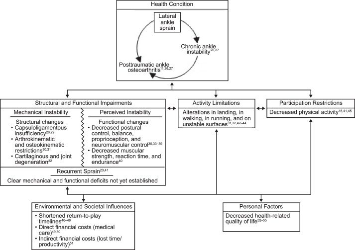
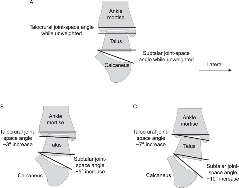
Copious research exists regarding ankle instability, yet lateral ankle sprains (LASs) persist in being among the most common recurrent musculoskeletal injuries. Key anatomical structures of the ankle include a triform articulating structure that includes the inferior tibiofibular, talocrural, and subtalar joints. Functionally, force absorption and propulsion through the ankle complex are necessary for any task that occurs in weight bearing. For optimal ankle performance and avoidance of injury, an intricate balance between stability and mobility is necessary to ensure that appropriate force transfer occurs during sports and activities of daily living. Consideration for the many structures that may be directly or indirectly involved in LASs will likely translate into advancements in clinical care. In this clinical review, we present the structure, function, and relevant pathologic states of the ankle complex to stimulate a better understanding of the prevention, evaluation, and treatment of LASs.

Keywords: anatomy; biomechanics; joint injury; lower extremity.


Figures

Figure 1
Figure 1
The effect of lateral ankle sprain in the context of the International Classification of Functioning, Disability, and Health.
Figure 2
Figure 2
The functional subtalar joint. The functional subtalar-joint complex is divided into the 2 compartments: the posterior compartment (talocalcaneal joint) and the anterior compartment (talocalcaneonavicular joint). Stability is provided by the extrinsic ligaments (the calcaneofibular and deltoid ligaments) and a series of broad, thick intrinsic ligaments situated in the tarsal canal.
Figure 3
Figure 3
The foot columns and subtalar-joint motion in the closed kinetic chain. These combined frontal-plane and transverse-plane motions occur simultaneously, resulting in a multiplanar rotation. This is different from single-plane, noncardinal motion and is a multiplanar rotary motion that is similar to the motion occurring at the knee. A, Supination. “Stacking” of the medial foot column laterally and vertically over the lateral foot column results in a higher, narrower foot. B, Pronation. “Unstacking” of the medial foot column medially away from the lateral foot column results in a wider, flatter foot.
Figure 4
Figure 4
The rockers. The 3 rockers of gait describe the foot and leg rotational motions during the absorption and propulsions stages of the stance phase of gait. A, Heel rocker. The foot is lowered to the ground around the calcaneus (heel). B, Ankle rocker. The leg advances over the foot around the talocrural joint. C, Forefoot rocker. The heel lifts off the ground around the metatarsophalangeal joints.
Figure 5
Figure 5
Ligaments relevant to stability of the ankle complex. A, Anterior view. B, Posterior view.
Figure 6
Figure 6
Ligaments relevant to stability of the ankle complex. A, Lateral view. B, Medial view.
Figure 7
Figure 7
The relevant neuromuscular structures of the ankle complex.
Figure 8
Figure 8
Sagittal-plane (medial) view of the talocrural and subtalar joints. A, Unweighted ankle. B, Displacement under stress of the stable ankle. C, Displacement under stress of the unstable ankle. Unstable ankles can have an average of 2 times the displacement of the talus on the mortise and the calcaneus on the talus. The anterior drawer stress test tends to be more specific to increased motion at the talocrural joint than the subtalar joint.
Figure 9
Figure 9
Frontal-plane (posterior) view of the talocrural and subtalar joints. A, Unweighted ankle. B, Joint displacement under the stress of the stable ankle. C, Joint displacement under the stress of the unstable ankle. Unstable ankles can have an average of twice the displacement of the talus on the mortise and the calcaneus on the talus. An inversion stress test tends to be more specific to increased motion at the subtalar joint than the talocrural joint.

References

    1. Kannus P, Renstrom P. Treatment for acute tears of the lateral ligaments of the ankle: operation, cast, or early controlled mobilization. J Bone Joint Surg Am. 1991;73(2):305–312. - PubMed
    1. Fernandez WG, Yard EE, Comstock RD. Epidemiology of lower extremity injuries among U.S. high school athletes. Acad Emerg Med. 2007;14(7):641–645. - PubMed
    1. Doherty C, Delahunt E, Caulfield B, Hertel J, Ryan J, Bleakley C. The incidence and prevalence of ankle sprain injury: a systematic review and meta-analysis of prospective epidemiological studies. Sports Med. 2014;44(1):123–140. - PubMed
    1. Swenson DM, Collins CL, Fields SK, Comstock RD. Epidemiology of U.S. high school sports-related ligamentous ankle injuries, 2005/06–2010/11. Clin J Sport Med. 2013;23(3):190–196. - PMC - PubMed
    1. de Bie RA, de Vet HC, van den Wildenberg FA, Lenssen T, Knipschild PG. The prognosis of ankle sprains. Int J Sports Med. 1997;18(4):285–289. - PubMed

MeSH terms