. 2019 Jun 11;5(14):e129781.
doi: 10.1172/jci.insight.129781.

Targeting ATGL to rescue BSCL2 lipodystrophy and its associated cardiomyopathy

Affiliations

Targeting ATGL to rescue BSCL2 lipodystrophy and its associated cardiomyopathy

Hongyi Zhou et al. JCI Insight. .

Abstract

Mutations in BSCL2 gene underlie human type 2 Berardinelli-Seip Congenital Lipodystrophy (BSCL2) disease. Global Bscl2-/- mice recapitulate human BSCL2 lipodystrophy and develop insulin resistance and hypertrophic cardiomyopathy. The pathological mechanisms underlying the development of lipodystrophy and cardiomyopathy in BSCL2 are controversial. Here we report that Bscl2-/- mice develop cardiac hypertrophy due to increased basal IGF1 receptor (IGF1R)-mediated PI3K/AKT signaling. Bscl2-/- hearts exhibited increased adipose triglyceride lipase (ATGL) protein stability and expression causing drastic reduction of glycerolipids. Excessive fatty acid oxidation was overt in Bscl2-/- hearts, partially attributing to the hyperacetylation of cardiac mitochondrial proteins. Intriguingly, pharmacological inhibition or genetic inactivation of ATGL could rescue adipocyte differentiation and lipodystrophy in Bscl2-/- cells and mice. Restoring a small portion of fat mass by ATGL partial deletion in Bscl2-/- mice not only reversed the systemic insulin resistance, but also ameliorated cardiac protein hyperacetylation, normalized cardiac substrate metabolism and improved contractile function. Collectively, our study uncovers novel pathways underlying lipodystrophy-induced cardiac hypertrophy and metabolic remodeling and pinpoints ATGL as a downstream target of BSCL2 in regulating the development of lipodystrophy and its associated cardiomyopathy.

Keywords: Adipose tissue; Cardiology; Heart failure; Metabolism.


Conflict of interest statement

Conflict of interest: The authors have declared that no conflict of interest exists.

Figures

Figure 1
Figure 1. Elevated basal IGF1R-mediated PI3K/AKT signaling in hypertrophic Bscl2–/– hearts.
(A and B) Transcriptome and pathway analyses of differentially expressed (DE) genes and heatmap from DE genes related to PI3K/AKT signaling in left ventricles of 10-week-old female Bscl2–/– mice after a 4-hour fast (n = 4 pooled from 3 animals each). (CG) Western blot and quantification of pAKT at Ser473, pGSK3β at Ser9, pS6K at Thr389, and pIGF1Rβ/IRβ at Tyr1158/Tyr1162/Tyr1163 in hearts (n = 5/group). (H) Immunoprecipitation of cardiac IGF1Rβ detects enhanced tyrosine phosphorylation of IGF1Rβ. Representative Western blot is shown. n = 5/group. (I) Plasma IGF1 levels (n = 11/group). For CI, ad libitum–fed 3-month-old male Bsc2+/+ and Bscl2–/– mice were used. *P < 0.05 by unpaired t test.
Figure 2
Figure 2. BSCL2 deficiency reduces intramyocellular glycerolipids and elevates ATGL stability and expression in the heart.
(A) Quantitative enzymatic analyses of ventricle triglyceride (TG) in 3- and 6-month-old (3M and 6M) nonfasting Bscl2+/+ (+/+) and Bscl2–/– (–/–) mice (male, n = 4–7/group). (B) Representative transmission electron microscopy of 6-month-old nonfasting male Bscl2+/+ and Bscl2–/– hearts. Arrows indicate lipid droplets. Scale bars: 2 μm. (C) Comparison of the total normalized ion abundances for glycerolipids including TG, diacylglycerol (DG), and monoacylglycerol (MG) identified by lipidomics in hearts of 6-month-old male Bscl2–/– mice fed ad libitum (n = 5 with each pooled from 3 animals). (D) RT-PCR analyses of Pnpla2 and Lipe gene expression in hearts of 3-month-old nonfasting mice (n = 5–7/group). (E and F) Representative Western blot and quantification of heart protein expression in 3-month-old male Bscl2+/+ and Bscl2–/– mice fed ad libitum (n = 3/group). (G) TG hydrolase activity in 3-month-old heart homogenates incubated with radiolabeled 3H-triolein. Free fatty acid (FFA) release was measured and normalized to protein (male, n = 3 in triplicate). (H) Viability and ATGL expression in primary adult mouse cardiomyocytes isolated from male C57BL/6J mice after incubation with vehicle (Veh) or 100 nM bortezomib (BZM) for 12 hours. (I and J) Cycloheximide (CHX) shutoff analysis of endogenous ATGL turnover in primary adult mouse cardiomyocytes isolated from 3-month-old male Bscl2+/+ and Bscl2–/– mice and in Bscl2+/+ and Bscl2–/– MEFs. Densitometry from Western blots was standardized to ATGL expression at 0 hours. *P < 0.05; **P < 0.005 by unpaired t test (C and G) or multiple t tests after correction using the Holm-Sidak method (A, D, F, I, and J).
Figure 3
Figure 3. Bscl2–/– mice display excessive cardiac fatty acid oxidation and hyperacetylation.
(A and B) CO2 and acid soluble metabolite (ASM) production after incubating heart crude mitochondrial fraction with 14C-palmitate (male, n = 6/group in triplicate). (C) Measurements of oxygen consumption rates (OCRs) to assess respiratory chain activities of mitochondrial complex I to IV (CI to CIV) by electron flow assays. Mitochondria were isolated from 3-month-old (3M) male Bscl2+/+ (+/+) and Bscl2–/– (–/–) mice. n = 4/group in triplicate. (D and E) Representative Western blot and quantification of total protein acetylation in hearts from 3- and 6-month-old male mice (n = 4/group). (F) Representative Western blot of protein acetylation in isolated mitochondria from hearts of 3- and 6-month-old male mice (n = 4/group). (G and H) Level of acyl-CoA dehydrogenase long chain (LCAD) acetylation. Total lysates from hearts of 6-month-old male Bscl2+/+ and Bscl2–/– mice were immunoprecipitated with anti–acetylated lysine (anti–Ac-Lysine) antibody and immunoblotted with antibody specific for LCAD (n = 4–5/group). (I) LCAD activity in hearts of 6-month-old male Bscl2+/+ and Bscl2–/– mice (n = 7/group). *P < 0.05; **P < 0.005 by multiple t tests after correction using the Holm-Sidak method (AC and E) or unpaired t test (H and I).
Figure 4
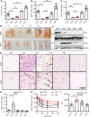
Figure 4. ATGL ablation rescues lipodystrophy and its associated insulin resistance in Bscl2–/– mice.
(AC) Percentage of fat mass assessed by NMR, and masses of gonadal white adipose tissue (gWAT) and subcutaneous WAT (sWAT) as normalized to body weight. (D and E) Representative images and H&E staining of gWAT and sWAT. Scale bar: 200 μm. (F) Representative Western blotting of mature adipose marker and lipid droplet protein (LDP). (G) Plasma insulin levels after 4-hour fast. For AG, 10-week-old female Atgl+/+Bscl2+/+ (AwBw), Atgl+/+Bscl2–/– (AwBk), Atgl+/–Bscl2–/– (AhBk), Atgl–/–Bscl2–/– (AkBk), and Atgl–/–Bscl2+/+ (AkBw) littermates were used for all experiments (n = 5–10/group). Limited numbers of male AkBk mice were obtained. However, a similar extent of rescue of lipodystrophy was observed in male AhBk and AkBk mice. (H and I) Insulin tolerance test and area under the curve (AUC) in mice other than AkBw (male and female, n = 5–7/group). *P < 0.05; **P < 0.005 by 1-way ANOVA with Dunnett’s multiple-comparisons correction.
Figure 5
Figure 5. ATGL inhibition partially rescues adipocyte differentiation of Bscl2–/– cells.
Bscl2+/+ and Bscl2–/– MEFs were subjected to a standard hormone cocktail DMI (dexamethasone, IBMX, and insulin) to induce adipocyte differentiation. Four days (D4) after differentiation, cells were treated with vehicle (Veh) or 10 μM Atglistatin and kept until D10. (A) Oil Red O and HCS LipidTOX Green neutral lipid staining. Scale bars: 200 μm. (B) Intracellular triglyceride (TG) content (2-way ANOVA with post hoc Tukey’s test) and (C) representative Western blot of PPARγ and PLIN1 at D10 after DMI induction. (DG) Stromal vascular cells isolated from Atgl+/+Bscl2+/+ (AwBw), Atgl+/+Bscl2–/– (AwBk), and Atgl–/– Bscl2–/– (AkBk) mice were subjected to DMI-induced adipocyte differentiation. (D) mRNA expression of Pnpla2, Bscl2, Pparγ, and Plin1 was measured at D0, D4, and D10 after DMI induction (2-way ANOVA with Dunnett’s multiple-comparisons post-hoc correction). (E) Representative protein expression, (F) Oil Red O staining, and (G) intracellular TG content at D10 after adipocyte induction (1-way ANOVA with Dunnett’s correction for multiple comparisons). *P < 0.05; **P < 0.005.
Figure 6
Figure 6. Partial ATGL inactivation rescues hypertrophy and cardiomyopathy in Bscl2–/– mice.
(A) Nonfasting plasma insulin levels, (B) ventricle weight (VW) normalized to tibia length (TL), and (C and D) fractional shortening (%) and left ventricle internal diameter at systole (LVIDs, mm). (E and F) Representative Western blotting (E) and fold changes of acetylated lysine (Ac-Lysine) as normalized to GAPDH (F) in whole heart. (G) Cardiac LCAD activity, (H) basal oxygen consumption rate (OCR) in isolated cardiac mitochondria, (I) CO2 production after incubating heart crude mitochondrial fraction with 14C-glucose. Six-month-old Atgl+/+Bscl2+/+ (AwBw), Atgl+/+Bscl2–/– (AwBk), and Atgl+/– Bscl2–/– (AhBk) male mice were used, n = 5–12/group. *P < 0.05, **P < 0.005 by one-way ANOVA with Dunnett’s multiple-comparisons correction.

Similar articles

Cited by

References

    1. Kolwicz SC, Tian R. Glucose metabolism and cardiac hypertrophy. Cardiovasc Res. 2011;90(2):194–201. doi: 10.1093/cvr/cvr071. - DOI - PMC - PubMed
    1. McGavock JM, et al. Cardiac steatosis in diabetes mellitus: a 1H-magnetic resonance spectroscopy study. Circulation. 2007;116(10):1170–1175. doi: 10.1161/CIRCULATIONAHA.106.645614. - DOI - PubMed
    1. Rijzewijk LJ, et al. Myocardial steatosis is an independent predictor of diastolic dysfunction in type 2 diabetes mellitus. J Am Coll Cardiol. 2008;52(22):1793–1799. doi: 10.1016/j.jacc.2008.07.062. - DOI - PubMed
    1. Joubert M, et al. The sodium-glucose cotransporter 2 inhibitor dapagliflozin prevents cardiomyopathy in a diabetic lipodystrophic mouse model. Diabetes. 2017;66(4):1030–1040. doi: 10.2337/db16-0733. - DOI - PubMed
    1. Liu S, et al. Development of hypertrophic cardiomyopathy in perilipin-1 null mice with adipose tissue dysfunction. Cardiovasc Res. 2015;105(1):20–30. doi: 10.1093/cvr/cvu214. - DOI - PubMed

Publication types

MeSH terms