. 2019 Jun 11;15(6):e1007809.
doi: 10.1371/journal.ppat.1007809. eCollection 2019 Jun.

Divergent roles for the RH5 complex components, CyRPA and RIPR in human-infective malaria parasites

Affiliations

Divergent roles for the RH5 complex components, CyRPA and RIPR in human-infective malaria parasites

Ellen Knuepfer et al. PLoS Pathog. .

Abstract

Malaria is caused by Plasmodium parasites, which invade and replicate in erythrocytes. For Plasmodium falciparum, the major cause of severe malaria in humans, a heterotrimeric complex comprised of the secreted parasite proteins, PfCyRPA, PfRIPR and PfRH5 is essential for erythrocyte invasion, mediated by the interaction between PfRH5 and erythrocyte receptor basigin (BSG). However, whilst CyRPA and RIPR are present in most Plasmodium species, RH5 is found only in the small Laverania subgenus. Existence of a complex analogous to PfRH5-PfCyRPA-PfRIPR targeting BSG, and involvement of CyRPA and RIPR in invasion, however, has not been addressed in non-Laverania parasites. Here, we establish that unlike P. falciparum, P. knowlesi and P. vivax do not universally require BSG as a host cell invasion receptor. Although we show that both PkCyRPA and PkRIPR are essential for successful invasion of erythrocytes by P. knowlesi parasites in vitro, neither protein forms a complex with each other or with an RH5-like molecule. Instead, PkRIPR is part of a different trimeric protein complex whereas PkCyRPA appears to function without other parasite binding partners. It therefore appears that in the absence of RH5, outside of the Laverania subgenus, RIPR and CyRPA have different, independent functions crucial for parasite survival.


Conflict of interest statement

The authors have declared that no competing interests exist.

Figures
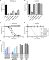
Fig 1
Fig 1. Role of basigin (BSG) as a receptor for erythrocyte invasion by P. falciparum, P. knowlesi and P. vivax.
(A) Merozoite invasion inhibition assay in the presence of anti-BSG mAb MEM-M6/6 or BSG protein for P. falciparum and (B) P. knowlesi. Late schizonts of P. falciparum and P. knowlesi were manually ruptured and the merozoites added to fresh erythrocytes, either with no additives, with heparin, MEM-M6/6, or BSG. Invasion was quantified after 12 h using flow cytometry. Three independent experiments were performed for P. falciparum and two for P. knowlesi in triplicate. Invasion is normalised to the RPMI medium alone control, and mean and standard deviation are displayed. (C) Growth inhibition assay (GIA) using schizont cultures of P. falciparum and (D) P. knowlesi in the presence of serial dilutions of anti-BSG mouse mAbs MEM-M6/6 and TRA-1-85, anti-BSG goat polyclonal IgG, anti-DARC CA111 nanobody as well as non-specific mouse and goat IgG (legend in box on the right of plot D). Parasitemias of three independent experiments in triplicate were measured 40 h after setup using flow cytometry. Mean and standard error are displayed. (E) Invasion inhibition assay using anti-BSG antibodies on field isolates of P. vivax. Blood samples were taken from patients in Peru, matured to schizont stage in vitro and incubated with anti-BSG mAb MEM-M6/6 and anti-BSG goat polyclonal IgG, as well as non-specific mouse IgG and goat IgG at 10 μg/ml. Ring stage parasitemia was determined by microscopy of Giemsa-stained blood smears. The relative parasitemia (in %) is displayed for each individual sample compared to control treatment of the same sample. Color-coding highlights the same patient samples treated with either of the anti-BSG antibodies.
Fig 2
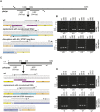
Fig 2. RIPR and CyRPA are essential for P. knowlesi parasite growth.
Schematics of disruption approaches for ripr (A) and cyrpa (C) in P. knowlesi, respectively. ORFs are depicted, indicating regions used as homology regions (HR) in repair plasmids. Black boxes represent endogenous DNA sequence (bp 1006–1054 (ripr) and 372–408 (cyrpa)) which was replaced either by recodonized DNA or by a triple HA-tag followed by a stop codon. Sequence details are given in the corresponding boxes below, of endogenous (wild type), recodonized DNA or HA-STOP regions. Primers used in PCR to genotype transfectants are indicated by arrows. B) Genotyping of PkA1-H.1 transfections with pGEMT-PkRIPRko or pGEMT-PkRIPRrecodonized with pDC596 and pDC645 Cas9/guide plasmids. D) Genotyping of PkA1-H.1 transfections using pGEMT-PkCyRPAko or pGEMT-PkCyRPArecodonized together with Cas9/guide plasmids pDC2915 or pDC2919. Primers for genotyping (S2 Table) were as follows: a, PKRIPRkoextFor; b, PKRIPRintRev; c, PKRIPRintFor; d, PKRIPRkoextRev; e, PKRIPRkorecRev; f, PKRIPRkorecodFor; g, HArev(Xma); h, HAfor(Xma); i, PKCyRPAextFor; j, PKCyRPAintRev; k, PKCyRPAintFor; l, PKCyRPAextRev; m, PKCyRPArecodRev; n, PKCyRPArecodFor. Expected PCR product sizes for Pkripr are: a/b = 426 bp; a/e = 434 bp; c/d = 412 bp; f/d = 418 bp; a/h = 509 bp; g/d = 499 bp. Expected PCR product sizes for Pkcyrpa are: i/j = 425 bp; i/m = 438 bp; k/l = 566 bp; n/l = 570 bp; i/h = 504 bp; g/l = 654 bp.
Fig 3
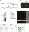
Fig 3. HA-tagging, localization and complex formation of PkRIPR.
(A) Schematic of single homologous recombination to generate C-terminally tagged parasite line, PkRIPR-HA. (B) Integration PCRs using genomic DNA from PkA1-H.1 (PK wt) parasites or PkRIPR-HA parasite clones I1 and G4. Using the primer pair PkRIPRextF1/PkRIPRutrRev, a 1595 bp fragment was amplified from wild type parasites, whereas primer pair PkRIPRextF1/HArev amplified a 1526 bp fragment only from transgenic PkRIPR-HA parasites. Positions of primer pairs are indicated in schematic (A) and size standards (kb) on the left of the gel (B). Primers used were PkRIPRextF1 (1), PkRIPRutrRev (2), HArev (3). (C) Southern blot of wildtype and transgenic parasite DNA digested with EcoRV/AvaI. Endogenous locus band, integration and episomal bands are indicated with arrows. (D) Immunoblot of purified schizont material solubilized in Laemmli sample buffer probed with anti-BiP and anti-HA (3F10) antibodies. Molecular mass standards are indicated on the left in kDa. (E) IFAs of schizonts and merozoites with micronemes not secreted (2nd row) or secreted (3rd row) were probed with anti-HA antibody (green), anti-PkAMA1 antibody, or anti-PkRhopH2 antibody (both red) and DAPI (Blue). The third panel shows an overlay of DAPI marking the nucleus with anti-HA and anti-PkAMA1; the fourth panel includes a differential interference contrast (DIC) image of the whole parasite. Scale bar = 2 μm. (F) Table of proteins interacting with PkRIPR-HA as identified by LC-MS/MS following immunoprecipitation. The three top-scoring, consistently identified proteins are listed. The number of peptides of three technical replicates of immunoprecipitates from PkRIPR-HA clone G4 or wild type parasites is displayed, as is the total peptide coverage, gene IDs and annotations (PlasmoDB release 25).
Fig 4
Fig 4. Newly identified complex components PkRIPR, PkCSS and PkPTRAMP are essential for human erythrocyte invasion, as is PkCyRPA.
Inducible knockouts of pkcss, pkptramp and pkcyrpa were produced in the P. knowlesi DiCre expressing PkpSKIP 9–10 parasite line using CRISP/Cas9 gene editing. (A) PCR screen of wild type (pSKIP) and two clones of each transgenic parasite line (ptrampiKO, cssiKO, cyrpaiKO) showing successful and complete recombination of floxed gene sequences after rapamycin addition (+). Primer pair 4/5 specific for the ptramp locus amplified a 1741 bp band in wild type parasites, a 1993 bp band in mock -treated (-), transgenic parasites and a reduced size band of 965 bp after excision induced by rapamycin (+). Primer pair 10/11 specific for the css locus amplified a 1761 bp band in wild type, a 2013 bp band in mock-treated, transgenic parasites and a 919 bp band after excision. Primer pair 16/17 specific for the cyrpa locus amplified a 1467 bp band in wild type, a 1559 bp band in mock-treated and a 891 bp band in rapamycin-treated, transgenic cyrpaiKO parasites. DNA marker sizes are shown on the left. (B) Immunoblot of schizonts purified 26 h after DMSO (-) or rapamycin (+) treatment of ring stage cultures from wild type (pSKIP) and inducible knockout parasite lines. Two clones are shown for each knockout parasite. Blots were probed with anti-HA and anti-BiP antibodies as indicated on the right of the panels. All protein signals correspond approximately with their predicted molecular masses (HA-tagged PkCSS has a predicted molecular mass of 40.6 kDa, HA-tagged PkPTRAMP a predicted molecular mass of 39.2 kDa and PkCyRPA-HA a predicted mass of 42.5 kDa). Molecular mass standards are indicated on the left. (C) Invasion assay of wild type and inducible knockout parasites lines over one growth cycle. Parasitemias were measured by flow cytometry 40 h after rapamycin / DMSO treatment. Means and standard errors of three independent experiments carried out in triplicate are displayed. (D) Giemsa-stained brightfield images of wild type and inducible knockout parasites 30 h after DMSO (-) or rapamycin (+) treatment. Arrowheads point to merozoites attached to erythrocytes which have failed to invade. Scale bars equal 2 μm. (E) Growth inhibition assay of PkA1-H.1 parasites over one cycle with serial dilutions of purified IgG raised against recombinantly produced PkPTRAMP, PkCSS, PkRIPR or control rabbit IgG. Parasitemias were determined by flow cytometry 18 h after assay setup at schizont stage. Means and standard errors of three independent experiments in triplicate are displayed.
Fig 5
Fig 5. Erythrocyte binding assay demonstrating the ability of PTRAMP but not CSS to bind to receptors on the erythrocyte surface.
Increasing amounts of recombinant proteins (in μg) were incubated with human erythrocytes, before repeated separation steps through oil and elution of bound protein. Immunoblots of eluted proteins are shown, probed with anti-His antibody. Recombinant RH5 and MSP3 were used as positive and negative controls respectively. Molecular mass standards are indicated on the left of each panel (in kDa).
Fig 6
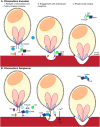
Fig 6. A cartoon depicting invasion of (A) P. knowlesi or (B) P. falciparum merozoites into human erythrocytes.
P. knowlesi lacks an RH5-like molecule, resulting in the secretion of RIPR complexed with PkPTRAMP and PkCSS. The precise role of this complex in the invasion process is currently unknown but we speculate that PkPTRAMP as a transmembrane protein is both displaying PkRIPR and PkCSS on the merozoite surface after microneme release and is recognizing a receptor on the human erythrocyte, which is an important step required for successful invasion. BSG is not used as a human erythrocyte receptor by P. knowlesi. In stark contrast, P. falciparum RH5 and the micronemal proteins PfRIPR and PfCyRPA form a trimeric complex following rhoptry neck and microneme secretion. This complex localizes to the merozoite surface, through an interaction between the N-terminus of RH5 and GPI-anchored protein P113, and connects the parasite via BSG to the host erythrocyte. This interaction between RH5 and BSG is speculated to result in rhoptry bulb secretion and Ca2+ influx into the host cell. RH5 and RIPR have been postulated to multimerize and integrate into the erythrocyte membrane, which by an unknown mechanism results in the influx of Ca2+ into the host cell. This is followed by the formation of the moving junction. The RH5 complex follows the moving junction, from the apical tip of the merozoite to its posterior end, setting in motion the irreversible process of active invasion of the host cell by the merozoite.

References

    1. World Health Organization. World malaria report 2018. 2018.
    1. World Health Organization. World malaria report 2017. 2017.
    1. Daneshvar C, Davis TME, Cox-Singh J, Rafa’ee MZ, Zakaria SK, Divis PCS, et al. Clinical and Laboratory Features of Human Plasmodium knowlesi Infection. Clin Infect Dis. 2009;49: 852–860. 10.1086/605439 - DOI - PMC - PubMed
    1. Weiss GE, Gilson PR, Taechalertpaisarn T, Tham W-H, de Jong NWM, Harvey KL, et al. Revealing the Sequence and Resulting Cellular Morphology of Receptor-Ligand Interactions during Plasmodium falciparum Invasion of Erythrocytes. PLOS Pathog. 2015;11: e1004670 10.1371/journal.ppat.1004670 - DOI - PMC - PubMed
    1. Boyle MJ, Richards JS, Gilson PR, Chai W, Beeson JG. Interactions with heparin-like molecules during erythrocyte invasion by Plasmodium falciparum merozoites. Blood. 2010;115: 4559–4568. 10.1182/blood-2009-09-243725 - DOI - PubMed

Publication types

MeSH terms