. 2019 Jun 11;19(1):120.
doi: 10.1186/s12862-019-1452-x.

Embryogenesis of flattened colonies implies the innovation required for the evolution of spheroidal colonies in volvocine green algae

Affiliations

Embryogenesis of flattened colonies implies the innovation required for the evolution of spheroidal colonies in volvocine green algae

Shota Yamashita et al. BMC Evol Biol. .

Abstract

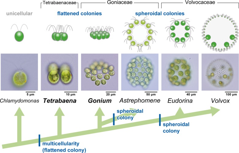
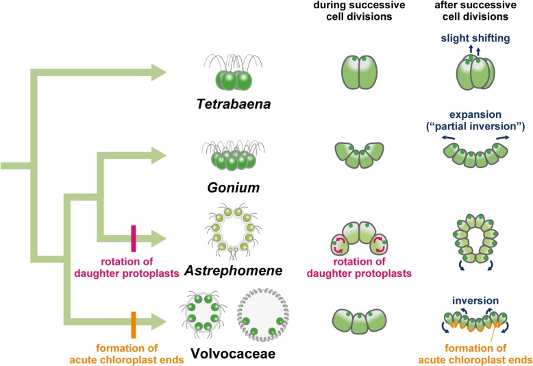
Background: Volvocine algae provide a suitable model for investigation of the evolution of multicellular organisms. Within this group, evolution of the body plan from flattened to spheroidal colonies is thought to have occurred independently in two different lineages, Volvocaceae and Astrephomene. Volvocacean species undergo inversion to form a spheroidal cell layer following successive cell divisions during embryogenesis. During inversion, the daughter protoplasts change their shape and develop acute chloroplast ends (opposite to basal bodies). By contrast, Astrephomene does not undergo inversion; rather, its daughter protoplasts rotate during successive cell divisions to form a spheroidal colony. However, the evolutionary pathways of these cellular events involved in the two tactics for formation of spheroidal colony are unclear, since the embryogenesis of extant volvocine genera with ancestral flattened colonies, such as Gonium and Tetrabaena, has not previously been investigated in detail.

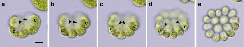
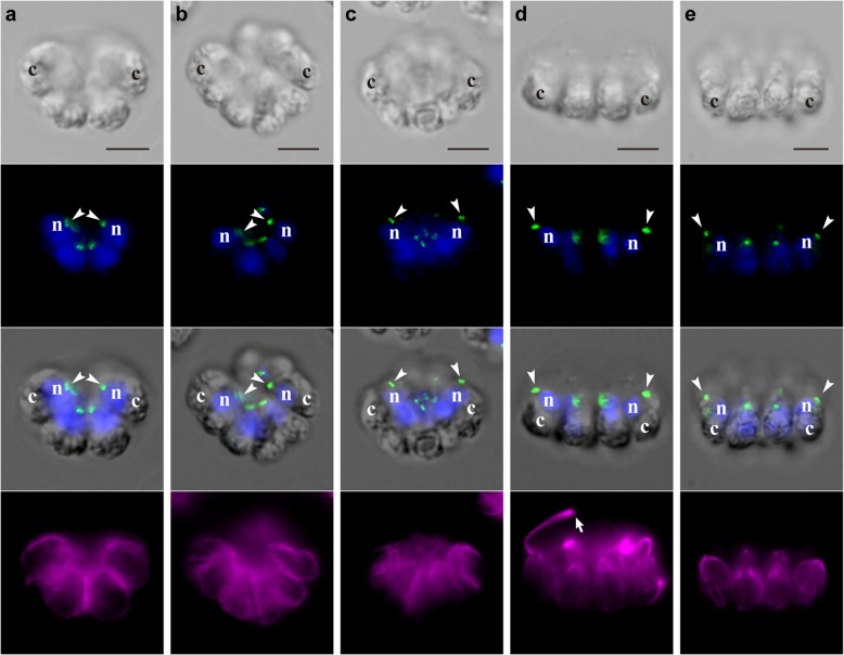
Results: We conducted time-lapse imaging by light microscopy and indirect immunofluorescence microscopy with staining of basal bodies, nuclei, and microtubules to observe embryogenesis in G. pectorale and T. socialis, which form 16-celled or 4-celled flattened colonies, respectively. In G. pectorale, a cup-shaped cell layer of the 16-celled embryo underwent gradual expansion after successive cell divisions, with the apical ends (position of basal bodies) of the square embryo's peripheral protoplasts separated from each other. In T. socialis, on the other hand, there was no apparent expansion of the daughter protoplasts in 4-celled embryos after successive cell divisions, however the two pairs of diagonally opposed daughter protoplasts shifted slightly and flattened after hatching. Neither of these two species exhibited rotation of daughter protoplasts during successive cell divisions as in Astrephomene or the formation of acute chloroplast ends of daughter protoplasts as in volvocacean inversion.

Conclusions: The present results indicate that the ancestor of Astrephomene might have newly acquired the rotation of daughter protoplasts after it diverged from the ancestor of Gonium, while the ancestor of Volvocaceae might have newly acquired the formation of acute chloroplast ends to complete inversion after divergence from the ancestor of Goniaceae (Gonium and Astrephomene).

Keywords: Body plan; Embryogenesis; Gonium; Multicellularity; Tetrabaena; Volvocine green algae.


Conflict of interest statement

Author HN is a member of the BMC Evolutionary Biology Editorial Board. The authors declare that they have no competing interests.

Figures

Fig. 1
Fig. 1
Schematic representation of the phylogenetic relationships of volvocine green algae and evolution of their body plans. Volvocine green algae consist of organisms of various complexities, ranging from unicellular Chlamydomonas reinhardtii to multicellular Volvox, which exhibits germ-soma differentiation. The evolution of spheroidal colonies is thought to have occurred twice, in the ancestors of Astrephomene and in those of Volvocaceae [–6]. The formation of spheroidal colonies during embryogenesis is based on different cellular mechanisms in the two lineages (Additional file 6: Figure S1) [7]. There are two extant lineages with ancestral flattened colonies, the genus Gonium and the family Tetrabaenaceae. The phylogeny is based on a previous report [5]. All drawings and photographs represent lateral views of individuals with anterior sides (the direction of swimming) oriented toward the top of the figure. The photographs of Tetrabaena, Astrephomene, and Volvox are from a previous study [7]. The other photographs are original
Fig. 2
Fig. 2
Successive cell divisions and expansion of the cell layer in embryogenesis of G. pectorale. Successive stages of an embryo observed by time-lapse analysis from the anterior-lateral view (Additional file 2). All images are at the same magnification. Scale bars: 5 μm. Note the longitudinal axis of each daughter protoplast, indicated by the positions of apical ends (arrowheads) and chloroplasts (letter c). a Early 8-celled stage. b Late 8-celled stage. c Early 16-celled stage. Rotation of daughter protoplasts is not observed during cell divisions (ac). d Mid 16-celled stage during partial inversion. The cup-shaped cell layer of the embryo has expanded and the apical ends of outer daughter protoplasts (arrowheads) have separated from one another and moved outwards. e Late 16-celled stage, just prior to hatching. The cell layer has increasingly expanded and rotated slightly to show an almost anterior view
Fig. 3
Fig. 3
Successive cell divisions and slight shifting of daughter protoplasts during embryogenesis of T. socialis. Successive stages of an embryo observed by time-lapse analysis from the anterior-lateral view (Additional file 5). All images are at the same magnification. Scale bars: 5 μm. Note the longitudinal axis of each daughter protoplast, indicated by the positions of apical ends (arrowheads) and chloroplasts (uppercase letters). Uppercase letters (A and B) correspond with those in Additional file 6: Figure S2 h. a Prior to embryogenesis. b Two-celled stage. c Early 4-celled stage. Daughter protoplasts do not rotate during cell divisions (ac). d Mid 4-celled stage. e Late 4-celled stage. One pair of diagonally opposed daughter protoplasts (A) has shifted slightly toward the anterior of the embryo relative to the other pair (B) (ce)
Fig. 4
Fig. 4
Indirect immunofluorescence microscopy showing successive stages of embryogenesis in G. pectorale. Each column shows a differential interference contrast (DIC) image (top row), a fluorescence image labeled with anti-SAS-6 antibody (green) and DAPI (blue) (second row), a merged DIC and fluorescence image of anti-SAS-6 antibody and DAPI (third row), and a fluorescence image labeled with anti-tubulin α antibody (magenta) of the same embryo. Positions of nuclei (letter n), chloroplasts (letter c), and basal bodies labeled with the anti-SAS-6 antibody (arrowheads) are shown. Scale bars: 5 μm. a Early 8-celled stage. b Late 8-celled stage. c Early 16-celled stage, just after the successive cell divisions. Basal bodies of daughter protoplasts are positioned in the center of the concave surface of the cell layer during successive cell divisions (ac). d Mid 16-celled stage during partial inversion showing emitted flagella (arrow). The basal bodies of peripheral daughter protoplasts (arrowheads) have become separated from each other and are located slightly outside of the position of the nuclei. e After hatching. The basal bodies of peripheral cells (arrowheads) point toward the outside of the daughter colony
Fig. 5
Fig. 5
Indirect immunofluorescence microscopy showing successive stages of embryogenesis in T. socialis. Each column shows a DIC image (top row), a fluorescence image labeled with anti-SAS-6 antibody (green) and DAPI (blue) (second row), a merged DIC and fluorescence image of anti-SAS-6 antibody and DAPI (third row), and a fluorescence image labeled with anti-tubulin α antibody (magenta) of the same embryo. Positions of nuclei (letter n), chloroplasts (letter c), and basal bodies labeled with anti-SAS-6 antibody (arrowheads) are shown. Scale bars: 5 μm. a Prior to embryogenesis. b Two-celled stage. c Early 4-celled stage. The angles of the longitudinal axes of daughter protoplasts, which are indicated by basal bodies and chloroplasts, did not change during successive cell divisions (ac). d Late 4-celled stage showing emitted flagella (arrows). The angles of longitudinal axes of daughter protoplasts did not change after successive cell divisions, though a pair of diagonally opposed daughter protoplasts shifted slightly toward the anterior of the embryo (“A” in Fig. 3c, e). e After hatching. The basal bodies of four cells are arranged in a square shape in the same plane
Fig. 6
Fig. 6
Schematic diagrams of the most likely evolutionary pathways of embryogenesis in volvocine green algae. Diagrams of stages of embryogenesis in Astrephomene and Volvocaceae (Eudorina) shown are based on a previous study [7], while those in Tetrabaena and Gonium are based on the present study (summarized in Additional file 6: Figure S5). Volvocacean species undergo the formation of acute chloroplast ends of daughter protoplasts, which is one of the principal factors producing the force for folding the cell layer during inversion after successive cell divisions. On the other hand, Astrephomene undergoes rotation of daughter protoplasts during successive cell divisions to form a spheroidal shape of the cell layer. Neither Gonium nor Tetrabaena showed the formation of acute chloroplast ends of daughter protoplasts after successive cell divisions or the rotation of daughter protoplasts during successive cell divisions. These results suggest that the ancestor of Astrephomene developed the rotation of daughter protoplasts after it diverged from the ancestors of Gonium, while the ancestor of Volvocaceae acquired the formation of acute chloroplast ends after it diverged from the ancestors of Goniaceae

Similar articles

Cited by

References

    1. Michod RE. On the transfer of fitness from the cell to the multicellular organism. Biol Philos. 2006;20(5):967–987. doi: 10.1007/s10539-005-9018-2. - DOI
    1. Grosberg RK, Strathmann RR. The evolution of multicellularity: a minor major transition? Annu Rev Ecol Evol Syst. 2007;38(1):621–654. doi: 10.1146/annurev.ecolsys.36.102403.114735. - DOI
    1. Rensing SA. (why) does evolution favour embryogenesis? Trends Plant Sci. 2016;21(7):562–573. doi: 10.1016/j.tplants.2016.02.004. - DOI - PubMed
    1. Nozaki H, Ito M. Phylogenetic relationships within the colonial Volvocales (Chlorophyta) inferred from cladistic analysis based on morphological data. J Phycol. 1994;30(2):353–365. doi: 10.1111/j.0022-3646.1994.00353.x. - DOI
    1. Nozaki H, Misawa K, Kajita T, Kato M, Nohara S, Watanabe MM. Origin and evolution of the colonial Volvocales (Chlorophyceae) as inferred from multiple, chloroplast gene sequences. Mol Phylogenet Evol. 2000;17(2):256–268. doi: 10.1006/mpev.2000.0831. - DOI - PubMed

Publication types

LinkOut - more resources