

Association of tumour-associated macrophages with cancer cell EMT, invasion, and metastasis of Kazakh oesophageal squamous cell cancer
- PMID: 31186031
- PMCID: PMC6560903
- DOI: 10.1186/s13000-019-0834-0
Association of tumour-associated macrophages with cancer cell EMT, invasion, and metastasis of Kazakh oesophageal squamous cell cancer
Abstract
Background: Tumour-associated macrophages (TAMs) play an important role in the growth, progression, and metastasis of tumours. Epithelial-mesenchymal transition (EMT) is a mechanism for tumour invasion and metastasis. In this study, we aimed to determine whether TAMs can induce EMT for the invasion and metastasis of Kazakh oesophageal squamous cell cancer (ESCC).
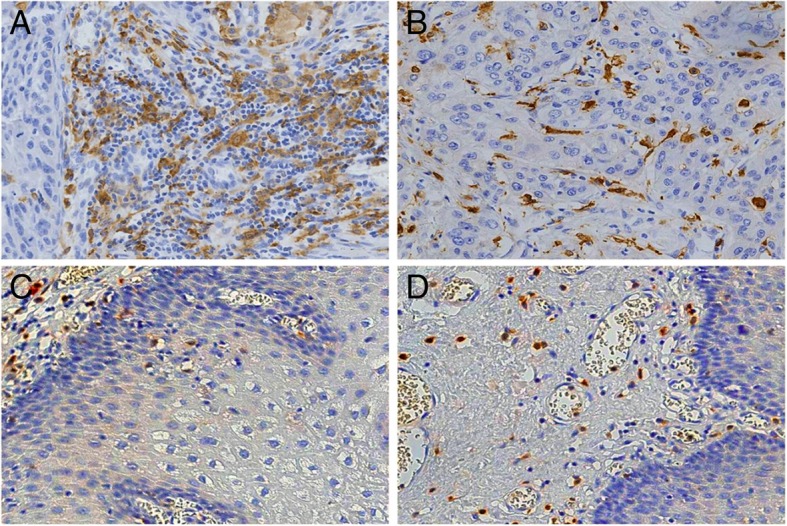
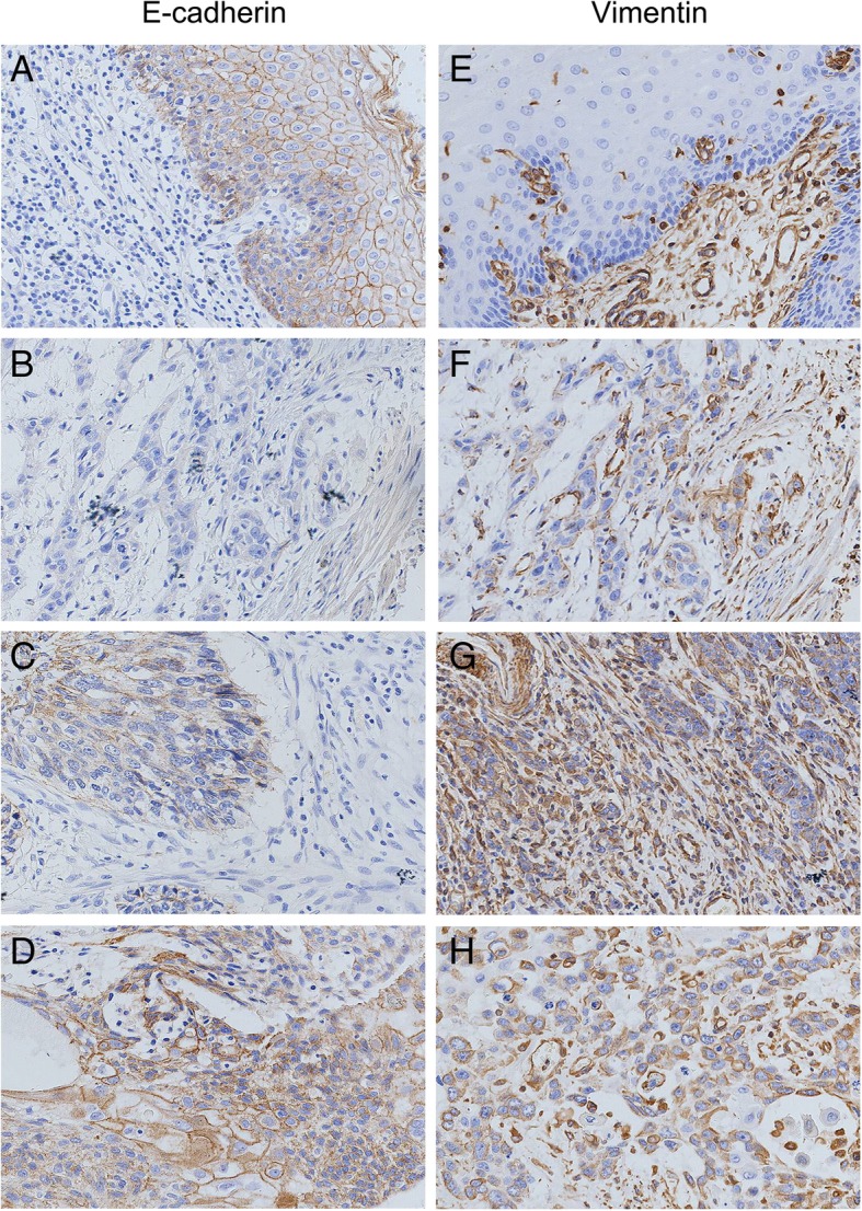
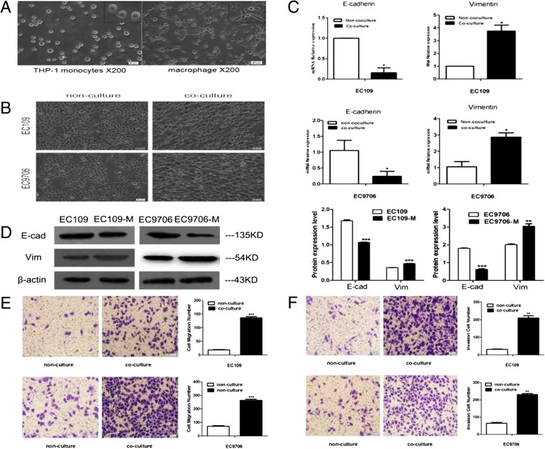
Methods: CD163 was used as a marker for TAMs, and the density of TAMs in tumour nest and surrounding stroma was quantified using immunohistochemistry (IHC). IHC staining was used to evaluate the expression of E-cadherin (epithelial marker) and vimentin (mesenchymal marker) in Kazakh ESCC and cancer-adjacent normal tissues (CANs). Additionally, 6-well transwell plates (0.4 μm) were used to establish the co-culture system of ESCC (EC109 or EC9706) cells and macrophages. Real-time quantitative polymerase chain reaction (qPCR) and western blot experiments were used to determine whether ESCC cells undergo EMT transformation after co-culture with macrophages. Transwell assays were used to detect the migration and invasion of the ESCC cells.
Results: The distribution of CD163-positive TAMs in cancer tissues was closely related to EMT in Kazakh ESCC. The expression of vimentin in the ESCC cells was significantly upregulated, the expression of E-cadherin was significantly downregulated, and the invasion and migration of the ESCC cells were significantly enhanced after tumour-associated macrophages were added to the co-culture.
Conclusions: Tumour-associated macrophages promote EMT in ESCC, which may be one of the important factors involved in the invasion and progression of Kazakh ESCC.
Keywords: Epithelial-mesenchymal transition; Kazakh; Migration and invasion; Oesophageal squamous cell carcinoma; Tumour-associated macrophages.
Conflict of interest statement
The authors declare that they have no competing interests.
Figures
Similar articles
-
The increased number of tumor-associated macrophage is associated with overexpression of VEGF-C, plays an important role in Kazakh ESCC invasion and metastasis.Exp Mol Pathol. 2017 Feb;102(1):15-21. doi: 10.1016/j.yexmp.2016.12.001. Epub 2016 Dec 7. Exp Mol Pathol. 2017. PMID: 27939650
-
TGF-β1/Smad signaling pathway regulates epithelial-to-mesenchymal transition in esophageal squamous cell carcinoma: in vitro and clinical analyses of cell lines and nomadic Kazakh patients from northwest Xinjiang, China.PLoS One. 2014 Dec 2;9(12):e112300. doi: 10.1371/journal.pone.0112300. eCollection 2014. PLoS One. 2014. PMID: 25464508 Free PMC article.
-
Infiltrated M2 tumour-associated macrophages in the stroma promote metastasis and poor survival in oesophageal squamous cell carcinoma.Histol Histopathol. 2019 May;34(5):563-572. doi: 10.14670/HH-18-061. Epub 2018 Nov 12. Histol Histopathol. 2019. PMID: 30417922
-
Similarities between wound re-epithelialization and Metastasis in ESCC and the crucial involvement of macrophages: A review.Cancer Treat Res Commun. 2022;32:100621. doi: 10.1016/j.ctarc.2022.100621. Epub 2022 Aug 17. Cancer Treat Res Commun. 2022. PMID: 36007473 Review.
-
Tumor associated macrophages in esophageal squamous carcinoma: Promising therapeutic implications.Biomed Pharmacother. 2023 Nov;167:115610. doi: 10.1016/j.biopha.2023.115610. Epub 2023 Sep 30. Biomed Pharmacother. 2023. PMID: 37783153 Review.
Cited by
-
An Extracellular Matrix-Based Signature Associated With Immune Microenvironment Predicts the Prognosis and Therapeutic Responses of Patients With Oesophageal Squamous Cell Carcinoma.Front Mol Biosci. 2021 Mar 18;8:598427. doi: 10.3389/fmolb.2021.598427. eCollection 2021. Front Mol Biosci. 2021. PMID: 33869274 Free PMC article.
-
Innate Immune Cells in the Esophageal Tumor Microenvironment.Front Immunol. 2021 Apr 28;12:654731. doi: 10.3389/fimmu.2021.654731. eCollection 2021. Front Immunol. 2021. PMID: 33995371 Free PMC article. Review.
-
Harnessing the tumor microenvironment: targeted cancer therapies through modulation of epithelial-mesenchymal transition.J Hematol Oncol. 2025 Jan 13;18(1):6. doi: 10.1186/s13045-024-01634-6. J Hematol Oncol. 2025. PMID: 39806516 Free PMC article. Review.
-
Multi-modal quantification of pathway activity with MAYA.Nat Commun. 2023 Mar 25;14(1):1668. doi: 10.1038/s41467-023-37410-2. Nat Commun. 2023. PMID: 36966153 Free PMC article.
-
Knockdown of lncRNA TUC338 inhibits esophageal cancer cells migration and invasion.J Thorac Dis. 2021 May;13(5):3061-3069. doi: 10.21037/jtd-21-563. J Thorac Dis. 2021. PMID: 34164197 Free PMC article.
References
-
- Ayxiam H, Ma H, Ilyar S, Zhang LW, Ablizi A, Batur M, et al. Metabonomic variation of esophageal cancer within different ethnic groups in Xinjiang, China. Zhonghua Yu Fang Yi Xue Za Zhi. 2009;43:591–596. - PubMed
MeSH terms
Substances
Grants and funding
LinkOut - more resources
Full Text Sources
Medical
Research Materials
