. 2019 Jun 11;7(1):150.
doi: 10.1186/s40425-019-0620-2.

Temporal changes within the (bladder) tumor microenvironment that accompany the therapeutic effects of the immunocytokine NHS-IL12

Affiliations

Temporal changes within the (bladder) tumor microenvironment that accompany the therapeutic effects of the immunocytokine NHS-IL12

Y Maurice Morillon 2nd et al. J Immunother Cancer. .

Abstract

Background: While significant strides in the treatment of metastatic bladder cancer have been made with immune checkpoint inhibitors, the treatment of carcinoma in situ and non-muscle invasive, non-metastatic (superficial) human urothelial carcinoma, also termed non-muscle invasive bladder cancer (NMIBC), remains intractable with bacillus Calmette-Guerin (BCG) employed as the standard of care. In this study, an immunocytokine, NHS-muIL12, which consists of two molecules of murine IL-12 fused to NHS76, a tumor necrosis-targeting human IgG1, was examined as an immunotherapeutic in an orthotopic MB49luc bladder tumor model.

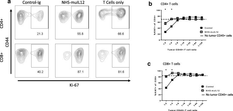
Methods: The antitumor activity of systemic administration of NHS-muIL12 was investigated on MB49luc tumors, an aggressive, bioluminescent orthotopic bladder cancer model. Temporal studies were carried out on MB49luc bladder tumors harvested during various time points during NHS-muIL12 treatment and cellular changes associated with the reduction in tumor burden following NHS-muIL12 were determined by flow cytometry. Effects of those changes on the proliferation/activation of lymphoid cells were also determined.

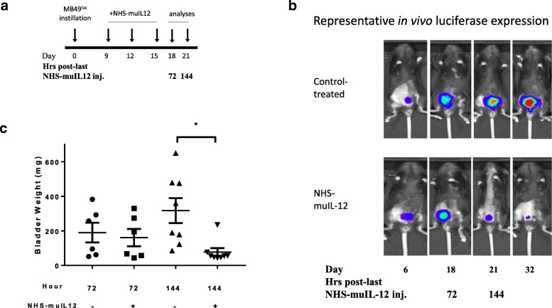
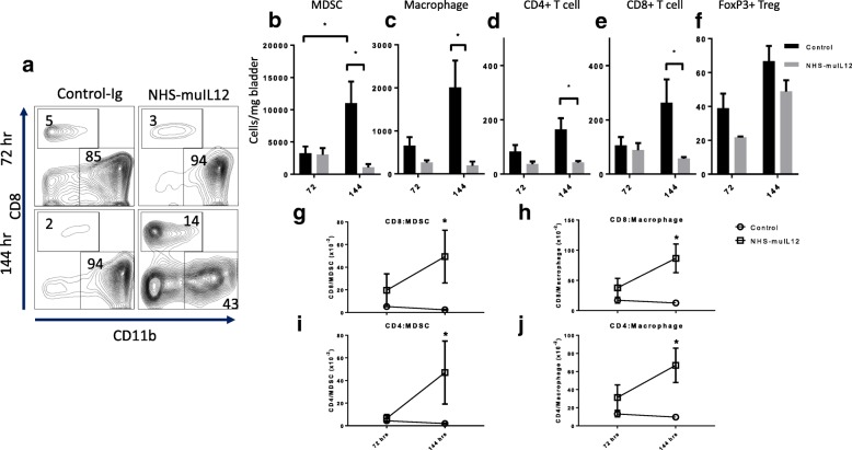
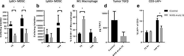
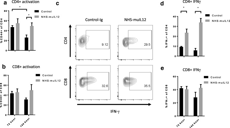
Results: Studies revealed a significant reduction in MB49luc bladder tumor burden occurring between days 3 and 6 after the third and final systemic administration of NHS-muIL12. Temporal analyses of the MB49luc bladder tumor microenvironment (TME) initially revealed a large accumulation of myeloid-derived suppressor cells (MDSCs) and macrophages that elicited potent immunosuppression. Immunosuppression was characterized by the inability of CD4+ and CD8+ T cells to respond to broad-based immune stimulants. NHS-muIL12 administration resulted in temporal-dependent reductions in the number of MDSCs, macrophages and tumor-associated TGF-β, which culminated in a re-ignition of CD4+ and CD8+ T cells to elicit potent antitumor responses against MB49luc bladder tumors.

Conclusions: These findings provide strong evidence that the systemic administration of an immunocytokine consisting of a tumor-targeting Ig through recognition of DNA and DNA-histone complexes coupled to muIL-12 can effectively target the bladder TME; this significantly reduces the myeloid cellular compartment and reverts an immunosuppressive to an immunopermissive TME, ultimately resulting in antitumor effects. These studies provide further rationale for the employment of NHS-IL12 as an immunomodulator and clinical immunotherapeutic for NMIBC.

Keywords: Immunotherapy; NHS-muIL12; Non-muscle invasive bladder cancer.


Conflict of interest statement

Authors from the National Cancer Institute do not have any competing interests to disclose. Author from EMD Serono is an employee/officer of said company.

Figures

Fig. 1
Fig. 1
Antitumor effects of NHS-muIL12 on MB49luc bladder tumors. a NHS-muIL12 treatment (days 9, 12 and 15) and analyses (days 18, 21) schedule of mice bearing MB49luc bladder tumors. b Representative in vivo luciferase expression images taken immediately prior to euthanasia at days 6, 18, 21, and 32 for control Ig– (top row) and NHS-muIL12–treated mice (bottom row). c Individual bladder weights from control Ig–treated (circles, triangles) and NHS-muIL12–treated (squares, inverted triangles) mice at 72 and 144 h post–final NHS-muIL12 injection (days 18 and 21). Horizontal lines represent the average bladder weight; error bars represent mean ± SEM, Student’s t-test; *P < 0.05. Data are from a representative experiment that was repeated with similar results
Fig. 2
Fig. 2
Cellular changes in the MB49luc bladder TME of control Ig– and NHS-muIL12– treated mice at 72 and 144 h after the final NHS-muIL12 treatment. a Representative FACS plots of cytotoxic T lymphocytes (CD8, y-axis) vs myeloid (CD11b, x-axis) cells from control Ig– (left column) and NHS-muIL12–treated (right column) mice bearing MB49luc bladder tumors. Plots are representative of live CD45+ tumor-derived cells at 72 (top row) and 144 h (bottom row) after the final NHS-muIL12 treatment. Numbers within each quadrant represent percent of total cells. In panels b-f, mice bearing MB49luc bladder tumors and treated with control Ig– (black bars) or NHS-muIL12 (grey bars) were euthanized at 72 and 144 h after the final NHS-muIL12 treatment and the lymphoid/myeloid cellular components within the TME were examined by flow cytometry. Absolute number of each cell type/mg bladder weight for (b) MDSCs: CD11b+Gr1+F4/80 (n = 9), (c) macrophages: CD11b+Gr1F4/80+ (n = 9), (d) CD4+ T cells (n = 9), (e) CD8+ T cells (n = 9), and (f) regulatory T cells (Tregs): CD3+CD4+FoxP3+ (n = 3) are shown. Panels g-j represent ratios of (g) CD8+/MDSCs, (h) CD8+/macrophages, (i) CD4+/MDSCs, and (j) CD4+/macrophages at 72 and 144 h after the final NHS-muIL12 treatment (n = 9). Circles and squares represent control Ig–treated and NHS-muIL12–treated mice, respectively. Error bars (panels b-j) represent mean + SEM, Student’s t-test; *P < 0.05. Data are from a representative experiment that was repeated twice with similar results
Fig. 3
Fig. 3
Temporal-dependent phenotypic (72 and 144 h after the final NHS-muIL12 treatment) changes in myeloid/lymphoid cell profiles in the MB49luc bladder TME of control Ig– (black bars) and NHS-muIL12–treated (grey bars) mice. Panels a-c represent differences in absolute number of cells/mg bladder weight of (a) Ly6C+ MDSCs, (b) Ly6G+ MDSCs and (c) in the frequencies of M1 of CD11b+Gr1F4/80+ macrophages. (d) Total tumor TGF-β from control Ig– (black bars) and NHS-muIL12–treated (grey bars) mice at the 144-h time point (n = 5). e Frequencies of LAP+ CD3 cells. Error bars represent mean + SEM, Student’s t-test; *P < 0.05, unless otherwise indicated, n = 3. Data are from a representative experiment that was repeated twice with similar results
Fig. 4
Fig. 4
Reversal of immunosuppression within the MB49luc bladder TME following NHS-muIL12 treatment. MB49luc bladder tumors (n = 3) from control Ig– and NHS-muIL12–treated mice were isolated 5 days post–final NHS-muIL12 treatment (day 20, see Fig. 1, panel a). CD45+ cells were purified from tumor homogenates and cocultured with CD4+ and CD8+ T cells (effectors) purified from the spleen of C57BL/6 mice in the presence of soluble αCD3 and αCD28 for 48 h as described in Methods. Following in vitro stimulation, cells were stained for T cell (CD3+, CD4+, CD8+), activation (CD44) and proliferative (Ki-67) markers. a Representative FACS plots showing the CD44 and Ki67-expressing CD4+ (top row) and CD8+ (bottom row) T cells after co-incubation with CD45+ tumor-derived cells (ratio 1:2) from MB49luc bladder from control Ig– and NHS-muIL12–treated mice. FACS plots indicated as “T cells only” were from cultures containing indicated splenic CD4+ or CD8+ T cells from naïve B6 mice (i.e., no addition of bladder tumor-derived CD45+ cells). Numbers within each FACS plot are the percentage of CD4+ and CD8+ T cells co-expressing CD44 and Ki67. b, c Frequency of CD4+ (b) and CD8+ (c) T cell proliferation reported as a percentage of Ki67-expressing cells using CD45+ cells from tumors of control Ig– (solid squares) and NHS-muIL12–treated mice (open squares) at ratios CD45+ tumor-derived cells to T cells of 1:2 to 1:128. Dashed line in b and c represents the average proliferation as measured by Ki67-expressing T cells in the absence of tumor-derived CD45+ cells. Student’s t-test; *P < 0.05 (vs. control Ig–treated mice). Data are from a single experiment
Fig. 5
Fig. 5
NHS-muIL12 treatment increases T cell activation (CD44 expression) and pro-inflammatory events (i.e., intracellular IFN-γ expression) within the MB49luc bladder TME. a, b MB49luc bladder tumors (n = 3) from control Ig– (black bars) and NHS-muIL12–treated (grey bars) mice were isolated at 72 and 144 h post–final NHS-muIL12 treatment and single cell suspensions were prepared (see Methods) and stained for CD4+, CD8+ T cells along with the CD44 activation marker. c Representative FACS plots for intracellular IFN-γ staining of CD4+ and CD8+ T cells after a 5-h in vitro stimulation (see Methods). d, e Frequencies of CD4+ and CD8+ T cells expressing intracellular IFN-γ from MB49luc bladder tumors from control Ig– (black bars) and NHS-muIL12–treated (grey bars) mice at 72 and 144 h post–final NHS-muIL12 administration. Error bars (panels a, b, d, e) represent mean + SEM. Student’s t-test; *P < 0.05 (vs. control-treated mice)

References

    1. Jemal A, Siegel R, Ward E, Hao Y, Xu J, Murray T, et al. Cancer statistics, 2008. CA Cancer J Clin. 2008;58(2):71–96. - PubMed
    1. Inman BA, Longo TA, Ramalingam S, Harrison MR. Atezolizumab: a PD-L1-blocking antibody for bladder cancer. Clin Cancer Res. 2017;23(8):1886–1890. - PubMed
    1. Sharma P, Callahan MK, Bono P, Kim J, Spiliopoulou P, Calvo E, et al. Nivolumab monotherapy in recurrent metastatic urothelial carcinoma (CheckMate 032): a multicentre, open-label, two-stage, multi-arm, phase 1/2 trial. Lancet Oncol. 2016;17(11):1590–1598. - PMC - PubMed
    1. Bellmunt J, de Wit R, Vaughn DJ, Fradet Y, Lee JL, Fong L, et al. Pembrolizumab as second-line therapy for advanced urothelial carcinoma. N Engl J Med. 2017;376(11):1015–1026. - PMC - PubMed
    1. Massard C, Gordon MS, Sharma S, Rafii S, Wainberg ZA, Luke J, et al. Safety and efficacy of durvalumab (MEDI4736), an anti-programmed cell death ligand-1 immune checkpoint inhibitor, in patients with advanced urothelial bladder cancer. J Clin Oncol. 2016;34(26):3119–3125. - PMC - PubMed

Publication types