. 2019 Jun 11;8(6):bio043182.
doi: 10.1242/bio.043182.

Cell recovery by reversal of ferroptosis

Affiliations

Cell recovery by reversal of ferroptosis

Ho Man Tang et al. Biol Open. .

Abstract

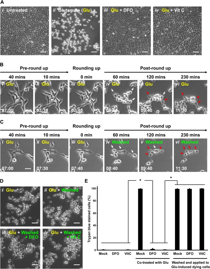
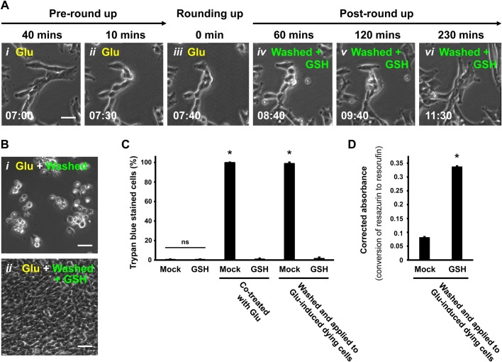
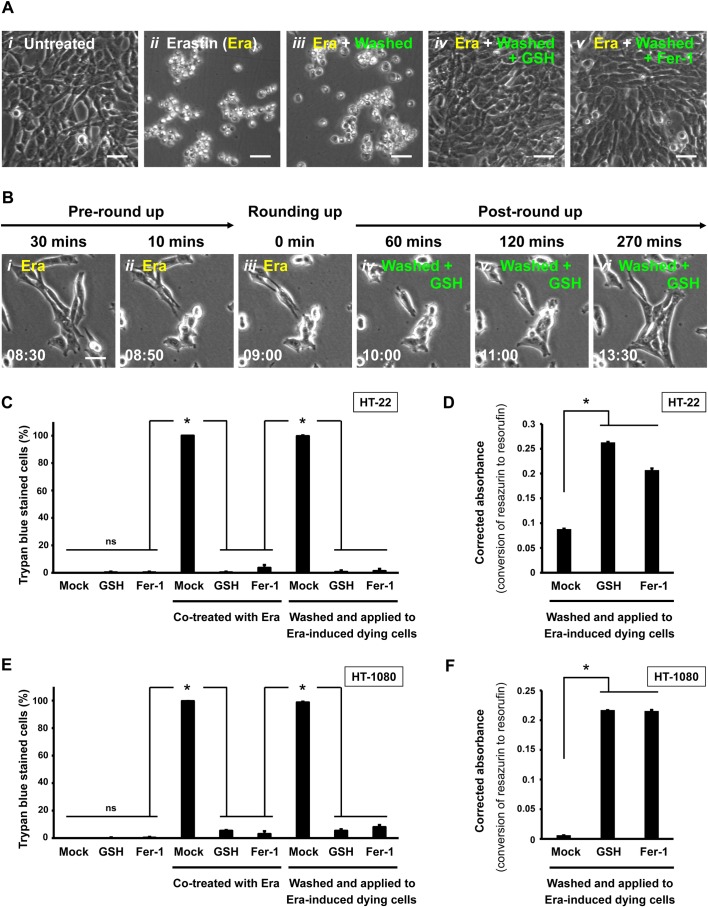
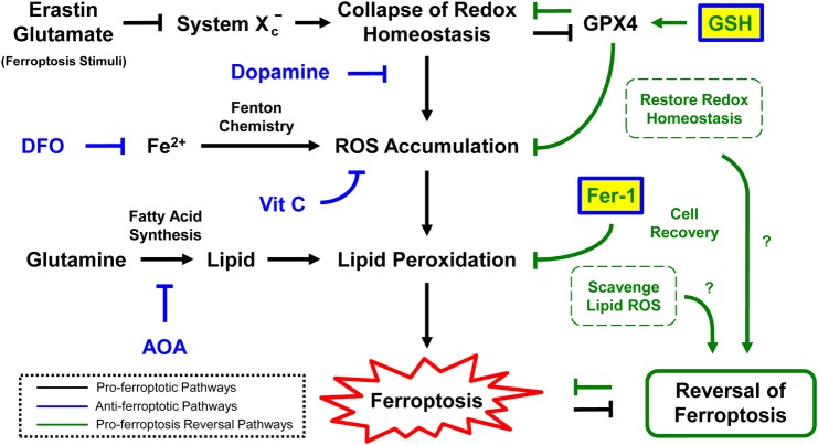
The classical view of cell death has long assumed that, once initiated, the dying process is irreversible. However, recent studies reveal that recovery of dying cells can actually occur, even after initiation of a cell suicide process called apoptosis. This discovery raised fundamental key questions about which forms of the cell death process could be reversible and how reversal is mediated. Here, we uncover an unanticipated reversibility of ferroptotic cell death process. Unlike apoptosis reversal, removal of ferroptosis inducers, such as erastin and glutamate, is insufficient to allow ferroptotic dying cells to escape the cell death process. However, by removing the cell death inducer and providing the reduced form of glutathione or the radical-trapping antioxidant ferrostatin-1, ferroptotic dying cells can be rescued and promoted to recover. Interestingly, although ferroptotic inhibitors such as aminooxyacetic acid, deferoxamine, dopamine and vitamin C can prevent initiation of ferroptosis, added alone they are unable to reverse the initiated ferroptosis, suggesting regulatory distinctions between preventing and reversing ferroptosis. Together, these results reveal the first evidence that ferroptosis is reversible and suggest strategies to enhance its reversibility, thereby providing a useful model for studying the physiological, pathological and therapeutic potentials of this cell recovery process.

Keywords: Anastasis; Ferrostatin-1; Glutamate; Glutathione; Reversal of apoptosis; Reversal of ferroptosis.


Conflict of interest statement

Competing interestsThe authors declare no competing or financial interests.

Figures

Fig. 1.
Fig. 1.
Demise of ferroptotic cells after removing cell death stimulus. (A) HT-22 cells were cultured in (i) cell medium, (ii) medium containing 10 mM glutamate (Glu) for 24 h, (iii) medium containing both 10 mM Glu and 100 µM deferoxamine (DFO) for 24 h, and (iv) medium containing both 10 mM Glu and 0.5 mM vitamin C (Vit C) for 24 h. Cell morphology was observed and recorded by phase contrast microscopy. Scale bars: 100 µm. (B) Time-lapse live-cell phase contrast microscopy of (i,ii) pre-rounding, (iii) rounding and (ivvi) post-rounding HT-22 cells exposed to 10 mM Glu. Same group of cells shown over time (h:min). Scale bar: 20 μm. White arrows indicate a few examples of rounded-up ferroptotic dying cells. Red arrows indicate collapsed ferroptotic cells with plasma membrane rupture. (C) Time-lapse live-cell phase contrast microscopy of HT-22 cells during 10 mM Glu induction and after being washed and incubated with fresh medium. Shown here is a group of cells in the culture after induction with Glu at 40 min (i) and 10 min (ii) prior to showing signs of rounding, at the time rounding began with Glu still in the medium (iii) and then at 60, 120 and 230 min after washing and continuing incubation in fresh medium containing no Glu (panels ivvi). Same group of cells shown over time (h:min). Scale bar: 20 μm. White arrows indicate rounded-up ferroptotic dying cells. Red arrows indicate collapsed ferroptotic cells with plasma membrane rupture. (D) Phase contrast microscopy of HT-22 cells treated with 10 mM Glu for 24 h (i), treated with 10 mM Glu for 7.5 h to initiate ferroptosis and then washed and incubated with fresh culture medium for 24 h (ii), treated as in ii but with 100 µM DFO added to the fresh culture medium (iii), treated as ii but with 0.5 mM Vit C added to the fresh culture medium (iv). Scale bars: 60 μm. (E) Percentage of HT-22 cells that displayed plasma membrane permeability in Trypan Blue exclusion assay, after the following treatments. First set of three show culture medium alone (Mock), medium containing 100 µM DFO or 0.5 mM Vit C for 24 h. Second set of three are as in the first set, but all cultures containing 10 mM Glu. Third set of three are as in the first set, but with all cultures induced with 10 mM Glu for 7.5 h to initiate ferroptosis prior to washing and then incubating the rounded-up ferroptotic cells for 24 h with fresh culture medium alone (Mock), or containing 100 mM DFO or 0.5 mM Vit C. Dead cells with plasma membrane rupture displayed full plasma membrane permeability as determined by the Trypan Blue exclusion assay. Data presented as means±s.d. of three independent experiments. Student's t-test: *P<0.001.
Fig. 2.
Fig. 2.
Reversal of ferroptosis by Fer-1 in glutamate-induced HT-22 dying cells. (A) Phase contrast images of HT-22 cells cultured in (i) medium containing 10 mM Glu for 24 h, (ii) medium containing both 10 mM Glu and 2 mM AOA for 24 h, (iii) medium containing both 10 mM Glu and 5 µM dopamine for 8 h or (iv) medium containing both 10 mM Glu and 10 µM Fer-1 for 24 h. Scale bars: 60 μm. (B) Phase contrast images of HT-22 cells treated with 10 mM Glu for 7.5 h to initiate ferroptosis (time for cell rounding), and then washed and incubated with (i) fresh culture medium alone (Mock), (ii) fresh medium containing 2 mM AOA, (iii) fresh medium containing 5 µM dopamine or (iv) fresh medium containing 10 µM Fer-1 for 24 h. Scale bars: 60 μm. (C) Time-lapse live-cell phase contrast microscopy of HT-22 cells showing reversal of ferroptosis by Fer-1. Cells were induced with 10 mM Glu and imaged 45 min and 10 min before showing signs of rounding (i,ii), the time rounding was first noted (iii), and 60, 120 and 300 min after washing to remove Glu and continuing incubation in medium containing 10 µM Fer-1 (ivvi). Same group of cells shown over time (h:min). Scale bar: 20 μm. (D) Percentage of HT-22 cells that displayed plasma membrane permeability in Trypan Blue exclusion assay (dead cells), after being treated as follows. First set of four cultures had nothing added to the medium (Mock), or contained 2 mM AOA, 5 µM dopamine (Dopa), or 10 µM Fer-1. The second set of cultures were like the first set, but also contained 10 mM Glu in the culture medium. The third set of cultures were induced for 7.5 h to initiate ferroptosis with 10 mM Glu and then washed and incubated 24 h with medium containing no additive (Mock), or with added 2 mM AOA, 5 µM Dopa or 10 µM Fer-1. Dead cells were scored as those showing full plasma membrane permeability to Trypan Blue in a viability assay. (E) Corrected absorbance for reduction of resazurin to resorufin in HT-22 cells induced with 10 mM glutamate for 7.5 h to initiate ferroptosis, and then washed and incubated 24 h with fresh medium containing either no additive (Mock) or with 10 µM Fer-1. Data presented as means±s.d. of three independent experiments. Student's t-test: *P<0.001.
Fig. 3.
Fig. 3.
Reversal of ferroptosis by GSH in glutamate-induced HT-22 dying cells. (A) Time-lapse live-cell phase contrast microscopy of HT-22 cells showing ferroptosis reversal by GSH. Cells were induced with 10 mM Glu and imaged 40 min and 10 min before showing signs of rounding (i,ii) and the time rounding was first noted (iii), and then 60, 120 and 230 min after washing to remove Glu and continuing incubation in medium containing 1.2 mM GSH (ivvi). Same group of cells shown over time (h:min). Scale bar: 20 μm. (B) Phase contrast microscopy of HT-22 cells induced with 10 mM Glu for 7.5 h to trigger ferroptosis, and then washed and incubated for 24 h with either (i) fresh culture medium alone or (ii) medium containing 1.2 mM GSH. (C) Percentage of HT-22 cells permeable to the vital stain Trypan Blue (dead cells), 24 h after the following treatments. The first set of cultures received no additions (Mock) or 1.2 mM GSH. The second set was treated with 10 mM Glu only (Mock) or with Glu+1.2 mM GSH. The third set was first induced by adding 10 mM Glu to the medium for 7.5 h (time of cell rounding) and then washed and incubated with fresh medium (Mock) or with 1.2 mM GSH. Dead cells were counted as those showing plasma membrane rupture with full permeability to the vital stain Trypan Blue. (D) Corrected absorbance for the reduction of resazurin to resorufin in HT-22 cells induced with 10 mM Glu for 7.5 h to initiate ferroptosis, and then washed and incubated 24 h with fresh medium containing either no additive (Mock) or 1.2 mM GSH. Data presented as means±s.d. of three independent experiments. Student's t-test: *P<0.001. ns, not significant.
Fig. 4.
Fig. 4.
Reversal of ferroptosis by GSH or Fer-1 in erastin-induced HT-22 and HT-1080 dying cells. (A) Phase contrast images of HT-22 cells 24 h after the following treatments. Nothing added to the culture medium (i), erastin (Era, 10 µM) added to the medium (ii), 10 µM Era in the medium for 9 h (cell rounding evident) followed by washing and incubation in medium with no additives (iii), like iii, but with 1.2 mM GSH added to the wash and incubation medium (iv), or like iii, but with 10 µM Fer-1 added to the wash and incubation medium (v). Scale bars: 60 μm. (B) Time-lapse live-cell phase contrast microscopy of HT-22 cells showing reversal of Era-induced ferroptosis promoted by GSH. Cells were induced with 10 µM Era and imaged 30 min and 10 min before showing signs of rounding (i,ii), at the time rounding was first noticed (iii), and 60, 120 and 270 min after washing to remove Era and continuing incubation in medium containing 1.2 mM GSH (ivvi). Same group of cells shown over time (h:min). Scale bar: 20 μm. (C) Percentage of HT-22 cells that displayed plasma membrane permeability and scored as dead in the Trypan Blue vital dye exclusion assay after the following treatments. The first set of cultures was incubated with no additives (Mock) or with 1.2 mM GSH or 10 µM Fer-1 in the culture medium. The second set was like those in the first set, but the culture medium also contained 10 µM Era. The third set was induced with 10 µM Era for 9 h (cell rounding evident) and then washed and incubated in medium with nothing added (Mock) or with 1.2 mM GSH or 10 µM Fer-1 added. Dead cells were counted as those showing plasma membrane rupture with full permeability to the vital stain Trypan Blue. (D) Corrected absorbance for the reduction of resazurin to resorufin in HT-22 cells induced with 10 µM Era for 9 h to initiate ferroptosis, and then washed and incubated for 24 h with fresh medium containing no additives (Mock) or containing 1.2 mM GSH or 10 µM Fer-1. (E) Percentage of HT-1080 cells showing plasma membrane permeability and scored as dead in the Trypan Blue vital dye exclusion assay after the following treatments. The first set of cultures was incubated with no additives (Mock) or with 1.2 mM GSH or 10 µM Fer-1 in the culture medium. The second set was like those in the first set, but the culture medium also contained 10 µM Era. The third set was induced with 10 µM Era for 7.5 h (cell rounding evident) and then washed and incubated in medium with nothing added (Mock) or with 1.2 mM GSH or 10 µM Fer-1 added. Dead cells were counted as those showing plasma membrane rupture with full permeability to the vital stain Trypan Blue. (F) Corrected absorbance for the reduction of resazurin to resorufin in HT-1080 cells induced with 10 µM erastin for 7.5 h to initiate ferroptosis, and then washed and incubated for 24 h with fresh medium with nothing added (Mock) or with 1.2 mM GSH or 10 µM Fer-1 added. Data presented as means±s.d. of three independent experiments. Student's t-test: *P<0.001; ns, not significant.
Fig. 5.
Fig. 5.
Proposed model for reversal of ferroptosis. Interactions between pro-ferroptosis reversal pathways and pro-ferroptosis pathways during reversal of ferroptosis.

References

    1. Alberts B., Johnson A., Lewis J., Raff M., Roberts K. and Walter P. (2002). The cell cycle and programmed cell death. In Molecular Biology of The Cell, pp. 983-1026, 4th edn New York, NY: Garland Science, Taylor and Francis Group.
    1. Alberts B., Johnson A., Lewis J., Morgan D., Raff M., Roberts K. and Walter P. (2014). Cell death. In Molecular Biology of The Cell, pp. 1021-1034, 6th edn New York, NY: Garland Science, Taylor and Francis Group.
    1. Ding A. X., Sun G., Argaw Y. G., Wong J. O., Easwaran S. and Montell D. J. (2016). CasExpress reveals widespread and diverse patterns of cell survival of caspase-3 activation during development in vivo. eLife 5, e10936 10.7554/eLife.10936 - DOI - PMC - PubMed
    1. Dixon S. J., Lemberg K. M., Lamprecht M. R., Skouta R., Zaitsev E. M., Gleason C. E., Patel D. N., Bauer A. J., Cantley A. M., Yang W. S. et al. (2012). Ferroptosis: an iron-dependent form of nonapoptotic cell death. Cell 149, 1060-1072. 10.1016/j.cell.2012.03.042 - DOI - PMC - PubMed
    1. Englard S. and Seifter S. (1986). The biochemical functions of ascorbic acid. Annu. Rev. Nutr. 6, 365-406. 10.1146/annurev.nu.06.070186.002053 - DOI - PubMed