. 2019 Sep 1;25(17):5351-5363.
doi: 10.1158/1078-0432.CCR-18-4192. Epub 2019 Jun 11.

Dissecting the Stromal Signaling and Regulation of Myeloid Cells and Memory Effector T Cells in Pancreatic Cancer

Affiliations

Dissecting the Stromal Signaling and Regulation of Myeloid Cells and Memory Effector T Cells in Pancreatic Cancer

Alex B Blair et al. Clin Cancer Res. .

Abstract

Purpose: Myeloid cells are a prominent immunosuppressive component within the stroma of pancreatic ductal adenocarcinoma (PDAC). Previously, targeting myeloid cells has had limited success. Here, we sought to target the myeloid cells through modifying a specific stromal component.

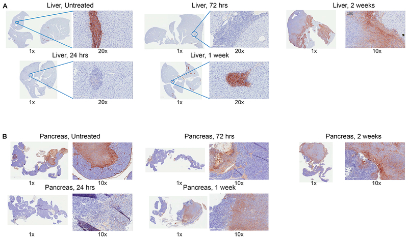
Experimental design: A murine model of metastatic PDAC treated with an irradiated whole-cell PDAC vaccine and PDAC specimens from patients treated with the same type of vaccine were used to assess the immune-modulating effect of stromal hyaluronan (HA) degradation by PEGPH20.

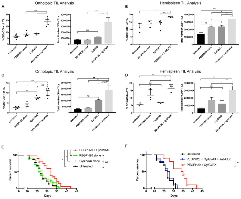
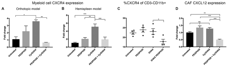
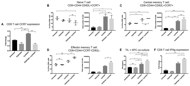
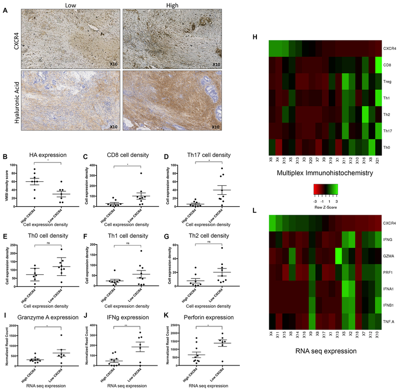
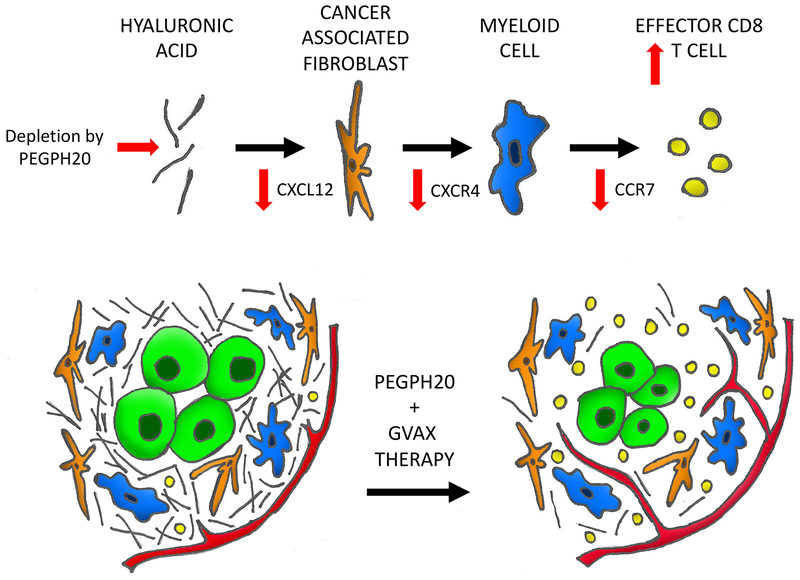
Results: Targeting stroma by degrading HA with PEGPH20 in combination with vaccine decreases CXCL12/CXCR4/CCR7 immunosuppressive signaling axis expression in cancer-associated fibroblasts, myeloid, and CD8+ T cells, respectively. This corresponds with increased CCR7- effector memory T-cell infiltration, an increase in tumor-specific IFNγ, and improved survival. In the stroma of human PDACs treated with the same vaccine, decreased stromal CXCR4 expression significantly correlated with decreased HA and increased cytotoxic activities, suggesting CXCR4 is an important therapeutic target.

Conclusions: This study represents the first to dissect signaling cascades following PDAC stroma remodeling via HA depletion, suggesting this not only overcomes a physical barrier for immune cell trafficking, but alters myeloid function leading to downstream selective increases in effector memory T-cell infiltration and antitumor activity.


Conflict of interest statement

Conflict of interests: LZ receives grant supports from Bristol-Meyer Squibb, Merck, iTeos, Amgen, Gradalis, NovaRock, and Halozyme. EMJ receives research funding from Roche and Aduro Biotech. EMJ and LZ received the royalty for licensing GVAX to Aduro Biotech. LZ is a paid consultant at Biosynergies, Merck, NovaRock, Foundation Medicine, Oncorus, Alphamab (stock owner), Mingruizhiyao (stock owner), and AstroZeneca. BB is an employee of Halozyme Therapeutics. NL is a former employee of Halozyme Therapeutics. LMC declares that she is a paid consultant for Cell Signaling Technologies and received reagent support from Plexxikon, Inc., Pharmacyclics, Inc., Acerta Pharma, Jansen R&D, Deciphera Pharmaceuticals, Genentech, Inc., Aduro Biotech., Eisai Inc., Abbott Labs, Roche Glycart AG, and NanoString Technologies. LMC is a member of the Scientific Advisory Boards of Syndax Pharmaceuticals, Inc., Carisma Therapeutics, Verseau Therapeutics, Inc., and Zymeworks Inc. ABB, VK, SM, MTS, TDA, TT, JH, RAB, CLW have no relevant conflict of interests to report.

Figures

Figure 1.
Figure 1.. Hyaluronan is expressed in peritumoral stroma and is degraded by PEGPH20 in murine liver and pancreas.
Pancreatic adenocarcinoma was established by either the hemispleen procedure or orthotopic implantation. One dose of IV PEGPH20 (40μg/kg) was given to mice on Day 6 following surgery. Mice were sacrificed at periodic intervals following the single PEGPH20 treatment with corresponding liver and pancreas sectioned and embedded in FFPE. Untreated mice were sacrificed on Day 5. Immunohistochemistry for hyaluronan was performed in FFPE sections in (A) livers of hemispleen model mice (n=9) and (B) pancreata of orthotopic implantation model mice (n=9).
Figure 2.
Figure 2.. Tumor stromal depletion of hyaluronan by PEGPH20 in combination with GVAX enhances CD8+ T-cell infiltration in PDAC and improves survival compared to single agent therapies.
Following the inoculation of KPC tumor cells by either orthotopic implantation or the hemispleen procedure, mice were treated with Cy (100 mg/kg) on Day 6, PEGPH20 (40μg/kg) on Day 6 and GVAX on Day 7. Flow cytometry was performed on tumor infiltrating lymphocytes (TIL) isolated from processed dissected orthotopic tumor or diffusely metastatic liver respectively. Each experimental group consisted of 3 or 4 mice analyzed individually. Relative percentage and total number of live CD3+CD8+ TIL in the combination PEPGH20/GVAX treatment group compared to single agent treatment groups in the (A) orthotopic model and (B) hemispleen model respectively. Relative percentage and total number of live CD3+CD4+ TIL in the combination PEGPH20/GVAX treatment group compared to single agent treatment groups in the (C) orthotopic model and (D) hemispleen model respectively. Data represent mean ± SEM from one representative experiment that was repeated three times. ns not significant, * p<0.05,** p<0.01, ***p<0.001. (E) Kaplan Meier survival curves of mice implanted with PDAC cells in untreated mice (n=22), mice treated with Cy/GVAX alone (n=18), PEGPH20 alone (n=18), or the combination PEGPH20/GVAX treatment (n=18). Data represent the combined data of two consecutive experiments. ns not significant, * p<0.05. (F) Kaplan Meier survival curves of mice implanted with PDAC cells in untreated mice (n=10) or mice treated with the combination PEGPH20/GVAX treatment with CD8+ T-cell depletion (n=10) and without CD8+ T-cell depletion (n=10). ns not significant,** p<0.01.
Figure 3.
Figure 3.. PEGPH20 in combination with GVAX decreases RNA expression within the CXCL12/CXCR4 signaling axis of cells in the tumor microenvironment.
Following the inoculation of KPC tumor cells by either orthotopic implantation or the hemispleen procedure, tumor-bearing mice were treated with Cy (100 mg/kg) on Day 6, PEGPH20 (40μg/kg) on Day 6 and GVAX on Day 7. Mice were sacrificed on Day 16. RNA was extracted from myeloid cells isolated with CD11b+ beads from the tumor microenvironment of (A) dissected orthotopic tumors or (B) diffusely metastatic livers of hemispleen mice, respectively. Quantitative real-time reverse transcription polymerase chain reaction (qPCR) reveals patterns of myeloid Cxcr4 expression in single agent treatments or the combination PEGPH20/GVAX treatment group (n=3 mice). (C) Flow cytometry was performed on tumor infiltrating lymphocytes (TIL) isolated from processed diffusely metastatic murine livers of hemispleen mice. Relative percentage of CD3CD11b+ cells expressing CXCR4 as determined by immune analysis of 4 mice analyzed individually by flow cytometry. RNA was extracted from cancer associated fibroblasts isolated with FAP+ beads from diffusely metastatic murine livers (n=3 mice). qPCR reveals patterns of CAF (C) Cxcl12 expression (n=3 mice). Data represent mean ± SEM from one representative experiment that was repeated twice. ns not significant, * p<0.05,** p<0.01, ***p<0.001.
Figure 4.
Figure 4.. Modulation of CXCL12/CXCR4/CCR7 axis by PEGPH20 in combination with GVAX is associated with increased number of effector memory CD8+ T-cells with enhanced antitumor IFNγ secretion and expression.
Following hemispleen implantation of KPC cells, tumor-bearing mice were treated with Cy (100 mg/kg) on Day 6, PEGPH20 (40μg/kg) on Day 6 and GVAX on Day 7. Mice were sacrificed on Day 16. (A) CD8+ cells were isolated from tumor infiltrating lymphocytes (TIL) by negative selection and RNA extracted (n=3 mice). qPCR reveals patterns of CD8+ cell CCR7 expression in single agent treatment and combination treatment groups. Flow cytometry was performed on TIL isolated from processed murine liver in 3 mice analyzed individually. Histograms show the percentage and total number of live (B) naïve T-cells as identified by the markers: CD8+CD44CD62L+CCR7+, (C) central memory T-cells: CD8+CD44+CD62L+CCR7+ and (D) effector memory T-cells: CD8+CD44+CCR7CD62L. (E) ELISA assays were performed to assess IFNγ secretion of isolated CD8+ T-cells in each treatment group (n=5 mice pooled). Autologous irradiated KPC tumor cells were utilized as antigenic targets and cocultured with CD8+ T-cells isolated from TIL by negative selection. (F) qPCR reveals IFNγ expression in CD8+ cells isolated from tumor infiltrating lymphocytes by negative selection (n=3 mice). Data represent mean ± SEM from one representative experiment that was repeated twice. ns not significant, * p<0.05,** p<0.01, ***p<0.001.
Figure 5.
Figure 5.. Relationships of hyaluronan and the CXCR4 axis are appreciated in human PDAC in the setting of GVAX.
Human PDAC tissue were collected from patients after one dose of neoadjuvant GVAX and subsequent surgical resection at our institution (n=22). (A) Immunohistochemistry (IHC) staining of CXCR4 (n=17) and hyaluronan (HA) expression was performed (n=15). (B) Analysis of HA expression in GVAX treated PDAC tumor with high stromal CXCR4 expression compared to tumor with low CXCR4 expression by IHC. The percentage of total cell subsets, as determined by multiplex IHC, were compared between high vs low CXCR4 expression groups: (C) CD8+ T-cells: [CD45+CD3+CD8+] (D) Th17+ T-cells: [CD45+CD3+CD8+Foxp3RORlt+] (E) Th0: [CD45+CD3+CD8Foxp3RORgtTbetGATA3] (F) Th1: [CD45+CD3+CD8Foxp3RORgtTbet+] (G) Th2: [CD45+CD3+CD8Foxp3RORgtGATA3+]. (H) Heat map generated to visualize the relationship of protein expression of CXCR4 and the density of immune cell infiltrates, particularly CD8+ T-cells, according to the quantification of IHC results. Whole exome RNA sequencing was performed on dissected pancreatic stroma of the same cohort of patients (n=17). (H) Granzyme A (I) IFNγ and (J) Perforin expression in PDAC tumor with high stromal CXCR4 expression were compared to tumor with low CXCR4 expression. (K) Heat map generated to visualize the relationship between the RNA expression of CXCR4 and the expression of markers of effector T-cell function. Data represent mean ± SEM. ns not significant, * p<0.05,** p<0.01
Figure 6.
Figure 6.. A working model of the mechanistic role of PEGPH20 in the microenvironment of PDAC.
The pancreatic cancer tumor microenvironment is dense, fibrotic and excluded from immune surveillance leading to a paucity of CD8 T-cells. PEGPH20 targets and depletes the extracellular matrix hyaluronic acid allowing vascular expansion and access to the tumor. An associated decrease in CXCL12 expression is appreciated in the cancer associated fibroblasts within the stroma. Downstream CXCR4 expression is diminished in the myeloid cells. CCR7 expression is then decreased in infiltrating CD8 cells leading to a skew towards the effector memory T-cell subtype.

Similar articles

Cited by

References

    1. Pardoll D. Cancer and the Immune System: Basic Concepts and Targets for Intervention. Semin Oncol 2015;42(4):523–38 doi 10.1053/j.seminoncol.2015.05.003. - DOI - PMC - PubMed
    1. Topalian SL, Hodi FS, Brahmer JR, Gettinger SN, Smith DC, McDermott DF, et al. Safety, activity, and immune correlates of anti-PD-1 antibody in cancer. N Engl J Med 2012;366(26):2443–54 doi 10.1056/NEJMoa1200690. - DOI - PMC - PubMed
    1. Wolchok JD, Kluger H, Callahan MK, Postow MA, Rizvi NA, Lesokhin AM, et al. Nivolumab plus ipilimumab in advanced melanoma. N Engl J Med 2013;369(2):122–33 doi 10.1056/NEJMoa1302369. - DOI - PMC - PubMed
    1. Brahmer JR, Tykodi SS, Chow LQ, Hwu WJ, Topalian SL, Hwu P, et al. Safety and activity of anti-PD-L1 antibody in patients with advanced cancer. N Engl J Med 2012;366(26):2455–65 doi 10.1056/NEJMoa1200694. - DOI - PMC - PubMed
    1. Royal RE, Levy C, Turner K, Mathur A, Hughes M, Kammula US, et al. Phase 2 trial of single agent Ipilimumab (anti-CTLA-4) for locally advanced or metastatic pancreatic adenocarcinoma. J Immunother 2010;33(8):828–33 doi 10.1097/CJI.0b013e3181eec14c. - DOI - PMC - PubMed

Publication types

MeSH terms