. 2019 Jun 11;4(4):e00257-19.
doi: 10.1128/mSystems.00257-19.

Multi-omics Signature of Candida auris, an Emerging and Multidrug-Resistant Pathogen

Affiliations

Multi-omics Signature of Candida auris, an Emerging and Multidrug-Resistant Pathogen

Daniel Zamith-Miranda et al. mSystems. .

Abstract

Candida auris is a recently described pathogenic fungus that is causing invasive outbreaks on all continents. The fungus is of high concern given the numbers of multidrug-resistant strains that have been isolated in distinct sites across the globe. The fact that its diagnosis is still problematic suggests that the spreading of the pathogen remains underestimated. Notably, the molecular mechanisms of virulence and antifungal resistance employed by this new species are largely unknown. In the present work, we compared two clinical isolates of C. auris with distinct drug susceptibility profiles and a Candida albicans reference strain using a multi-omics approach. Our results show that, despite the distinct drug resistance profile, both C. auris isolates appear to be very similar, albeit with a few notable differences. However, compared to C. albicans both C. auris isolates have major differences regarding their carbon utilization and downstream lipid and protein content, suggesting a multifactorial mechanism of drug resistance. The molecular profile displayed by C. auris helps to explain the antifungal resistance and virulence phenotypes of this new emerging pathogen.IMPORTANCE Candida auris was first described in Japan in 2009 and has now been the cause of significant outbreaks across the globe. The high number of isolates that are resistant to one or more antifungals, as well as the high mortality rates from patients with bloodstream infections, has attracted the attention of the medical mycology, infectious disease, and public health communities to this pathogenic fungus. In the current work, we performed a broad multi-omics approach on two clinical isolates isolated in New York, the most affected area in the United States and found that the omic profile of C. auris differs significantly from C. albicans In addition to our insights into C. auris carbon utilization and lipid and protein content, we believe that the availability of these data will enhance our ability to combat this rapidly emerging pathogenic yeast.

Keywords: Candida auris; antifungal resistance; fluconazole; multi-omics.


Figures

FIG 1
FIG 1
Abundance of proteins in C. auris and C. albicans. Proteins are listed in the heatmap with enriched KEGG pathways separated into two clusters based on the protein abundance between the two Candida species. For a complete list of proteins, their relative abundances, and P values (determined by t test), see Table S3 in the supplemental material.
FIG 2
FIG 2
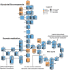
Central carbon metabolism of C. auris and C. albicans. The figure shows the relative abundance of proteins (blue boxes) and the production of metabolites (orange boxes) involved in the central carbon metabolism in both C. albicans and C. auris. Paralog proteins were grouped and posted side by side in the map. *, Genes that were only annotated in the C. albicans genome. P ≤ 0.05 indicates statistically significant hits determined by t test in any of the three comparisons. For complete comparisons between the different samples and abundances of each analyte, see Table S4 in the supplemental material.
FIG 3
FIG 3
Ergosterol biosynthesis pathway in C. auris and C. albicans. The bar graphs represent the relative abundances of proteins (blue boxes) and metabolites (orange boxes) of the pathway. Note that Erg28 is not an enzyme but a scaffold protein that docs Erg26 and Erg27 close together. P ≤ 0.05 indicates statistically significant hits determined by t test in any of the three comparisons. For complete comparisons between the different samples and abundances of each analyte, see Table S4 in the supplemental material.
FIG 4
FIG 4
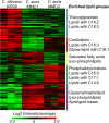
Lipid species found in C. auris and C. albicans. The abundance of all detected lipids is shown above in the heatmap. Lipids were grouped in clusters based on their abundance between different species/isolates. The enrichment of lipid intrinsic features (head group, fatty acid length, fatty acid unsaturation, etc.) is listed by the side of each cluster. For a complete list of proteins, their relative abundances, and P values (determined by t test), see Table S5 in the supplemental material.
FIG 5
FIG 5
Fatty acids and sphingoid bases analyzed by GC-MS. The graph indicates the abundance of lipids containing odd-chain fatty acids and phytosphingosine for both Candida species/isolates. t test determinations: **, P ≤ 0.01; ns, P > 0.05.
FIG 6
FIG 6
Cell wall integrity pathway. The heatmap includes signaling and major cell wall polysaccharides synthesis/degradation enzymes found in C. auris and C. albicans. For a complete list of proteins, their relative abundances, and P values (determined by t test), see Table S6 in the supplemental material.
FIG 7
FIG 7
Protein abundance profile of drug resistance-related transporters. The heatmap shows the detected transporters involved with drug resistance and their abundances in both Candida species/isolates. For a complete list of proteins, their relative abundances, and P values (determined by t test), see Table S3 in the supplemental material.

References

    1. Satoh K, Makimura K, Hasumi Y, Nishiyama Y, Uchida K, Yamaguchi H. 2009. Candida auris sp. nov., a novel ascomycetous yeast isolated from the external ear canal of an inpatient in a Japanese hospital. Microbiol Immunol 53:41–44. doi: 10.1111/j.1348-0421.2008.00083.x. - DOI - PubMed
    1. Lee WG, Shin JH, Uh Y, Kang MG, Kim SH, Park KH, Jang H-C. 2011. First three reported cases of nosocomial fungemia caused by Candida auris. J Clin Microbiol 49:3139–3142. doi: 10.1128/JCM.00319-11. - DOI - PMC - PubMed
    1. Jeffery-Smith A, Taori SK, Schelenz S, Jeffery K, Johnson EM, Borman A, Manuel R, Brown CS. 2018. Candida auris: a review of the literature. Clin Microbiol Rev 31:e00029-17. - PMC - PubMed
    1. Kean R, Sherry L, Townsend E, McKloud E, Short B, Akinbobola A, Mackay WG, Williams C, Jones BL, Ramage G. 2018. Surface disinfection challenges for Candida auris: an in-vitro study. J Hosp Infect 98:433–436. doi: 10.1016/j.jhin.2017.11.015. - DOI - PubMed
    1. Sarma S, Upadhyay S. 2017. Current perspective on emergence, diagnosis, and drug resistance in Candida auris. Infect Drug Resist 10:155–165. doi: 10.2147/IDR.S116229. - DOI - PMC - PubMed