. 2019 Jul 26;294(30):11568-11578.
doi: 10.1074/jbc.RA119.009037. Epub 2019 Jun 11.

Cardiolipin-induced activation of pyruvate dehydrogenase links mitochondrial lipid biosynthesis to TCA cycle function

Affiliations

Cardiolipin-induced activation of pyruvate dehydrogenase links mitochondrial lipid biosynthesis to TCA cycle function

Yiran Li et al. J Biol Chem. .

Abstract

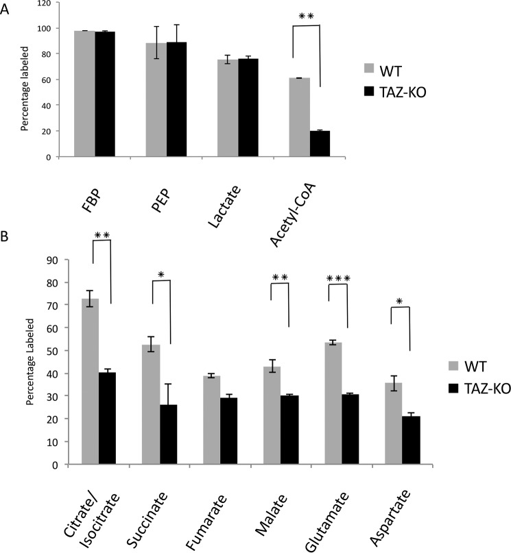
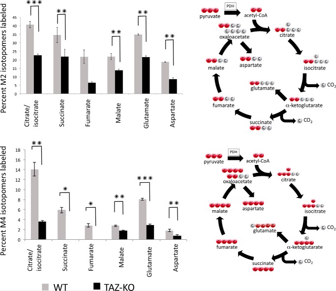
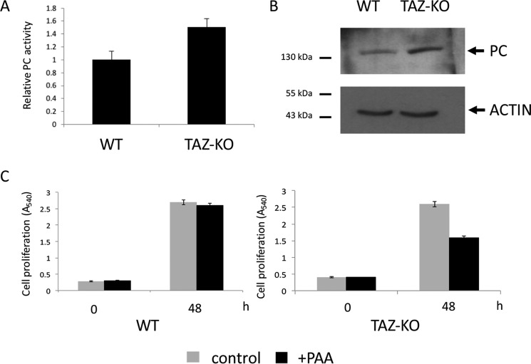
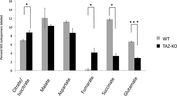
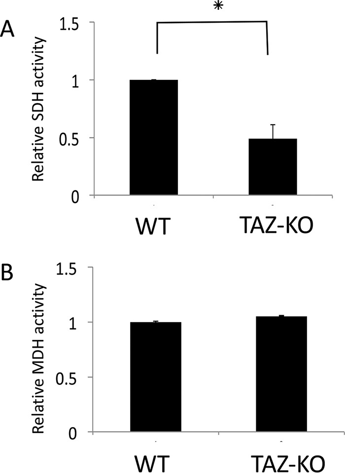
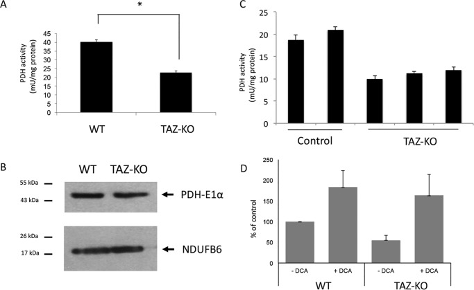
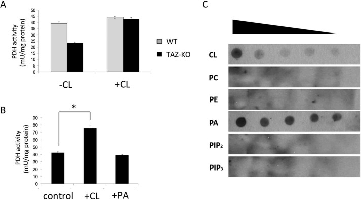
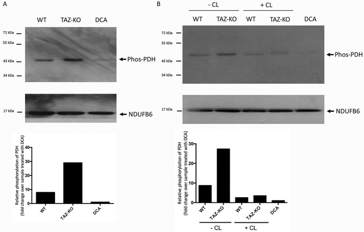
Cardiolipin (CL) is the signature phospholipid of mitochondrial membranes. Although it has long been known that CL plays an important role in mitochondrial bioenergetics, recent evidence in the yeast model indicates that CL is also essential for intermediary metabolism. To gain insight into the function of CL in energy metabolism in mammalian cells, here we analyzed the metabolic flux of [U-13C]glucose in a mouse C2C12 myoblast cell line, TAZ-KO, which is CL-deficient because of CRISPR/Cas9-mediated knockout of the CL-remodeling enzyme tafazzin (TAZ). TAZ-KO cells exhibited decreased flux of [U-13C]glucose to [13C]acetyl-CoA and M2 and M4 isotopomers of tricarboxylic acid (TCA) cycle intermediates. The activity of pyruvate carboxylase, the predominant enzyme for anaplerotic replenishing of the TCA cycle, was elevated in TAZ-KO cells, which also exhibited increased sensitivity to the pyruvate carboxylase inhibitor phenylacetate. We attributed a decreased carbon flux from glucose to acetyl-CoA in the TAZ-KO cells to a ∼50% decrease in pyruvate dehydrogenase (PDH) activity, which was observed in both TAZ-KO cells and cardiac tissue from TAZ-KO mice. Protein-lipid overlay experiments revealed that PDH binds to CL, and supplementing digitonin-solubilized TAZ-KO mitochondria with CL restored PDH activity to WT levels. Mitochondria from TAZ-KO cells exhibited an increase in phosphorylated PDH, levels of which were reduced in the presence of supplemented CL. These findings indicate that CL is required for optimal PDH activation, generation of acetyl-CoA, and TCA cycle function, findings that link the key mitochondrial lipid CL to TCA cycle function and energy metabolism.

Keywords: cardiolipin; mitochondria; pyruvate carboxylase (PC); pyruvate dehydrogenase complex (PDC); tricarboxylic acid cycle (TCA cycle) (Krebs cycle).


Conflict of interest statement

The authors declare that they have no conflicts of interest with the contents of this article. The content is solely the responsibility of the authors and does not necessarily represent the official views of the National Institutes of Health

Figures

Figure 1.
Figure 1.
Flux of [U-13C]glucose in TAZ-KO cells relative to WT. A and B, the percentage of 13C-labeled metabolites was measured after 1-h incubation with [U-13C]glucose. Metabolites from glycolysis (A) and the TCA cycle (B) were analyzed by LC-MS and GC-MS. Data shown are mean ± S.D. (n = 3). *, p < 0.05; **, p < 0.01; ***, p < 0.001.
Figure 2.
Figure 2.
Mass-isotopomer distribution (MID) of M2 and M4 metabolites. MID of M2 (top panel) and M4 (bottom panel) isotopomers was determined by LC-MS and GC-MS after 1-h incubation with [U-13C]glucose. Data shown are mean ± S.D. (n = 3). *, p < 0.05; **, p < 0.01; ***p < 0.001.
Figure 3.
Figure 3.
Increased PC activity in TAZ-KO cells. A, PC activity was assayed in isolated mitochondria as described under “Experimental procedures.” Data shown are mean ± S.D. (n = 3). B, levels of PC from cell extracts were determined by Western blot analysis with anti-PC antibody. 50 μg of total protein from each sample was loaded onto an SDS gel under reducing conditions, and actin was used as a loading control. C, cells were treated with 4 mm phenylacetic acid (PAA, a PC inhibitor, pH adjusted to 7.4). Cell viability was measured by MTT viability assay as described under “Experimental procedures.”
Figure 4.
Figure 4.
MID of M3 metabolites. MID of M3 isotopomers was determined by LC-MS and GC-MS after 1-h incubation with [U-13C]glucose. Data shown are mean ± S.D. (n = 3). *, p < 0.05; ***, p < 0.001.
Figure 5.
Figure 5.
TAZ-KO cells exhibit decreased SDH activity. A and B, the activity of SDH (A) and MDH (B) was assayed in isolated mitochondria as described under “Experimental procedures.” Data shown are mean ± S.D. (n = 3). *, p < 0.05.
Figure 6.
Figure 6.
Decreased activity of PDH in tafazzin-deficient cells. A, PDH activity was assayed in isolated mitochondria as described under “Experimental procedures.” Data shown are mean ± S.D. (n = 3). *, p < 0.05. B, levels of PDH-E1α in mitochondrial extracts were determined by Western blot analysis with anti-PDH-E1α antibody. 50 μg of total protein from each sample was loaded onto an SDS gel under reducing conditions, and the mitochondrial protein Ndufb6 was used as a loading control. C, PDH activity was assayed in heart tissue extracted from WT and TAZ-KO mice. Each column represents a separate mouse. Data shown are mean ± S.D. (n = 2). D, WT and TAZ-KO cells were seeded in DMEM (without glucose, pyruvate, or glutamine/1% FCS), followed by 3-h incubation in Dulbecco's PBS (Life Technologies) supplemented with 100 μm [1-14C]pyruvate, and the PDH flux assay was performed in the presence or absence of 5 mm DCA. PDH activity was assayed by measuring the release of 14CO2 from [1-14C]pyruvate as described under “Experimental procedures.” Data shown are mean ± S.D. (n = 3). *, p < 0.05.
Figure 7.
Figure 7.
CL binds to PDH and increases its activity. A, digitonin-solubilized mitochondria from WT and TAZ-KO cells were treated with or without CL. PDH activity was assayed as described under “Experimental procedures.” B, purified PDH complex (Sigma) was incubated with exogenous CL or phosphatidic acid (PA). PDH activity was assayed as described under “Experimental procedures”. Data shown are mean ± S.D. (n = 3). *, p < 0.05. C, the indicated lipids CL, phosphatidylethanolamine (PE), phosphatidylcholine (PC), phosphatidylinositol 4,5-bisphosphate (PIP2), phosphatidylinositol 3,4,5-trisphosphate (PIP3), and phosphatidic acid were serially diluted and spotted onto a nitrocellulose membrane, which was incubated overnight in buffer containing 10 μg of PDH complex. Interactions were detected by immunoblotting with an antibody against the PDH complex.
Figure 8.
Figure 8.
CL decreases levels of phosphorylated PDH. Phosphorylated PDH was identified by Western blot analysis with an antibody against the phosphorylated enzyme (Phos-PDH). The mitochondrial protein NDUFB6 was included as a loading control. For DCA-treated samples, 5 mm DCA was added to cultured WT cells for 24 h before mitochondrial isolation. A, levels of phosphorylated PDH in mitochondrial protein from WT and TAZ-KO cells. B, levels of phosphorylated PDH in digitonin-solubilized mitochondria from WT and TAZ-KO cells incubated for 2 h on ice with or without CL. Bottom panels, the signal intensities of protein bands and surrounding background were scanned and quantified using ImageJ. The background-subtracted value for each protein band was normalized to that of NDUFB6 and quantified relative to the DCA-treated sample.

References

    1. Planas-Iglesias J., Dwarakanath H., Mohammadyani D., Yanamala N., Kagan V. E., and Klein-Seetharaman J. (2015) Cardiolipin interactions with proteins. Biophys. J. 109, 1282–1294 10.1016/j.bpj.2015.07.034 - DOI - PMC - PubMed
    1. Ren M., Phoon C. K., and Schlame M. (2014) Metabolism and function of mitochondrial cardiolipin. Prog. Lipid Res. 55, 1–16 10.1016/j.plipres.2014.04.001 - DOI - PubMed
    1. Paradies G., Paradies V., De Benedictis V., Ruggiero F. M., and Petrosillo G. (2014) Functional role of cardiolipin in mitochondrial bioenergetics. Biochim. Biophys. Acta 1837, 408–417 10.1016/j.bbabio.2013.10.006 - DOI - PubMed
    1. Houtkooper R. H., and Vaz F. M. (2008) Cardiolipin, the heart of mitochondrial metabolism. Cell Mol. Life Sci. 65, 2493–2506 10.1007/s00018-008-8030-5 - DOI - PMC - PubMed
    1. Joshi A. S., Zhou J., Gohil V. M., Chen S., and Greenberg M. L. (2009) Cellular functions of cardiolipin in yeast. Biochim. Biophys. Acta 1793, 212–218 10.1016/j.bbamcr.2008.07.024 - DOI - PMC - PubMed

Publication types