. 2019 Jun 25;116(26):12980-12985.
doi: 10.1073/pnas.1902165116. Epub 2019 Jun 11.

Endothelial cell Piezo1 mediates pressure-induced lung vascular hyperpermeability via disruption of adherens junctions

Affiliations

Endothelial cell Piezo1 mediates pressure-induced lung vascular hyperpermeability via disruption of adherens junctions

Emily E Friedrich et al. Proc Natl Acad Sci U S A. .

Abstract

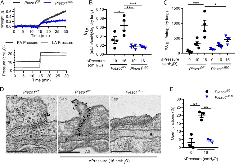
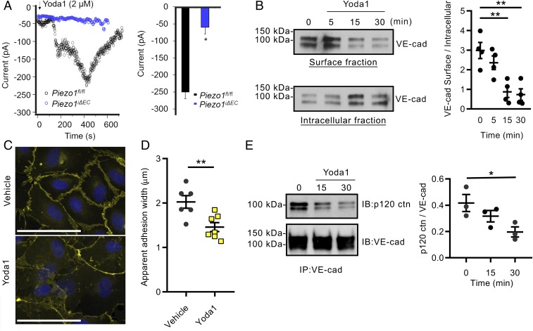
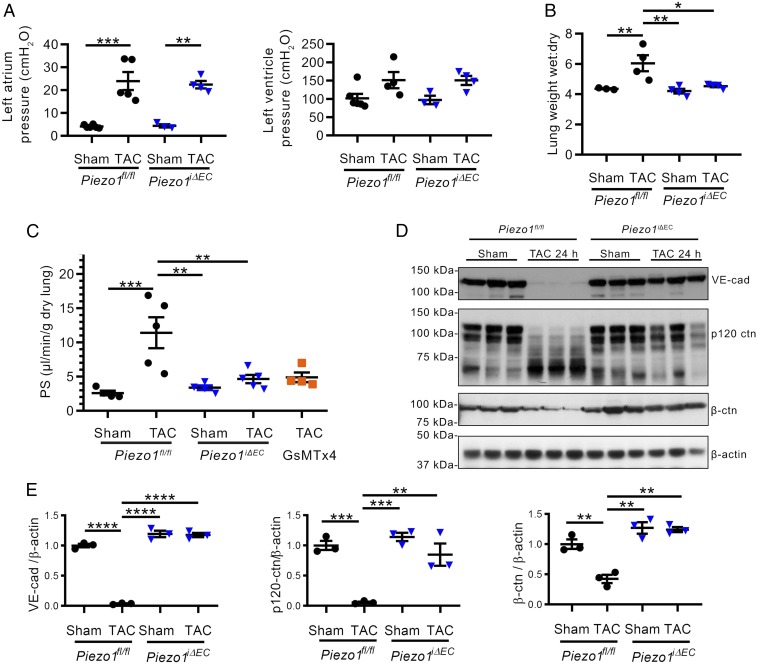
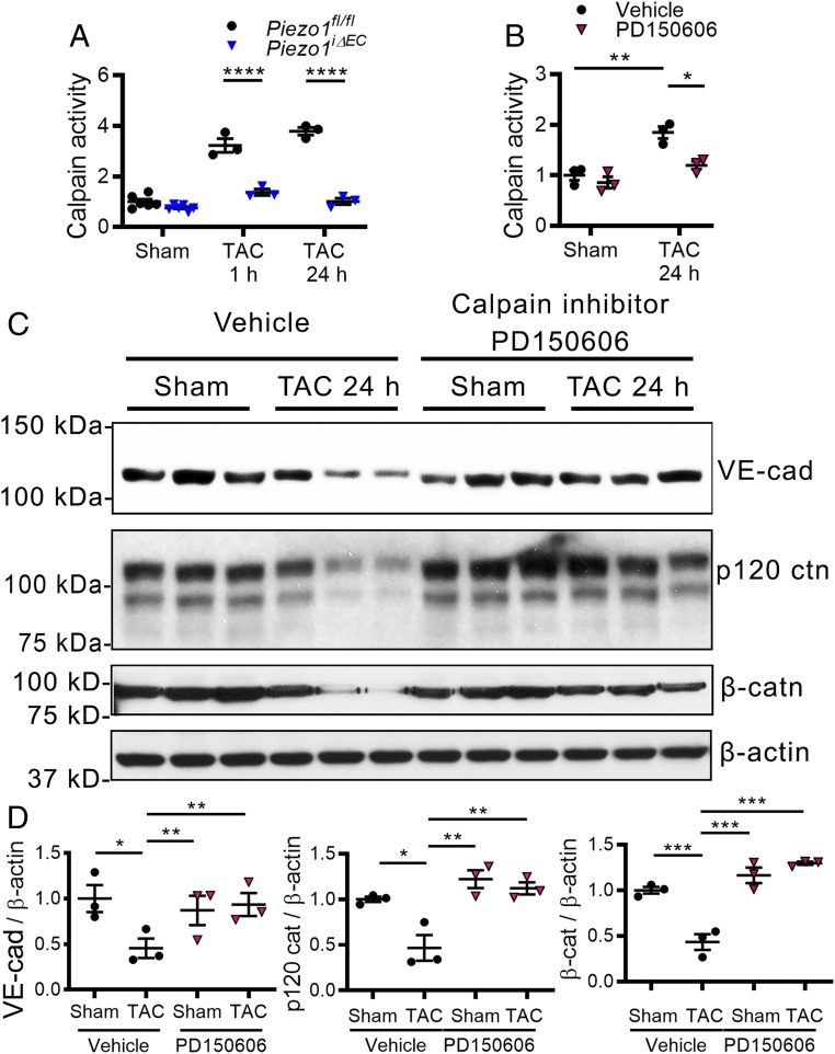
Increased pulmonary microvessel pressure experienced in left heart failure, head trauma, or high altitude can lead to endothelial barrier disruption referred to as capillary "stress failure" that causes leakage of protein-rich plasma and pulmonary edema. However, little is known about vascular endothelial sensing and transduction of mechanical stimuli inducing endothelial barrier disruption. Piezo1, a mechanosensing ion channel expressed in endothelial cells (ECs), is activated by elevated pressure and other mechanical stimuli. Here, we demonstrate the involvement of Piezo1 in sensing increased lung microvessel pressure and mediating endothelial barrier disruption. Studies were made in mice in which Piezo1 was deleted conditionally in ECs (Piezo1iΔEC ), and lung microvessel pressure was increased either by raising left atrial pressure or by aortic constriction. We observed that lung endothelial barrier leakiness and edema induced by raising pulmonary microvessel pressure were abrogated in Piezo1iΔEC mice. Piezo1 signaled lung vascular hyperpermeability by promoting the internalization and degradation of the endothelial adherens junction (AJ) protein VE-cadherin. Breakdown of AJs was the result of activation of the calcium-dependent protease calpain and degradation of the AJ proteins VE-cadherin, β-catenin, and p120-catenin. Deletion of Piezo1 in ECs or inhibition of calpain similarly prevented reduction in the AJ proteins. Thus, Piezo1 activation in ECs induced by elevated lung microvessel pressure mediates capillary stress failure and edema formation secondary to calpain-induced disruption of VE-cadherin adhesion. Inhibiting Piezo1 signaling may be a useful strategy to limit lung capillary stress failure injury in response to elevated vascular pressures.

Keywords: Piezo1; endothelial; permeability.


Conflict of interest statement

The authors declare no conflict of interest.

Figures

Fig. 1.
Fig. 1.
Elevated pulmonary microvessel pressure increases lung vascular permeability by a Piezo1-dependent mechanism. (A) Changes in lung wet weight and pulmonary artery pressure in response to increased left atrial pressure in lungs from Piezo1fl/fl and Piezo1iΔEC mice. (B and C) Increases in capillary filtration coefficient (Kf,c) (B) and pulmonary transvascular permeability (PS) to albumin tracer (C) in response to increased left atrial pressure in lungs from Piezo1fl/fl and Piezo1iΔEC mice. (D) Transmission electron microscopy of interendothelial junctions in lung capillaries of Piezo1fl/fl and Piezo1iΔEC mice subjected to increased left atrial pressure; representative images from n = 3 mice. Lung vessels were subjected to the same rise in left atrial pressure as in A. Cap, capillary lumen; AS, alveolar space; #, basement membrane. (Scale bar, 0.2 µm.) (E) Percentage of junctions with gold–albumin particles for each condition. Data are shown as mean ± SEM; *P ≤ 0.05; **P ≤ 0.01; ***P ≤ 0.001.
Fig. 2.
Fig. 2.
Activation of Piezo1 disrupts VE-cadherin junctions in EC monolayers. (A) Time course of whole-cell inward current development (Left) and summary of the peak inward current (Right) induced by extracellular application of Yoda1 in lung ECs from both Piezo1fl/fl and Piezo1iΔEC mice. Inward currents were elicited by −110 mV voltage. (B) Activation of Piezo1 with 5 µM Yoda1 induces VE-cadherin internalization in human lung microvascular ECs. Western blot analysis of biotinylated VE-cadherin at plasma membrane and in cytosol (Left), and quantified as a ratio of surface-to-intracellular fractions (Right). (C) Decreased accumulation of VE-cadherin (yellow) at AJs in EC monolayers treated with Yoda1 for 30 min. (Scale bar, 100 µm.) (D) Quantification of data in C. (E) Yoda1 activation of Piezo1 decreased p120-catenin association with VE-cadherin as shown by coimmunoprecipitation assay (Left) and quantified as a ratio of p120-catenin to VE-cadherin (Right). Individual data points shown with mean ± SEM; *P ≤ 0.05; **P ≤ 0.01.
Fig. 3.
Fig. 3.
Increased lung microvessel pressure induces Piezo1-dependent lung vascular hyperpermeability secondary to reduced expression of AJ proteins. (A) Effects of 50% constriction of aorta (TAC) in Piezo1fl/fl and Piezo1iΔEC mice on left atrial pressure (Left) and left ventricular systolic pressure (Right) recorded at 24 h post-TAC. (B and C) Accumulation of lung water (B) and increased lung transvascular albumin permeability (PS) (C) were blocked in Piezo1iΔEC mice and in Piezo1fl/fl mice treated with GsMTx4 at 24 h post-TAC. (D and E) Loss of VE-cadherin, β-catenin, and p120-catenin at 24 h post-TAC. Individual data points are shown as well as mean ± SEM; *P ≤ 0.05; **P ≤ 0.01; ***P ≤ 0.001; ****P ≤ 0.0001.
Fig. 4.
Fig. 4.
Piezo1-dependent activation of calpain cleaves junctional proteins in lung ECs of mice in response to elevated lung microvessel pressure. (A) Increased activity of calpain was blocked in Piezo1iΔEC mice at 1 and 24 h post-TAC. (BD) Treatment of mice with calpain inhibitor PD150606 reduced calpain activity after TAC (B) and prevented the loss of VE-cadherin, β-catenin, and p120-catenin in lung vessels (C and D). Individual data points are shown as well as mean ± SEM; *P ≤ 0.05; **P ≤ 0.01; ***P ≤ 0.001; ****P ≤ 0.0001.

References

    1. Mehta D., Malik A. B., Signaling mechanisms regulating endothelial permeability. Physiol. Rev. 86, 279–367 (2006). - PubMed
    1. West J. B., Mathieu-Costello O., Vulnerability of pulmonary capillaries in heart disease. Circulation 92, 622–631 (1995). - PubMed
    1. Vestweber D., Winderlich M., Cagna G., Nottebaum A. F., Cell adhesion dynamics at endothelial junctions: VE-cadherin as a major player. Trends Cell Biol. 19, 8–15 (2009). - PubMed
    1. Corada M., et al. , Vascular endothelial-cadherin is an important determinant of microvascular integrity in vivo. Proc. Natl. Acad. Sci. U.S.A. 96, 9815–9820 (1999). - PMC - PubMed
    1. Malik A. B., Selig W. M., Burhop K. E., Cellular and humoral mediators of pulmonary edema. Lung 163, 193–219 (1985). - PubMed

Publication types

MeSH terms

LinkOut - more resources