. 2019 Jun 11;10(6):454.
doi: 10.1038/s41419-019-1685-z.

MIR17HG-miR-18a/19a axis, regulated by interferon regulatory factor-1, promotes gastric cancer metastasis via Wnt/β-catenin signalling

Affiliations

MIR17HG-miR-18a/19a axis, regulated by interferon regulatory factor-1, promotes gastric cancer metastasis via Wnt/β-catenin signalling

Jingsheng Yuan et al. Cell Death Dis. .

Abstract

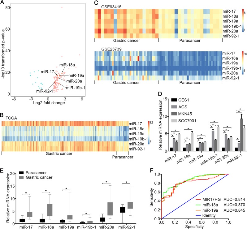
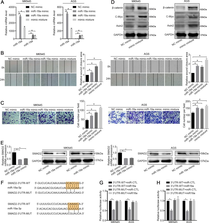
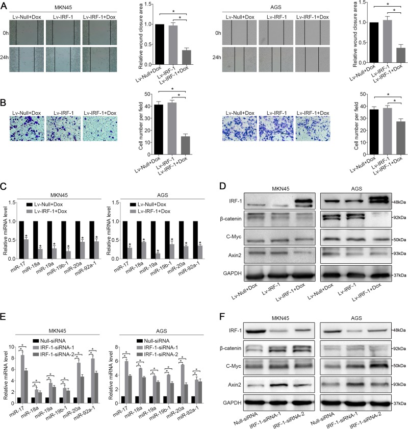
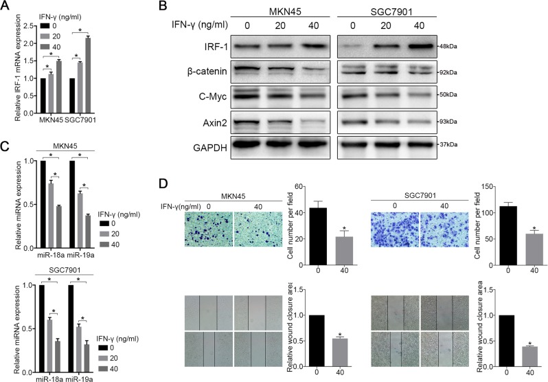
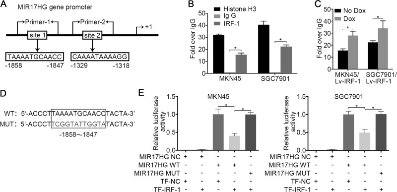
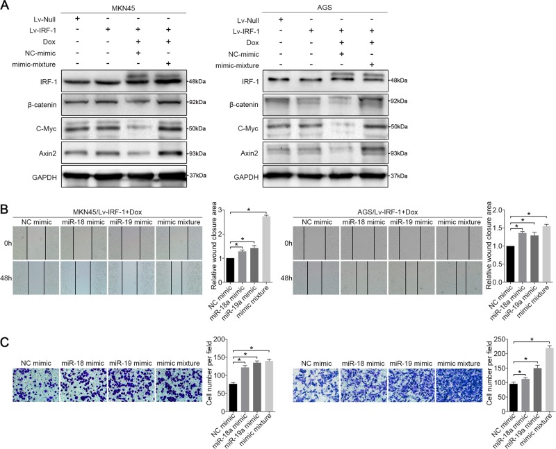
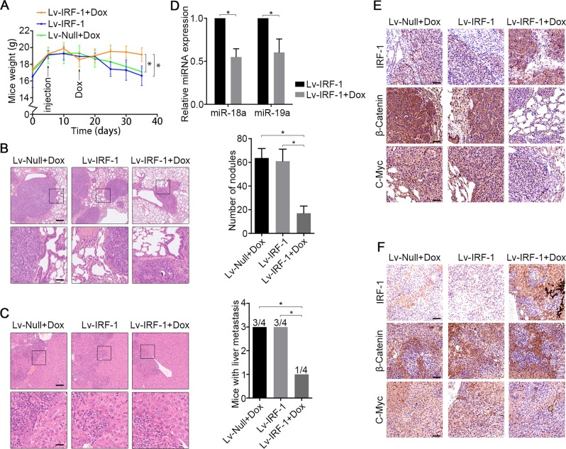
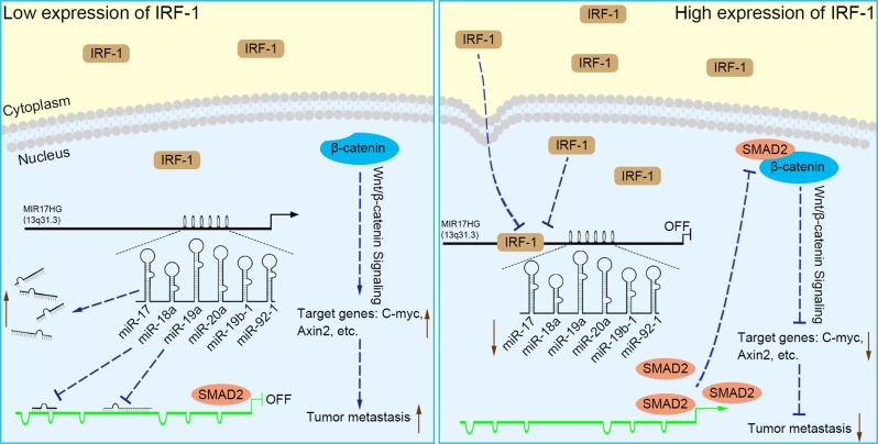
MIR17HG, located on chromosome 13, is a class of Pri-miRNAs that generates six miRNAs: miR-17, miR-18a, miR-19a, miR-20a, miR-19b-1 and miR-92-1. These miRNAs are ubiquitously overexpressed in diverse tumour types and exhibit complex biological links to tumour metastasis. We demonstrated that MIR17HG-derived miR-18a and miR-19a coordinately mediate gastric cancer cell metastasis by directly inhibiting SMAD2 expression and upregulating Wnt/β-catenin signalling. Based on previous studies, we hypothesised that an investigation of MIR17HG inhibition would be beneficial to clinical gastric cancer treatment, and systematically coupled bioinformatics analyses brought interferon regulatory factor-1 (IRF-1) to our attention. We then established stable clones in gastric cancer cells containing a doxycycline-inducible IRF-1 expression system and found that the expression of IRF-1 downregulates the embedded miRNAs of MIR17HG in gastric cancer cells and inhibits gastric cancer cell metastasis by attenuating Wnt/β-catenin signalling. Further rescue assays confirmed the crucial roles of miR-18a and miR-19a in the IRF-1-mediated inhibition of Wnt/β-catenin signalling. We also demonstrated that IRF-1 binds to the transcriptional site in the MIR17HG promoter and inhibits MIR17HG expression. Moreover, IFN-γ induced the IRF-1-mediated downregulation of MIR17HG in gastric cancer cells. Our hypothesis was supported by the results of immunohistochemistry analyses of clinical gastric cancer samples, and we also demonstrated the role of IRF-1 in inhibiting MIR17HG expression and tumour metastasis in vivo. We conclude that IRF-1 inhibits gastric cancer metastasis by downregulating MIR17HG-miR-18a/miR-19a axis expression and attenuating Wnt/β-catenin signalling.


Conflict of interest statement

The authors declare that they have no conflict of interest.

Figures

Fig. 1
Fig. 1. Expression profiles of miRNAs in GC cell lines and GC tissues.
a The differentially expressed miRNAs in 387 GC samples and 41 adjacent gastric mucosal tissues are visualised in a volcano plot. The red and green colours indicate high and low expression (|log2(fold change) | > 1, FDR < 0.05), respectively. b Heatmap of six polycistronic miRNAs derived from MIR17HG in the TCGA data set. c Heatmap of six polycistronic miRNAs derived from MIR17HG in the GEO verification data set (GSE93415 and GSE23739). b, c the columns represent the clinical samples, while the rows show the relative expression of individual miRNAs. The red and blue colours indicate high and low expression, respectively. d qRT-PCR analysis of the expression of six polycistronic miRNAs derived from MIR17HG in GC cell lines (AGS, MKN45 and SGC7901) and a normal gastric epithelial cell line (GES1). e qRT-PCR analysis of the expression of six polycistronic miRNAs derived from MIR17HG in 10 pairs of cancerous and adjacent tissues harvested from patients with GC. d, e U6 snRNA served as the internal control. N = 3 independent experiments performed in triplicate. *P < 0.05, as demonstrated by paired Student’s t test. The data are presented as the means ± standard deviations (SDs). f Receiver operating characteristic (ROC) curve of MIR17HG, miR-18a and miR-19a among GC patients
Fig. 2
Fig. 2. miR-18a and miR-19a cooperate to drive GC cell metastasis viaWnt/β-catenin signalling pathways.
a At 48 hours after transfection, the expression levels of miR-18a and miR-19a in the MKN45 and AGS cell lines were examined by qRT-PCR. U6 snRNA served as the internal control. b Wound-healing assay and c migration assay of MKN45 and AGS cells treated with an NC mimic, a miR-18a mimic, a miR-19a mimic and a mimic mixture. d Western blot analysis of β-catenin, C-Myc and Axin2 in MKN45 and AGS cells treated with an NC mimic and a miR-18a/19a mimic mixture. e The mRNA and protein levels of SMAD2 in cells treated with miR-18a mimic, miR-19a mimic and NC mimic were examined. f Predicted miR-18a and miR-19a-binding sites in the 3’ UTRs of human SMAD2. g, h Dual luciferase assays of SMAD2 that were predicted to be regulated by miR-18a or miR-19a. All the above experiments were independently performed in triplicate (N = 3). The data a, b, c, e, g and h represent the means ± SDs. *P < 0.05, as demonstrated by paired Student’s t test
Fig. 3
Fig. 3. IRF-1 regulates MIR17HG expression.
a Wound-healing and b migration assays of Lv-IRF-1 Dox-treated, Lv-IRF-1 Dox-untreated, and Lv-Null Dox-treated MKN45 and AGS cells were performed. c After 48 hours of Dox induction, the expression of six polycistronic miRNAs derived from MIR17HG in the MKN45 and AGS cell lines transfected with Lv-IRF-1 and Lv-Null was analysed by qRT-PCR. d After 48 hours of Dox induction, IRF-1, β-catenin, C-Myc and Axin2 expression in the MKN45 and AGS cell lines transfected with Lv-IRF-1 and Lv-Null was analysed by western blot analysis. e qRT-PCR analysis of the expression of six polycistronic miRNAs derived from MIR17HG in the MKN45 and AGS cell lines after IRF-1 knockdown. f Western blot analysis of IRF-1, β-catenin, C-Myc and Axin2 expression in MKN45 and AGS cell lines after IRF-1 knockdown. All the above experiments were independently performed in triplicate (N = 3). *P < 0.05, as determined by paired Student’s t test. The data a, b, c and e are presented as the means ± SDs
Fig. 4
Fig. 4. IFN-γ inhibits MIR17HG expression through IRF-1.
MKN45 and SGC7901 cells were treated with the indicated amount of IFN-γ for 48 hours, and a the mRNA expression of IRF-1 was determined by qRT-PCR. b The expression of IRF-1, β-catenin, C-Myc and Axin2 was analysed by western blotting. c the expression of miR-18a and miR-19a was analysed by qRT-PCR, and d migration and wound-healing assays were performed. Three independent experiments were performed in triplicate (N = 3). *P < 0.05, as demonstrated by paired Student’s t test. The data in a, c and d are presented as the means ± SDs
Fig. 5
Fig. 5. IRF-1 suppresses MIR17HG promoter activity.
a Two putative IRF-1 transcriptional binding sites in the MIR17HG gene promoter region. b ChIP assays of MKN45 and SGC7901 cells were performed to characterise the recruitment of IRF-1 to the MIR17HG gene promoter. c MKN45/Lv-IRF-1 and SGC7901/Lv-IRF-1 cells were treated with 4 μg/ml Dox for 24 hours, and ChIP analyses were performed to assess the binding of IRF-1 to the MIR17HG gene promoter. d Schematic diagram of the reporter constructs of the wild-type (WT) and mutant (MUT) MIR17HG promoter binding site-1 fragment. e The indicated MIR17HG reporter constructs were co-transfected into MKN45 and SGC7901 cells with IRF-1 plasmids (TR-IRF-1) or normal control plasmids (TR-NC), and the luciferase activities were then measured and analysed. Three independent experiments were performed in triplicate (N = 3). *P < 0.05, as determined by paired Student’s t test. The data b, c and e are presented as the means ± SDs
Fig. 6
Fig. 6. miR-18a and miR-19a reverse the inhibitory effect of IRF-1.
a After 48 hours of Dox induction, the expression levels of IRF-1, β-catenin, C-Myc and Axin2 in MKN45/Lv-IRF-1 and AGS/Lv-IRF-1 cells treated with a mimic mixture and an NC mimic compared with those in MKN45 and AGS cell lines transfected with Lv-IRF-1 and Lv-Null, respectively, were analysed by western blotting. b Wound-healing assay and c migration assay of MKN45/Lv-IRF-1 and AGS/Lv-IRF-1 cells treated with an NC mimic, a miR-18a mimic, a miR-19a mimic and a mimic mixture. All the above experiments were independently performed in triplicate (N = 3). *P < 0.05, as determined by paired Student’s t test. The data in b and c are presented as the means ± SDs
Fig. 7
Fig. 7. IRF-1 inhibits tumour metastases in vivo by affecting MIR17HG expression and Wnt/β-catenin signalling.
a Average body weights of mice per group throughout the experiment. b H&E staining of lung tissue and number of nodules per mouse with lung metastases in each group. Scale bars, 200 μm (above) and 50 μm (below). c H&E staining of liver tissue and number of mice with liver metastases in each group. Scale bars, 100 μm (above) and 25 μm (below). d The lung metastasis nodules of the Lv-IRF-1 and Lv-IRF-1 + Dox groups were carefully stripped, and the expression levels of miR-18a and miR-19a were analysed by qRT-PCR. *P < 0.05, as determined by two-tailed unpaired Student’s t test. The data in a, b, c and d are presented as the means ± SDs. e Lung and f liver tissue, the expression of IRF-1, β-catenin and C-Myc were analysed by immunohistochemistry. N = 4 biological replicates of each experiment. Scale bars, 100 μm
Fig. 8
Fig. 8. Schematic diagram of the results of our study.
When IRF-1 expression in GC is low, large numbers of miR-17, miR-18a, miR-19a, miR-20a, miR-19b-1 and miR-92–1 are derived from MIR17HG, of which miR-18a and miR-19a decrease SMAD2 protein expression by inhibiting its promoter activity, attenuating its inhibitory effect on β-catenin. The increased expression of Wnt/β-catenin signalling elements, such as C-Myc, Axin2, etc., increases the metastasis of GC. When IRF-1 expression is increased, MIR17HG promoter activity is inhibited and the expression of miRNAs from MIR17HG is reduced. Subsequently, the inhibitory effect of miR-18a and miR-19a on SMAD2 promoter is attenuated. The increased expression of SMAD2 leads to it binding β-catenin and inhibiting its activity, thereby attenuating Wnt/β-catenin signalling and inhibiting GC metastasis

Similar articles

Cited by

References

    1. Siegel RL, Miller KD, Jemal A. Cancer statistics, 2018. CA Cancer J. Clin. 2018;68:7–30. doi: 10.3322/caac.21442. - DOI - PubMed
    1. Bray F, et al. Global Cancer Statistics 2018: GLOBOCAN Estimates of Incidence and Mortality Worldwide for 36 Cancers in 185 Countries. CA Cancer J Clin. 2018;68:394–424. doi: 10.3322/caac.21492. - DOI - PubMed
    1. Pasechnikov V, Chukov S, Fedorov E, Kikuste I, Leja M. Gastric cancer: prevention, screening and early diagnosis. World J. Gastroenterol. 2014;20:13842–13862. doi: 10.3748/wjg.v20.i38.13842. - DOI - PMC - PubMed
    1. Ferlay J, et al. Cancer incidence and mortality patterns in Europe: estimates for 40 countries in 2012. Eur. J. Cancer. 2013;49:1374–1403. doi: 10.1016/j.ejca.2012.12.027. - DOI - PubMed
    1. Gu H, et al. The prognostic efficacy and improvements of the7th edition Union for International Cancer Control tumor-node-metastasis classifications for Chinese patients with gastric cancer: results based on a retrospective three-decade population study. Tumour Biol. 2017;39:1010428317694548. doi: 10.1177/1010428317694548. - DOI - PubMed

Publication types

MeSH terms