. 2019 Jun 11;9(1):8456.
doi: 10.1038/s41598-019-44620-6.

Bone mineral: new insights into its chemical composition

Affiliations

Bone mineral: new insights into its chemical composition

Stanislas Von Euw et al. Sci Rep. .

Abstract

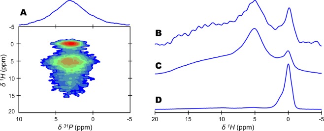
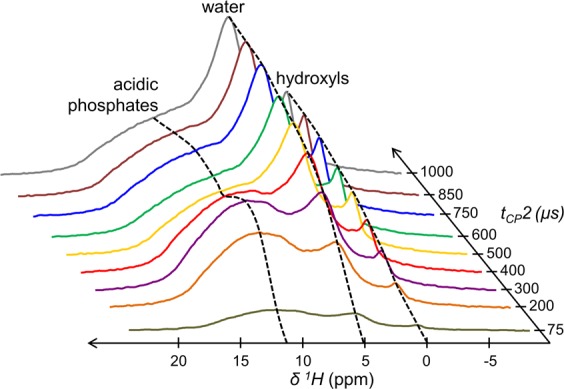
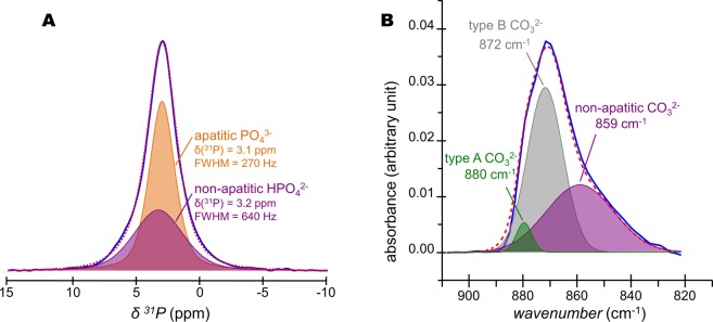
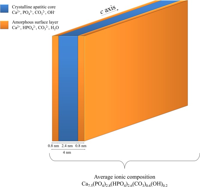
Some compositional and structural features of mature bone mineral particles remain unclear. They have been described as calcium-deficient and hydroxyl-deficient carbonated hydroxyapatite particles in which a fraction of the PO43- lattice sites are occupied by HPO42- ions. The time has come to revise this description since it has now been proven that the surface of mature bone mineral particles is not in the form of hydroxyapatite but rather in the form of hydrated amorphous calcium phosphate. Using a combination of dedicated solid-state nuclear magnetic resonance techniques, the hydrogen-bearing species present in bone mineral and especially the HPO42- ions were closely scrutinized. We show that these HPO42- ions are concentrated at the surface of bone mineral particles in the so-called amorphous surface layer whose thickness was estimated here to be about 0.8 nm for a 4-nm thick particle. We also show that their molar proportion is much higher than previously estimated since they stand for about half of the overall amount of inorganic phosphate ions that compose bone mineral. As such, the mineral-mineral and mineral-biomolecule interfaces in bone tissue must be driven by metastable hydrated amorphous environments rich in HPO42- ions rather than by stable crystalline environments of hydroxyapatite structure.


Conflict of interest statement

The authors declare no competing interests.

Figures

Figure 1
Figure 1
Detection of hydrogen-bearing species in bone mineral. 1H-31P cross polarization (CP) based magic angle spinning (MAS) solid-state Nuclear Magnetic Resonance (ssNMR) spectra of a dry 2-year-old sheep bone tissue sample. (A) two-dimensional (2D) {1H}31P Heteronuclear Correlation (HetCor) spectrum (contact time, tCP = 1000 µs). Signal intensity increases from blue to red. (B) 1H projection of the vertical (F1) dimension of the 2D {1H}31P HetCor spectrum shown in (A). {1H-31P}1H double CP MAS spectra recorded with the following contact times: (C) tCP1 = tCP2 = 1000 µs; and, (D) tCP1 = tCP2 = 15000 µs. The total experimental time was the same in each experiment (i.e., 9 hours).
Figure 2
Figure 2
Cross-polarization dynamics of the 1H resonances from bone mineral. {1H-31P}1H double cross polarization (CP) magic angle spinning (MAS) solid-state Nuclear Magnetic Resonance (ssNMR) dynamics of a dry 2-year-old sheep bone tissue sample. Contact time 1 (tCP1) was fixed at 1000 µs, while contact time 2 (tCP2) was varied from 75 µs to 1000 µs. Black dashed lines are guidelines for the eyes.
Figure 3
Figure 3
Determination of 31P-1H internuclear distances within the acidic phosphate species present in bone mineral. (A) {1H-31P}1H double cross polarization (CP) magic angle spinning (MAS) solid-state Nuclear Magnetic Resonance (ssNMR) spectrum of a dry 2-year-old sheep bone tissue sample (blue line) and its corresponding fitting (red dashed line). Contact times 1 (tCP1) was 1000 µs, while contact time 2 (tCP2) was 500 µs. (B) Numerical modelling of the evolution of the magnetization of the δ(1H) = 0 (purple, hydroxyl ions); 5.2 (grey, structural water molecules); 9.8 (blue, acidic phosphate species); and, 14.0 (green, acidic phosphate species) ppm peaks shown in (A). The acidic phosphate species peaks were simulated according to Eq. (1), while the hydroxyl ions and structural water molecules resonances were simulated according to Eq. (3). Numerical modelling according to Eq. (1) of the magnetization evolution of the δ(1H) = 14.0 (C) and 9.8 (D) ppm peaks attributed to acidic phosphate species; together with the calculations of their respective dipolar constant (DPH) and P•••H distance (dPH).
Figure 4
Figure 4
Implication of octacalcium phosphate (OCP) in the non-apatitic environments of bone mineral. (A) Two-dimensional (2D) {1H}31P Heteronuclear Correlation (HetCor) magic angle spinning (MAS) solid-state Nuclear Magnetic Resonance (ssNMR) spectrum of a fresh 2-year-old sheep bone tissue sample (contact time, tCP = 1000 µs). Signal intensity increases from blue to red. (B) One-dimensional (1D) individual 31P NMR signal of the H2O and HPO42−-containing non-apatitic environments attributed to the amorphous surface layer that coats bone mineral particles (blue line); and 1D individual 31P NMR signal of the OH-containing apatitic environments that compose the internal crystalline core of bone mineral particles (orange line). These individual 31P NMR signals were generated from the 2D {1H}31P HetCor ssNMR spectrum shown in (A). To do so, the F2 slices taken at the bound water molecules position [from δ(1H) = 3 to 7 ppm, blue area] and hydroxyl ions position [from δ(1H) = −2 to 2 ppm, orange area] in F1 have been summed. (C) 1D 31P CP MAS ssNMR spectrum (tCP = 1000 µs) of a synthetic octacalcium phosphate (OCP) sample. P1 to P6 correspond to the six different phosphate groups present in the OCP crystal lattice according to the work of Davies et al.. The red dashed-line marks the most intense resonance in the signal of OCP which is not detected in bone mineral (B).
Figure 5
Figure 5
Quantification of HPO42− and CO32− ions present in bone mineral. (A) Quantitative 31P single pulse (SP) magic angle spinning (MAS) solid-state Nuclear Magnetic Resonance (ssNMR) spectrum of a fresh 2-year-old sheep bone tissue sample (blue line) and its corresponding fitting (red dashed line) with two peaks. Those two peaks, whose lineshape and linewidth were revealed in Fig. 4B, correspond to the PO43−-containing internal crystalline core in the form of hydroxyapatite (orange peak) and the HPO42−-containing non-apatitic environments in the form of an amorphous surface layer (purple peak). (B) Fourier Transform-Infrared (FT-IR) spectrum of the ν2(CO3) vibration mode for a 2-year-old sheep bone tissue sample (blue line) and its corresponding fitting (red dashed line). Type B CO32− ions occupy the PO43− sites within the hydroxyapatite’s crystal lattice; type A CO32− ions occupy the OH sites within the hydroxyapatite’s crystal lattice; whereas non-apatitic CO32− are present within the amorphous surface layer that coats bone mineral particles.
Figure 6
Figure 6
Chemical and structural model of mature bone mineral particles. Schematic representation of platelet-shaped mature bone mineral particles including dimensions and ionic composition according to the results obtained from our 2-year-old sheep bone tissue sample. They are composed by an internal crystalline core in the form of carbonated hydroxyapatite coated by an amorphous layer in which the HPO42− ions are concentrated.

References

    1. Reznikov N, Bilton M, Lari L, Stevens MM, Kröger R. Fractal-like hierarchical organization of bone begins at the nanoscale. Science. 2018;360:eaao2189. doi: 10.1126/science.aao2189. - DOI - PMC - PubMed
    1. Grandfield K, Vuong V, Schwarcz HP. Ultrastructure of Bone: Hierarchical Features from Nanometer to Micrometer Scale Revealed in Focused Ion Beam Sections in the TEM. Calcif. Tissue Int. 2018;103:606–616. doi: 10.1007/s00223-018-0454-9. - DOI - PubMed
    1. Glimcher MJ. Bone: Nature of the Calcium Phosphate Crystals and Cellular, Structural, and Physical Chemical Mechanisms in Their Formation. Rev. Mineral. Geochem. 2006;64:223–282. doi: 10.2138/rmg.2006.64.8. - DOI
    1. Delmas PD, Tracy RP, Riggs BL, Mann KG. Identification of the noncollagenous proteins of bovine bone by two-dimensional gel electrophoresis. Calcif. Tissue Int. 1984;36:308–316. doi: 10.1007/BF02405335. - DOI - PubMed
    1. Boskey AL. Noncollagenous matrix proteins and their role in mineralization. Bone Miner. 1989;6:111–123. doi: 10.1016/0169-6009(89)90044-5. - DOI - PubMed

Publication types