. 2019 Jun 11;9(1):8495.
doi: 10.1038/s41598-019-44839-3.

Deep Learning for the Radiographic Detection of Periodontal Bone Loss

Affiliations

Deep Learning for the Radiographic Detection of Periodontal Bone Loss

Joachim Krois et al. Sci Rep. .

Abstract

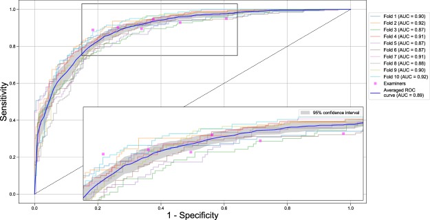
We applied deep convolutional neural networks (CNNs) to detect periodontal bone loss (PBL) on panoramic dental radiographs. We synthesized a set of 2001 image segments from panoramic radiographs. Our reference test was the measured % of PBL. A deep feed-forward CNN was trained and validated via 10-times repeated group shuffling. Model architectures and hyperparameters were tuned using grid search. The final model was a seven-layer deep neural network, parameterized by a total number of 4,299,651 weights. For comparison, six dentists assessed the image segments for PBL. Averaged over 10 validation folds the mean (SD) classification accuracy of the CNN was 0.81 (0.02). Mean (SD) sensitivity and specificity were 0.81 (0.04), 0.81 (0.05), respectively. The mean (SD) accuracy of the dentists was 0.76 (0.06), but the CNN was not statistically significant superior compared to the examiners (p = 0.067/t-test). Mean sensitivity and specificity of the dentists was 0.92 (0.02) and 0.63 (0.14), respectively. A CNN trained on a limited amount of radiographic image segments showed at least similar discrimination ability as dentists for assessing PBL on panoramic radiographs. Dentists' diagnostic efforts when using radiographs may be reduced by applying machine-learning based technologies.


Conflict of interest statement

The authors declare no competing interests.

Figures

Figure 1
Figure 1
Receiver operating characteristic (ROC) curves for the base-case model (reference test defined by average of three independent measurements of the % PBL, cut-off 20%, all teeth included). The CNN was evaluated against the reference test with respect to sensitivity (the proportion of positives that are correctly identified as such) and specificity (the proportion of negatives that are correctly identified as such). The colored curves indicate the discrimination ability in each validation fold. The bold blue line represents the mean discrimination ability; the gray area corresponds to the 95% confidence interval (CI), respectively. The discrimination ability is further summarized by the AUC. A single examiner’s discrimination ability is represented by a magenta marker (operating point). If the operating point for a particular examiner lies outside the 95% CI, the model’s discriminative ability is significantly different (superior, inferior) from the examiner’s. Insert: magnified area. Sensitivity analyses with variations in reference test construction, cut-off and tooth subsets can be found in Table 1 and the appendix.

References

    1. Papapanou PN, et al. Periodontitis: Consensus report of workgroup 2 of the 2017 World Workshop on the Classification of Periodontal and Peri‐Implant Diseases and Conditions. Journal of clinical periodontology. 2018;45:S162–S170. doi: 10.1111/jcpe.12946. - DOI - PubMed
    1. Persson RE, et al. Comparison between panoramic and intra-oral radiographs for the assessment of alveolar bone levels in a periodontal maintenance population. Journal of clinical periodontology. 2003;30:833–839. doi: 10.1034/j.1600-051X.2003.00379.x. - DOI - PubMed
    1. Walsh TF, al-Hokail OS, Fosam EB. The relationship of bone loss observed on panoramic radiographs with clinical periodontal screening. Journal of clinical periodontology. 1997;24:153–157. doi: 10.1111/j.1600-051X.1997.tb00484.x. - DOI - PubMed
    1. Akesson L, Hakansson J, Rohlin M. Comparison of panoramic and intraoral radiography and pocket probing for the measurement of the marginal bone level. Journal of clinical periodontology. 1992;19:326–332. doi: 10.1111/j.1600-051X.1992.tb00654.x. - DOI - PubMed
    1. Choi IGG, Cortes ARG, Arita ES, Georgetti MAP. Comparison of conventional imaging techniques and CBCT for periodontal evaluation: A systematic review. Imaging science in dentistry. 2018;48:79–86. doi: 10.5624/isd.2018.48.2.79. - DOI - PMC - PubMed

Publication types