. 2019 Jun 11;9(1):8511.
doi: 10.1038/s41598-019-44838-4.

Dynamic biochemical tissue analysis detects functional selectin ligands on human cancer tissues

Affiliations

Dynamic biochemical tissue analysis detects functional selectin ligands on human cancer tissues

Eric W Martin et al. Sci Rep. .

Abstract

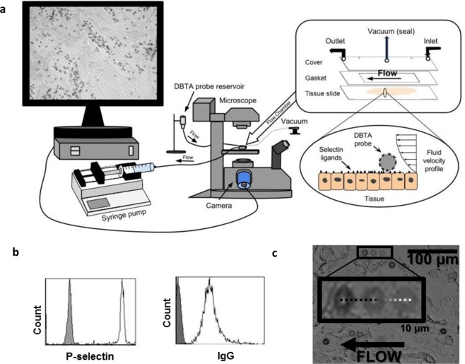
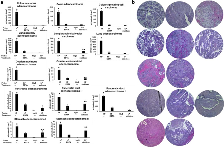
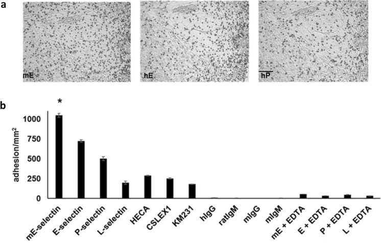
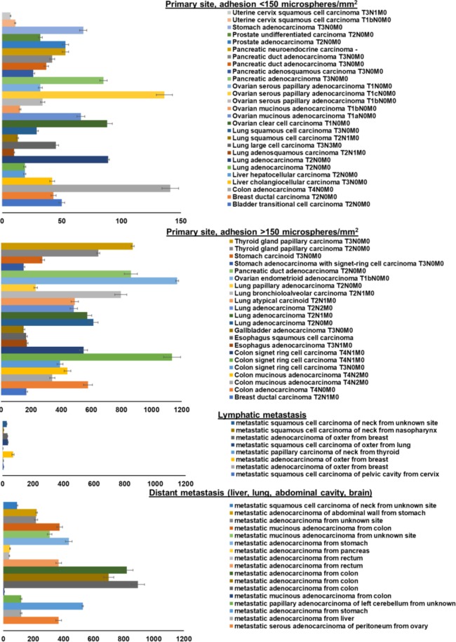
Cell adhesion mediated by selectins (expressed by activated endothelium, activated platelets, and leukocytes) binding to their resepective selectin ligands (expressed by cancer cells) may be involved in metastasis. Therefore, methods of characterizing selectin ligands expressed on human tissue may serve as valuable assays. Presented herein is an innovative method for detecting functional selectin ligands expressed on human tissue that uses a dynamic approach, which allows for control over the force applied to the bonds between the probe and target molecules. This new method of tissue interrogation, known as dynamic biochemical tissue analysis (DBTA), involves the perfusion of molecular probe-coated microspheres over tissues. DBTA using selectin-coated probes is able to detect functional selectin ligands expressed on tissue from multiple cancer types at both primary and metastatic sites.


Conflict of interest statement

The authors declare no competing interests.

Figures

Figure 1
Figure 1
Introduction to the DBTA (dynamic biochemical tissue analysis) (a) DBTA probes (microspheres conjugated with a molecular probe such as a selectin or antibody) are perfused over tissue at defined wall shear stresses using a syringe pump and parallel plate flow chamber. Schematics are not to scale. (b) DBTA probe conjugation with the desired molecular probe (e.g., P-selectin or human IgG) can be verified with flow cytometry. (c) Images from three time points have been combined to show a sample P-selectin DBTA probe rolling on a colon carcinoma tissue.
Figure 2
Figure 2
DBTA signal is specific, quantifiable, and discernible from SBTA. (a) P-selectin DBTA probes adhesion at 0.50 dyne/cm2 to colon tissue (from left to right: SRCC T4N1M0 22 y/o, adenocarcinoma T4N0M0, SRCC T4N1M0 48 y/o, mucinous adenocarcinoma T4N2M0, noncancerous colon tissue). DBTA probes appear as black circles. (b) P-selectin SBTA conducted on serial sections of the tissues examined in (a) with DBTA revealed detection of ligands that are not in complete agreement with the purported functional ligands detected via DBTA (a). Order of tissues is the same as (a). (c) P-selectin DBTA probe adhesion to colon tissue was specific and significantly greater than control probes. DBTA probes were perfused at 250,000 probes/mL and 0.50 dyne/cm2. Specificity of interaction was confirmed using 10 mM EDTA (divalent cation chelator, Ca2+ is required for selectin/selectin-ligand binding) and hIgG DBTA probes as negative controls. Data shown are mean adhesion ± SD of three technical replicates and are representative of independent experiments conducted on tissue sections from >10 independent cases of colon cancer and >3 independent noncancerous cases. #P < 0.001, $P < 0.01, and &P < 0.05, compared to all others intragroup. Scale bar = 100 µm.
Figure 3
Figure 3
DBTA probe adhesion to signet ring cell colon carcinoma (SRCC) tissue is force-dependent. (a) Position profile of a P-selectin DBTA probe rolling on a tissue section (SRCC 48 y/o case) at 0.75 dyne/cm2. Video frame rate = 175 fps (Supplementary Video S8). (b) Velocity profile of the P-selectin DBTA probe tracked in (a). (c) Increasing levels of applied force initially increased then decreased the lifetime of P-selectin DBTA probe adhesion to SRCC tissue. Data shown are mean adhesion duration in seconds ± SD of 30 separate pauses at each wall shear stress collected in a 100 μm × 100 μm region. $F = 12.59 > F0.01, 6,273 = 2.80 (d) Increasing levels of applied force initially decreased then increased the off-rate (koff) of P-selectin DBTA probe from the SRCC tissue. Data shown represent the estimation of P-selectin DBTA probe koff using the method of maximum likelihood from 30 separate pauses at each wall shear stress. Error bars represent 95% CI. #Levene’s test = 4.0818 (equivalent to P = 0.0014).
Figure 4
Figure 4
The presence of sLeX/A does not dictate a functional P-selectin ligand. (a) SBTA with the HECA-452 monoclonal antibody (green pseudocolor), which detects both sLeX and sLeA, has been combined with an image showing the adhesion patterns of P-selectin DBTA probes (Fig. 2). HECA-452 SBTA was conducted on serial sections. For the adenocarcinoma and SRCC 22 y/o tissues, but not the SRCC 48 y/o and mucinous adenocarcinoma tissues, there was an agreement between P-selectin DBTA and SBTA with HECA-452 in which the regions of tissue that express sLeX or sLeA mediate adhesion to P-selectin DBTA probes. However, with regard to the SRCC 48 y/o and mucinous adenocarcinoma tissues, not all regions that express sLeX or sLeA, as detected by HECA-452 SBTA, appeared to mediate binding to P-selectin DBTA probes (Supplementary Videos S1, S4, and S7). (b) HECA-452 DBTA probes (microspheres coated with HECA-452) adhered to tissue at 0.50 dyne/cm2 (Supplementary Videos S9 and S10). (c) The greatest amount of adhesion to the tissue occurred with the P-selectin DBTA probes. Probes were perfused at 250,000 probes/mL and 0.50 dyne/cm2. Specificity of interaction was validated using hIgG DBTA probes, 10 mM EDTA, and rIgM DBTA probes as controls. Data shown are mean adhesion ± SD of three technical replicates and are representative of independent experiments conducted on tissue sections from >3 independent cases of colon cancer. *P < 0.0025, compared to all others intragroup. Scale bar = 100 µm.
Figure 5
Figure 5
Adhesion in regions not detected by SBTA implies the presence of potentially distinct P-selectin ligands. (a) In the same regions of tissue that displayed P-selectin DBTA probe adhesion (Supplementary Video S11), examination of serial sections with SBTA (immunostaining) revealed no detectable levels of CD24 or PSGL-1 epitopes. CD44 expression was detected, but not all regions displaying specific reactivity with the P-selectin DBTA probes used in DBTA were recognized with the CD44 antibody in SBTA. Follow-up CD45 SBTA ruled out the possibility of DBTA probe interaction with infiltrated leukocytes, in agreement with the lack of PSGL-1 detection. (b) P-selectin DBTA probes specifically adhered to the tissue, with respect to the negative controls. Data shown are mean adhesion ± SD of three technical replicates and are representative of independent experiments conducted on tissue sections from >3 independent cases of colon cancer. *P < 0.0025 compared to all other conditions. (c) A direct comparison using a composite image of P-selectin DBTA probe adhesion (microspheres) and SBTA detection of CD44 (green pseudocolor) on colon adenocarcinoma tissue. Red ellipses indicate regions analyzed in (d,e). (d) Characterization of the adhesion of P-selectin DBTA probes to functional selectin ligands that were and were not detected with SBTA using CD44 antibody in (a). Data shown are mean pause duration ± SD of 30 separate pauses (n = 30). $P = 0.0068. (e) Data shown represent the estimation of P-selectin DBTA probe koff using the method of maximum likelihood from 30 separate pauses (n = 30). Error bars represent 95% CI. #Mann-Whitney U-value = 1152.5 (equivalent to P < 0.001 for t-test for normal distribution). All probes were perfused at 500,000 probes/mL and 0.50 dyne/cm2. Specificity of interaction was confirmed using 10 mM EDTA and hIgG DBTA probes as negative controls. Scale bar = 100 µm. These results imply the presence of potentially distinct P-selectin ligands in the tissue sections.
Figure 6
Figure 6
P-selectin DBTA probes specifically adhere to consecutive serial sections of colon, lung, ovarian, pancreatic, and stomach cancer tissues. (a) Functional P-selectin ligands are expressed on colon, lung, ovarian, pancreatic, and stomach cancer tissues. DBTA probes were perfused over three serial sections at 0.50 dyne/cm2 and 500,000 probes/mL. Specificity of interaction was confirmed using 10 mM EDTA, hIgG DBTA probes, and human P-selectin (hP) DBTA probes perfused over sialidase-treated tissue as negative controls. Data shown are mean adhesion ± SEM of three technical replicates from each of three independent serial sections. * and **P < 0.05 for P-selectin (hP) with respect to the three controls (hP to hP + EDTA, hIgG, and hP + sialidase) and the sialidase-treated tissue with respect to hIgG and hP + EDTA, respectively. See Supplementary Table S1 for P-values and Supplementary Videos S12–S17 for adhesion patterns. (b) Corresponding H&E images on the right are shown in the same order as (a). Core diameter is 2.5 mm.
Figure 7
Figure 7
The greatest amount of specific adhesion to SRCC occurs with the mE-selectin DBTA probe. mE-, E-, P-, L-selectin, HECA-452, CLSEX-1, and KM231 DBTA probes adhered to SRCC 22 y/o tissue. (a) Representative images of the adhesion patterns for mE-, E-, and P-selectin are shown. (b) The greatest amount of specific DBTA probe adhesion occurred with the mE-selectin probe. Probes were perfused at 250,000 probes/mL and 0.50 dyne/cm2. Specificity of interaction was confirmed using 10 mM EDTA (divalent cation chelator, Ca2+ is required for selectin/selectin-ligand binding) as well as with hIgG, rat IgM, mouse IgG, and mouse IgM DBTA probes as negative controls. Data shown are mean adhesion ± SD of three technical replicates and are representative of independent experiments conducted on tissue sections from >3 independent cases colon cancer. Scale bar = 100 µm. *P < 0.001 compared to all other conditions. See Supplementary Fig. S2 for validation of DBTA probe conjugation. Scale bar = 100 µm.
Figure 8
Figure 8
DBTA detects functional selectin ligands expressed on tissue from multiple solid tumors at both primary and metastatic sites. mE-selectin DBTA probes adhesion to cancerous tissues derived from primary and metastatic sites. DBTA probes were perfused at 500,000 probes/mL and 0.50 dyne/cm2. Data shown are mean adhesion per mm2 ± SD of three technical replicates and include the deduction of control probe adhesion (viz. mean of P-selectin adhesion minus the greater mean value of either P-selectin + EDTA or IgG control probes).

Similar articles

Cited by

References

    1. Kim YJ, Borsig L, Varki NM, Varki A. P-selectin deficiency attenuates tumor growth and metastasis. Proc. Natl. Acad. Sci. USA. 1998;95:9325–9330. doi: 10.1073/pnas.95.16.9325. - DOI - PMC - PubMed
    1. Köhler S, Ullrich S, Richter U, Schumacher U. E-/P-selectins and colon carcinoma metastasis: first in vivo evidence for their crucial role in a clinically relevant model of spontaneous metastasis formation in the lung. Br. J. Cancer. 2010;102:602–609. doi: 10.1038/sj.bjc.6605492. - DOI - PMC - PubMed
    1. Borsig L, et al. Heparin and cancer revisited: mechanistic connections involving platelets, P-selectin, carcinoma mucins, and tumor metastasis. Proc. Natl. Acad. Sci. USA. 2001;98:3352–3357. doi: 10.1073/pnas.061615598. - DOI - PMC - PubMed
    1. Ludwig RJ, et al. Endothelial P-Selectin as a target of heparin action in experimental melanoma lung metastasis. Cancer Res. 2004;64:2743–2750. doi: 10.1158/0008-5472.CAN-03-1054. - DOI - PubMed
    1. Stevenson JL, Varki A, Borsig L. Heparin attenuates metastasis mainly due to inhibition of P- and L-selectin, but non-anticoagulant heparins can have additional effects. Thromb. Res. 2007;120:S107–11. doi: 10.1016/S0049-3848(07)70138-X. - DOI - PubMed

Publication types