

Neutrophils mediate early cerebral cortical hypoperfusion in a murine model of subarachnoid haemorrhage
- PMID: 31186479
- PMCID: PMC6560094
- DOI: 10.1038/s41598-019-44906-9
Neutrophils mediate early cerebral cortical hypoperfusion in a murine model of subarachnoid haemorrhage
Abstract
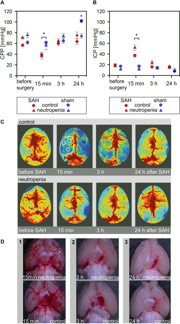
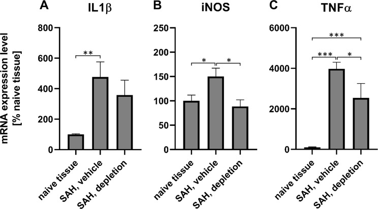
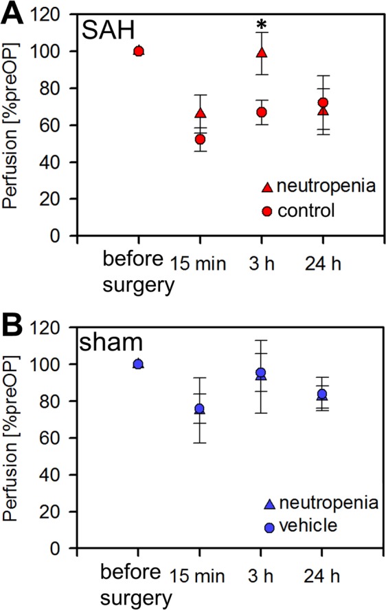
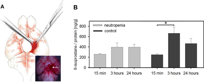
Cerebral hypoperfusion in the first hours after subarachnoid haemorrhage (SAH) is a major determinant of poor neurological outcome. However, the underlying pathophysiology is only partly understood. Here we induced neutropenia in C57BL/6N mice by anti-Ly6G antibody injection, induced SAH by endovascular filament perforation, and analysed cerebral cortical perfusion with laser SPECKLE contrast imaging to investigate the role of neutrophils in mediating cerebral hypoperfusion during the first 24 h post-SAH. SAH induction significantly increased the intracranial pressure (ICP), and significantly reduced the cerebral perfusion pressure (CPP). At 3 h after SAH, ICP had returned to baseline and CPP was similar between SAH and sham mice. However, in SAH mice with normal neutrophil counts cortical hypoperfusion persisted. Conversely, despite similar CPP, cortical perfusion was significantly higher at 3 h after SAH in mice with neutropenia. The levels of 8-iso-prostaglandin-F2α in the subarachnoid haematoma increased significantly at 3 h after SAH in animals with normal neutrophil counts indicating oxidative stress, which was not the case in neutropenic SAH animals. These results suggest that neutrophils are important mediators of cortical hypoperfusion and oxidative stress early after SAH. Targeting neutrophil function and neutrophil-induced oxidative stress could be a promising new approach to mitigate cerebral hypoperfusion early after SAH.
Conflict of interest statement
The authors declare no competing interests.
Figures
Similar articles
-
Effect of decompressive craniectomy on outcome following subarachnoid hemorrhage in mice.Stroke. 2015 Mar;46(3):819-26. doi: 10.1161/STROKEAHA.114.007703. Epub 2015 Jan 15. Stroke. 2015. PMID: 25593134
-
Clazosentan, an endothelin receptor antagonist, prevents early hypoperfusion during the acute phase of massive experimental subarachnoid hemorrhage: a laser Doppler flowmetry study in rats.J Neurosurg. 2008 Dec;109(6):1134-40. doi: 10.3171/JNS.2008.109.12.1134. J Neurosurg. 2008. PMID: 19035733
-
Acute hypoperfusion immediately after subarachnoid hemorrhage: a xenon contrast-enhanced CT study.J Neurotrauma. 2009 Dec;26(12):2225-31. doi: 10.1089/neu.2009.0924. J Neurotrauma. 2009. PMID: 19929373
-
Hypoperfusion in the acute phase of subarachnoid hemorrhage.Acta Neurochir Suppl. 2011;110(Pt 1):35-8. doi: 10.1007/978-3-7091-0353-1_6. Acta Neurochir Suppl. 2011. PMID: 21116911 Review.
-
Use of 31P magnetic resonance spectroscopy to study the effect of cortical magnesium and energy metabolism after subarachnoid hemorrhage.Cerebrovasc Dis. 2008;26(3):223-30. doi: 10.1159/000147448. Epub 2008 Jul 23. Cerebrovasc Dis. 2008. PMID: 18648193 Review.
Cited by
-
Regulation of nuclear factor erythroid-2-related factor 2 as a potential therapeutic target in intracerebral hemorrhage.Front Mol Neurosci. 2022 Sep 29;15:995518. doi: 10.3389/fnmol.2022.995518. eCollection 2022. Front Mol Neurosci. 2022. PMID: 36245922 Free PMC article. Review.
-
Inflammation and Oxidative Stress: Potential Targets for Improving Prognosis After Subarachnoid Hemorrhage.Front Cell Neurosci. 2021 Sep 24;15:739506. doi: 10.3389/fncel.2021.739506. eCollection 2021. Front Cell Neurosci. 2021. PMID: 34630043 Free PMC article. Review.
-
The Role of the Blood Neutrophil-to-Lymphocyte Ratio in Aneurysmal Subarachnoid Hemorrhage.Front Neurol. 2021 Jun 3;12:671098. doi: 10.3389/fneur.2021.671098. eCollection 2021. Front Neurol. 2021. PMID: 34149601 Free PMC article. Review.
-
The dynamic changes of peripheral blood cell counts predict the clinical outcomes of aneurysmal subarachnoid hemorrhage.Heliyon. 2024 Apr 16;10(8):e29763. doi: 10.1016/j.heliyon.2024.e29763. eCollection 2024 Apr 30. Heliyon. 2024. PMID: 38681624 Free PMC article.
-
The role of TLR4 and HO-1 in neuroinflammation after subarachnoid hemorrhage.J Neurosci Res. 2020 Mar;98(3):549-556. doi: 10.1002/jnr.24515. Epub 2019 Aug 29. J Neurosci Res. 2020. PMID: 31468571 Free PMC article. Review.
References
-
- van Lieshout Jasper H., Dibué-Adjei Maxine, Cornelius Jan F., Slotty Philipp J., Schneider Toni, Restin Tanja, Boogaarts Hieronymus D., Steiger Hans-Jakob, Petridis Athanasios K., Kamp Marcel A. An introduction to the pathophysiology of aneurysmal subarachnoid hemorrhage. Neurosurgical Review. 2017;41(4):917–930. doi: 10.1007/s10143-017-0827-y. - DOI - PubMed
Publication types
MeSH terms
Substances
LinkOut - more resources
Full Text Sources
