. 2019 Jun 11;9(1):8462.
doi: 10.1038/s41598-019-44934-5.

Regulation of the Hepatitis B virus replication and gene expression by the multi-functional protein TARDBP

Affiliations

Regulation of the Hepatitis B virus replication and gene expression by the multi-functional protein TARDBP

Grace Naswa Makokha et al. Sci Rep. .

Erratum in

Abstract

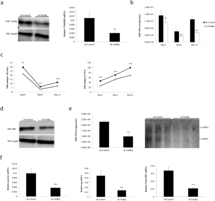
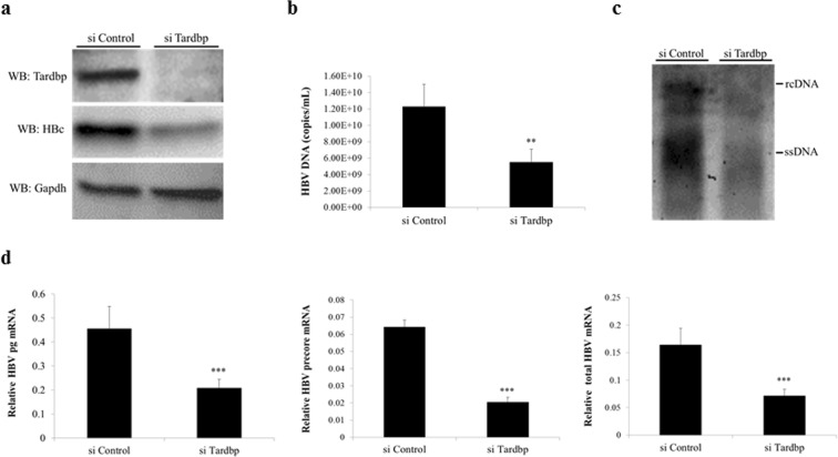
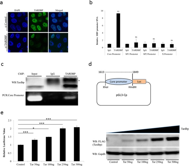
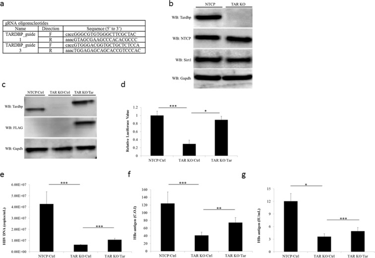
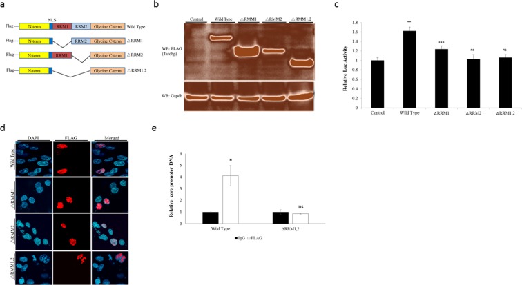
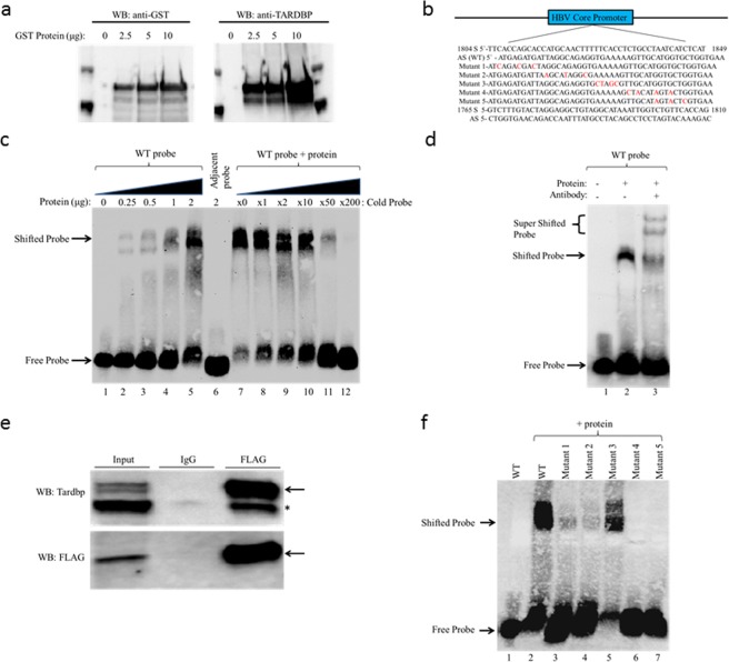
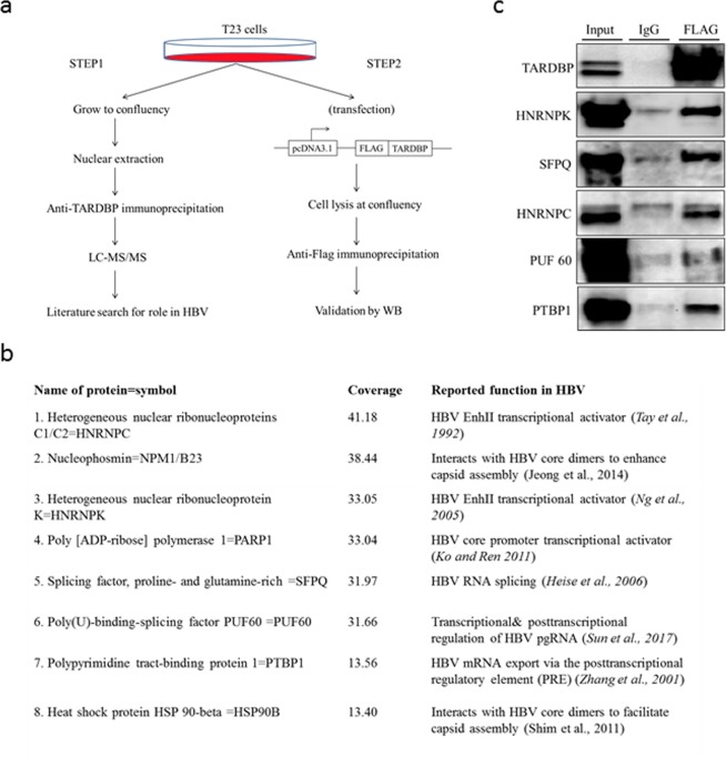
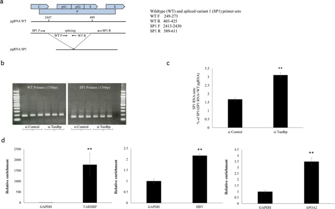
Hepatitis B virus (HBV) infects the liver and is a key risk factor for hepatocellular carcinoma. Identification of host factors that support viral replication is important to understand mechanisms of viral replication and to develop new therapeutic strategies. We identified TARDBP as a host factor that regulates HBV. Silencing or knocking out the protein in HBV infected cells severely impaired the production of viral replicative intermediates, mRNAs, proteins, and virions, whereas ectopic expression of TARDBP rescued production of these products. Mechanistically, we found that the protein binds to the HBV core promoter, as shown by chromatin precipitation as well as mutagenesis and protein-DNA interaction assays. Using LC-MS/MS analysis, we also found that TARDBP binds to a number of other proteins known to support the HBV life cycle, including NPM1, PARP1, Hsp90, HNRNPC, SFPQ, PTBP1, HNRNPK, and PUF60. Interestingly, given its key role as a regulator of RNA splicing, we found that TARDBP has an inhibitory role on pregenomic RNA splicing, which might help the virus to export its non-canonical RNAs from the nucleus without being subjected to unwanted splicing, even though mRNA nuclear export is normally closely tied to RNA splicing. Taken together, our results demonstrate that TARDBP is involved in multiple steps of HBV replication via binding to both HBV DNA and RNA. The protein's broad interactome suggests that TARDBP may function as part of a RNA-binding scaffold involved in HBV replication and that the interaction between these proteins might be a target for development of anti-HBV drugs.


Conflict of interest statement

The authors declare no competing interests.

Figures

Figure 1
Figure 1
Endogenous TARDBP regulates HBV gene expression in a HBV infection model. (a) NTCP-HepG2 C4 cells were transduced with a lentiviral vector allowing the expression of a control shRNA (shControl) or shRNA directed against TARDBP (shTardbp). After selection with puromycin, reduction of TARDBP mRNA and protein level by shRNA was confirmed by qPCR and western blotting, respectively. (b) The cells in (a) were then infected with HBV in duplicate sets for 12 days. Supernatants were collected at the indicated time points for extracellular HBV DNA analysis. (c) A separate portion of supernatants from (b) were also analyzed by CLEIA for measurement of HBs and HBe antigen levels. (d) To detect the level of the HBV core protein, one set of the cells were harvested at the end of the 12-day infection period for protein lysis and subjected to western blotting using the anti-HBc antibody. (e) For detection of the core-associated HBV DNA, the lysates in (d) were subjected to immunoprecipitation by an anti-HBc antibody followed by qPCR (left panel) and Southern blotting (right panel). (f) Total RNA was extracted from the other set of cells from after the 12-day infection period in (b) and subjected to RT qPCR to detect HBV precore, pregenomic (pg) and total mRNAs. Gene expression was normalized to that of GAPDH. The data is shown as the mean ± SD (n = 3 per bar), **P < 0.01, ***P < 0.001 and ns, non-significant.
Figure 2
Figure 2
Silencing of TARDBP represses HBV transcription in a HBV-producing cell line. (a) The T23 cell line, which stably expresses HBV plasmid, was transfected with the negative control siRNA (siControl) or the TARDBP siRNA (siTARDBP) in duplicate sets for 7 days. One set of the cells was lysed, and the TARDBP protein and core protein were quantified by western blotting using specific antibodies. (b) The supernatants were harvested from one set of the cells in (a) and analyzed for extracellular HBV DNA. (c) For the intracellular HBV DNA, protein lysates harvested in A were immunoprecipated with an anti-HBc antibody, followed by Southern blotting. (d) The total RNA was extracted from the second set of cells and subjected to RT qPCR to detect the HBV mRNAs (pregenomic-pg, precore, and total). The mRNA values were normalized against the GAPDH RNA internal control. The data is shown as the mean ± SD (n = 3 per bar), **P < 0.01, ***P < 0.001.
Figure 3
Figure 3
TARDBP activates the HBV core promoter. (a) Endogenous TARDBP was detected by immunofluorescence of the HBV expressing cell line T23. The protein was visualized using the anti-TARDBP polyclonal antibody and anti-Rabbit IgG (Alexa Fluor® 488) secondary antibody. Cells were treated with control siRNA or siRNA specific to TARDBP to confirm specificity of the antibody. (b) The T23 cells were harvested at confluency and analyzed by ChIP assay using the antibody against TARDBP or the control rabbit IgG. Immunoprecipitated DNA was analyzed in triplicate by qPCR with primers specific for each of the HBV promoter DNA sequences (Core, Sp1, Sp2 and X). The results are displayed as the ratio of the amount of DNA bound to the TARDBP antibody to that bound to the control antibody, with the amount bound to the control antibody set to one. (c) To confirm precipitation of TARDBP and core promoter by the antibody, western blotting was performed using the TARDBP antibody, whereas the presence of core promoter DNA was detected by PCR using the core specific primers. The asterisk (*) indicates the location of the IgG heavy chain bands. (d) A schematic representation of the luciferase reporter plasmid pGL3-Cp that contains the luciferase reporter whose expression is controlled by the core promoter. (e) The pGL3-CP plasmid was co-transfected with the control vector pcDNA3.1/FLAG or increasing concentrations of pcDNA3.1/TARDBP-FLAG into NTCP-HepG2 cells as indicated. The pRL-TK plasmid, which expresses Renilla luciferase, was also included to monitor the transfection efficiency. Cells were lysed at 48 hours after transfection for the luciferase assay. Firefly luciferase values were normalized with control Renilla luciferase activity. The results are expressed as relative luciferase value, which refers to differences (n-fold) from the control value, which is set at one. The protein expression of TARDBP in the lysates was confirmed by western blotting using the anti-FLAG antibody. Error bars represent the SD of three independent experiments. *P < 0.05, **P < 0.01, ***P < 0.001 and ns-non significant.
Figure 4
Figure 4
Establishment of TARDBP-knockout (KO) cells by a CRISPR/Cas9 system. (a) A TARDBP-deficient NTCP-HepG2 cell line (TARDBP KO) was established by the CRISPR/Cas9 system using the two RNA guide sequences shown. (b) Western blot analysis of the NTCP-HepG2 parent cell and the TARDBP knockout NTCP-HepG2 cell line using the antibodies against NTCP, TARDBP, SIRT1 and the control GAPDH is shown. (c) A TARDBP plasmid was transfected into the KO cells for exogenous protein expression. The expression was confirmed by western blotting using the anti-TARDBP and anti-FLAG antibodies. (d) NTCP-HepG2 parent cell, TARDBP KO and TARDBP KO cells complemented with exogenous TARDBP were transfected with the pGL3-CP plasmid for comparison of the core promoter activities. Cell lysates were harvested after 48 hours for the dual luciferase assay as in Fig. 3e. (e,f,g) HBV infection was carried out in the three cells treated as in (c) for 12 days, similar to the experiment in Fig. 1. The supernatants were harvested at the end of the infection period for measurement of extracellular HBV DNA, HBe antigen and HBs antigen levels, respectively. The data is shown as the mean ± SD, *P < 0.05 **P < 0.01, ***P < 0.001.
Figure 5
Figure 5
The RNA recognition motifs (RRMs) of TARDBP are crucial for activation of the core promoter. (a) A schematic structure of the wild type (WT) TARDBP showing the location of the FLAG tag, the two RRMs (RRM1 and RRM2) and the nuclear localization signal (NLS). Alongside are the three mutants with deletion of RRM1 (ΔRRM1), RRM2 (ΔRRM1) or both RRM1 and 2 (ΔRRM1, 2). (b) Each of the four plasmids or the control empty vector was transfected into NTCP-HepG2 cells, and after 48 hours protein expression was determined by western blotting using an anti-FLAG antibody. (c) To compare the effect of these proteins on the core promoter activity, the pGL3-CP plasmid was transfected alongside equal amounts of the control plasmid, WT TARDBP, or each of the three RRM mutant plasmids into NTCP-HepG2 cells. Luciferase activities were measured at 48 hours after transfections. Relative values were calculated as described in Fig. 3e. (d) For intracellular localization of the proteins, the plasmids were transfected as in (b) above. After 48 hours, the cells were treated with the anti-FLAG antibody and visualized by immunofluorescence microscopy. The nucleus was visualized with DAPI. (e) To confirm that the two RRMs play a role in interaction of TARDBP with the core promoter, the FLAG tagged WT and the ΔRRM1, 2 mutant proteins were expressed in T23 cells, followed by a ChIP assay as in Fig. 3b using control and anti-FLAG antibodies. The resulting DNA was analyzed with the core promoter primers by qPCR. The relative values were normalized to the amount of DNA precipitated by the control which, was set at one. Results are reported as mean ± SD, *P < 0.05, **P < 0.01, ***P < 0.001 and ns-non significant.
Figure 6
Figure 6
Detection of TARDBP-core promoter interaction in an electrophoretic mobility shift assay (EMSA). (a) Western blot analysis of the recombinant GST-fused TARDBP protein purified using glutathione agarose beads. The protein was detected using antibodies directed against the GST tag and TARDBP protein. (b) The nucleotide sequence of TARDBP binding site within the HBV core promoter region is shown (nt1804-nt1849). The sense (S) and antisense (AS) probes of the sequence, together with the five AS mutant probes (Mutant 1–5), and the S and AS sequence of the adjacent probe (nt1765-nt1810) used for the EMSA assay are indicated. The nucleotide changes in the mutants are marked in red. (c) The AS (WT) probe was incubated alone (lane 1) or with increasing concentrations of TARDBP protein (lanes 2–5); the adjacent AS probe was incubated with 2 μg of TARDBP (lane 6); the AS (WT) probe was incubated with 2 μg of TARDBP alongside increasing concentrations of an unlabeled specific competitor at 0, 1, 2, 10, 50 and 200 fold respectively, (lanes 7–12). (d) A supershift assay was performed with an anti-TARDBP antibody; lane 1, AS (WT) probe alone; lane 2, AS (WT) probe with TARDBP protein; lane 3 AS (WT) probe with TARDBP protein along with 2 μg rabbit anti-TARDBP antibody. The specific TARDBP-DNA complex (shifted probe arrow), and the antibody-TARDBP-DNA complex (super shifted probe arrow) is indicated. (e) The HBV-producing T23 cells were transfected with 5 μg of FLAG tagged TARDBP, immunoprecipated with an anti-FLAG antibody and blotted with anti-FLAG and anti-TARDBP antibodies. The arrow shows the location of the exogenous protein, and the asterisk (*) shows the endogenous protein which was also precipitated by the FLAG-antibody. (f) Starting at the N-terminus, mutations were introduced at five sites in the AS (WT) probe to make five different mutant probes, Mutants 1–5, as shown in (b). The WT probe was incubated alone (lane 1) or in the presence of 2 μg of TARDBP protein (lane 2), while each of the five mutant probes was also incubated with the equivalent amount of TARDBP protein (lanes 3–7). The location of the free probe and the specific TARDBP-DNA complex (shifted probe) are indicated by arrows.
Figure 7
Figure 7
TARDBP assembles protein complexes that support the lifecycle of HBV. (a) An experimental design for the identification of TARDBP-interacting proteins. In the first step, nuclear lysates were prepared from the HBV producing cell line T23, immunoprecipitated by a TARDBP antibody and subjected to LC-MS/MS analysis. A literature search was done on the top scorers to ascertain which of them likely played a role in the HBV life cycle. In the second step, interactions of the candidate proteins in vivo were validated by western blotting using the same cell line as in (a) but with an exogenously expressed TARDBP protein. (b) The set of 8 proteins that scored a coverage of >10 on the LC/MS-MS analysis and were found from literature to have a role in HBV replication. The figure includes the specified role and the literature associated with each protein. (c) Nuclear lysates of the T23 cells expressing FLAG-tagged TARDBP were precipitated by the anti-FLAG antibody or the control mouse IgG and subjected to western blotting for each protein using their specific antibodies as shown.
Figure 8
Figure 8
TARDBP regulates HBV pgRNA splicing. (a) A schematic representation of the HBV genome indicating the site of splicing for the major splice variant, SP1 (intron 2447/489), and the primers designed for PCR amplification of the wild type (WT) and SP1 pgRNA. (b) The T23 cells, which stably express HBV, were transfected in triplicate with siRNAs against TARDBP or control. Total RNA was extracted from the cells, followed by reverse transcription. The resulting cDNAs were analyzed by PCR followed by agarose gel electrophoresis with primers specific for WT pgRNA or SP1 RNA. The results confirmed that there was no non-specific amplification of WT products with SP1 primers and that the two products were at the expected sizes. (c) The primers in (b) were henceforth utilized in a quantitative PCR of the samples to determine the proportion of SP1 RNA, which was calculated as SP1 RNA/(SP1 pgRNA + WT pgRNA). (d) Whole cell lysate from FLAG-TARDBP or FLAG-transfected T23 cells was immunoprecipitated with an anti-FLAG antibody and subjected to isolation of RNA and qPCR analysis using primers for TARDBP, APOA2 and total HBV mRNAs. The amount of mRNA that was precipitated from the TARDBP transfected cells was calculated as a ratio relative to that bound to the control-transfected cells for each of the primers. Relative mRNA values are presented as a ratio with respect to the amount of GAPDH mRNA, which was set at one. Results are reported as mean ± SD, while **P < 0.01.

Similar articles

Cited by

References

    1. Schweitzer A, Horn J, Mikolajczyk RT, Krause G, Ott JJ. Estimations of worldwide prevalence of chronic hepatitis B virus infection: A systematic review of data published between 1965 and 2013. Lancet. 2015;386:1546–1555. doi: 10.1016/S0140-6736(15)61412-X. - DOI - PubMed
    1. Lavanchy D. Hepatitis B virus epidemiology, disease burden, treatment, and current and emerging prevention and control measures. J. Viral Hepat. 2009;3:1–17. - PubMed
    1. Zhang Z, et al. Heparin sulphate d-glucosaminyl 3-O-sulfotransferase 3B1 plays a role in HBV replication. Virology. 2010;406:280–285. doi: 10.1016/j.virol.2010.07.030. - DOI - PubMed
    1. Zoulim F, Locarnini S, Hepatitis B. Virus Resistance to Nucleos(t)ide Analogues. Gastroenterology. 2009;137:1593–1608.e2. doi: 10.1053/j.gastro.2009.08.063. - DOI - PubMed
    1. Yan H, et al. Sodium taurocholate cotransporting polypeptide is a functional receptor for human hepatitis B and D virus. Elife. 2012;2012:1–28. - PMC - PubMed

Publication types