. 2019 Nov;27(11):1668-1676.
doi: 10.1038/s41431-019-0444-z. Epub 2019 Jun 11.

DEGS1 variant causes neurological disorder

Affiliations

DEGS1 variant causes neurological disorder

Vadim Dolgin et al. Eur J Hum Genet. 2019 Nov.

Abstract

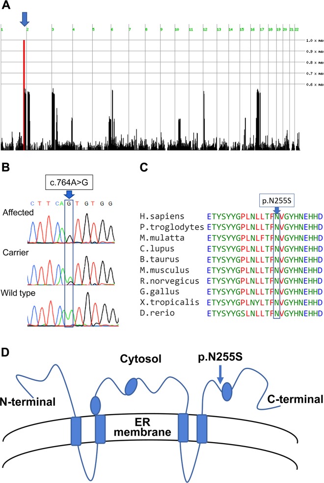
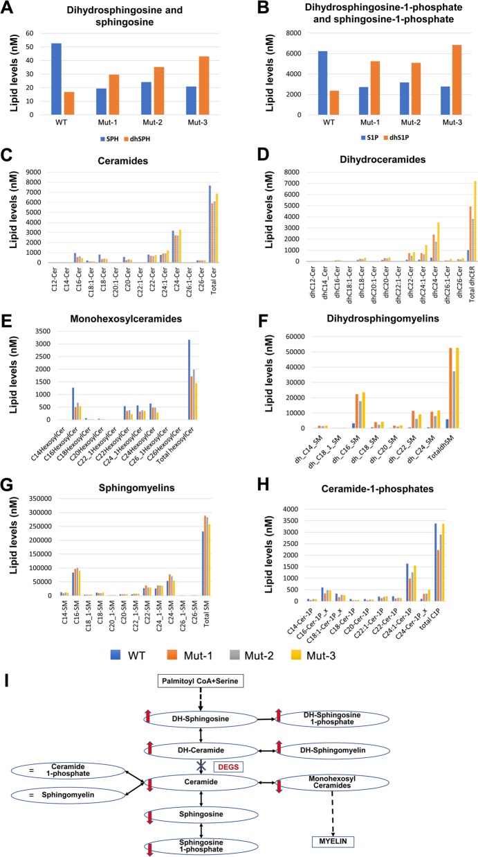
Sphingolipidoses are monogenic lipid storage diseases caused by variants in enzymes of lipid synthesis and metabolism. We describe an autosomal recessive complex neurological disorder affecting consanguineous kindred. All four affected individuals, born at term following normal pregnancies, had mild to severe intellectual disability, spastic quadriplegia, scoliosis and epilepsy in most, with no dysmorphic features. Brain MRI findings were suggestive of leukodystrophy, with abnormal hyperintense signal in the periventricular perioccipital region and thinning of the body of corpus callosum. Notably, all affected individuals were asymptomatic at early infancy and developed normally until the age of 8-18 months, when deterioration ensued. Homozygosity mapping identified a single 8.7 Mb disease-associated locus on chromosome 1q41-1q42.13 between rs1511695 and rs537250 (two-point LOD score 2.1). Whole exome sequencing, validated through Sanger sequencing, identified within this locus a single disease-associated homozygous variant in DEGS1, encoding C4-dihydroceramide desaturase, an enzyme of the ceramide synthesis pathway. The missense variant, segregating within the family as expected for recessive heredity, affects an evolutionary-conserved amino acid of all isoforms of DEGS1 (c.656A>G, c.764A>G; p.(N219S), p.(N255S)) and was not found in a homozygous state in ExAC and gnomAD databases or in 300 ethnically matched individuals. Lipidomcs analysis of whole blood of affected individuals demonstrated augmented levels of dihydroceramides, dihydrosphingosine, dihydrosphingosine-1-phosphate and dihydrosphingomyelins with reduced levels of ceramide, sphingosine, sphingosine-1-phosphate and monohexosylceramides, as expected in malfunction of C4-dihydroceramide desaturase. Thus, we describe a sphingolipidosis causing a severe regressive neurological disease.


Conflict of interest statement

The authors declare that they have no conflict of interest.

Figures

Fig. 1
Fig. 1
The studied kindred and disease phenotype: a The studied kindred. b Brain MRI of IV-1 at age 3.5 years and of IV-2 at age 7 years demonstrated moderate dilatation of the lateral ventricles with abnormal hyperintense signal in the periventricular perioccipital region, as well as thinning of the body of the corpus callosum
Fig. 2
Fig. 2
Identification of the DEGS1 variant: a Homozygosity mapping: SNPs colored red, blue and dashed red represent homozygosity for major allele, heterozygosity and homozygosity for the minor allele, respectively; arrow marks a single chromosome 1 homozygous locus shared by affected individuals (Homozygosity-Mapper). b The c.764A>G DEGS1 variant (Sanger sequencing). c evolutionary conservation of the p.(N255S) variant. d Location of the variant within the cytosolic domain of DEGS1
Fig. 3
Fig. 3
Biochemical consequences of the DEGS1 variant in patients’ whole blood: af Compared to healthy control (WT, IV-5), affected individuals (Mut-1, 2, 3 are IV-1, IV-6, IV-9, respectively) had higher levels of dihydroceramides, dihydrosphingosine, dihydrosphingosine-1-phosphate, and dihydrosphingomyelins, and reduced levels of ceramides, sphingosine, sphingosine-1-phosphate and monohexosylceramides. g, h Levels of sphingomyelins and ceramide-1-phosphates were similar between the healthy control and the patients. (i) Schema demonstrating that these results are consistent with inhibition of conversion of dihydroceramide to ceramide due to the DEGS1 variant

References

    1. Olsen ASB, Faergeman NJ. Sphingolipids: membrane microdomains in brain development, function and neurological diseases. Open Biol. 2017;7:1–17. doi: 10.1098/rsob.170069. - DOI - PMC - PubMed
    1. Grassi S, Chiricozzi E, Mauri L, Sonnino S, Prinetti A. Sphingolipids and neuronal degeneration in lysosomal storage disorders. J Neurochem. 2019;148:600–11. doi: 10.1111/jnc.14540. - DOI - PubMed
    1. Sandhoff K. Neuronal sphingolipidoses: Membrane lipids and sphingolipid activator proteins regulate lysosomal sphingolipid catabolism. Biochimie. 2016;130:146–51. doi: 10.1016/j.biochi.2016.05.004. - DOI - PubMed
    1. Hannun YA, Obeid LM. Sphingolipids and their metabolism in physiology and disease. Nat Rev Mol Cell Biol. 2018;19:175–91. doi: 10.1038/nrm.2017.107. - DOI - PMC - PubMed
    1. Perez Y, Menascu S, Cohen I, Kadir R, Basha O, Shorer Z, et al. RSRC1 mutation affects intellect and behavior through aberrant splicing and transcription, downregulating IGFBP3. Brain. 2018;141:961–70. doi: 10.1093/brain/awy045. - DOI - PubMed

Publication types

MeSH terms