Review
. 2019 May 2:2019:7079604.
doi: 10.1155/2019/7079604. eCollection 2019.

The Role of Nonerythroid Spectrin α II in Cancer

Affiliations
Review

The Role of Nonerythroid Spectrin α II in Cancer

Anne Ackermann et al. J Oncol. .

Abstract

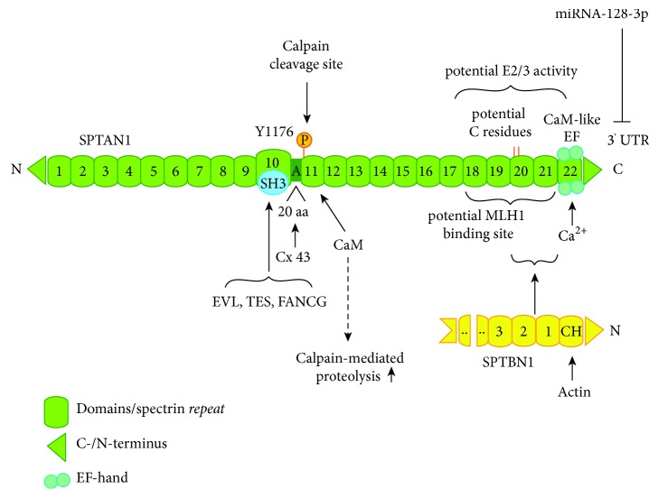
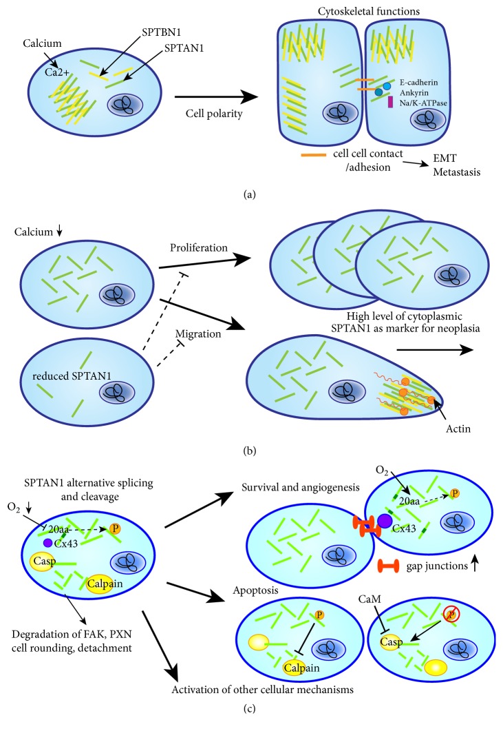
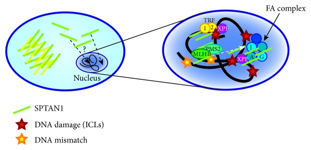
Nonerythroid spectrin αII (SPTAN1) is an important cytoskeletal protein that ensures vital cellular properties including polarity and cell stabilization. In addition, it is involved in cell adhesion, cell-cell contact, and apoptosis. The detection of altered expression of SPTAN1 in tumors indicates that SPTAN1 might be involved in the development and progression of cancer. SPTAN1 has been described in cancer and therapy response and proposed as a potential marker protein for neoplasia, tumor aggressiveness, and therapeutic efficiency. On one hand, the existing data suggest that overexpression of SPTAN1 in tumor cells reflects neoplastic and tumor promoting activity. On the other hand, nuclear SPTAN1 can have tumor suppressing effects by enabling DNA repair through interaction with DNA repair proteins. Moreover, SPTAN1 cleavage products occur during apoptosis and could serve as markers for the efficacy of cancer therapy. Due to SPTAN1's multifaceted functions and its role in adhesion and migration, SPTAN1 can influence tumor growth and progression in both positive and negative directions depending on its specific regulation. This review summarizes the current knowledge on SPTAN1 in cancer and depicts several mechanisms by which SPTAN1 could impact tumor development and aggressiveness.


Figures

Figure 1
Figure 1
Structure of SPTAN1. SPTAN1 harbors 22 domains, which are presented in green. Shown are the following: characteristic spectrin repeats (green boxes); C- and N-terminus (green rectangle); domain 10 with a SH3 domain (blue circle), which allows binding of EVL, TES, and FANCG; a 20 amino acid (aa) motif and alternatively spliced region between domains 10 and 11, which allows specific binding of Connexin 43 (Cox43); phosphorylation site (orange) at residue Y1176 in domain 11 and behind that the calpain cleavage site of SPTAN1 and the calmodulin (CaM) binding site, which regulates calpain-mediated proteolysis; a potential MLH1 binding site between domains 18 and 22; potential cystein (C) residues (orange lines) in domain 20, which might mediate a potential E2/E3 ubiquitin-protein-conjugating or -ligating activity; domains 20 and 21, which mediate dimerization of SPTAN1 and SPTBN1 by binding to the N-terminal first two spectrin repeats of SPTBN1 (yellow) [5] and C-terminal the CaM-like domain 22, which can bind calcium through two EF-hand motifs. Translational inhibition by miRNA-128-3p targets the 3′ untranslated region (3′UTR) of SPTAN1 [16]. SPTBN1 can bind actin through its N-terminal Calponin homology (CH) domain [4]. Spectrin heterodimers formed by antiparallel lateral dimerization of SPTAN1 and SPTBN1 then form tetramers by head to head assembly [3, 4]. Modified after Bennett and Baines (2001) and Baines (2010) [4, 17].
Figure 2
Figure 2
Localization and functional relevance of cytoplasmic SPTAN1. SPTAN1 can have diverse functions in the cell depending on its localization. (a) SPTAN1 (green bars) serves as cytoskeletal scaffolding protein and, under physiological calcium levels, together with SPTBN1 (yellow bars) and other proteins, it forms a stabilizing mesh beneath the cell membrane allowing cell polarity. Shown are epithelial cells expressing SPTAN1 apically and laterally in the cell. Upon cell-cell contact (orange bars), SPTAN1 interacts with E-cadherin, ankyrin, and Na/K-ATPase and thus might influence EMT and metastasis. (b) SPTAN1 can affect tumor growth and outcome by enhancing cell proliferation and migration (upper panel), which are impaired in case of reduced SPTAN1 levels (lower panel). High levels of cytoplasmic SPTAN1 in proliferating cells could be used as marker for neoplasia. Moreover, migration might be influenced by the interaction of SPTAN1 and SPTBN1 with actin filaments. (c) SPTAN1 has also a role in survival, angiogenesis, apoptosis, and other cellular mechanisms through expression of alternative spliced forms and cleavage products. A SPTAN1 spliceform including a 20 amino acid (aa) motif contributes to gap junctions (orange dumbbells) through association of this motif with Connexin 43 (Cx43). This association is regulated by JNK-mediated phosphorylation. Expression of this spliceform is repressed during hypoxia, thus leading to a decrease in gap junctions. SPTAN1 is involved in apoptosis if cleaved by calpain and caspases. This is regulated by phosphorylation of Y1176 and dephosphorylation enhances the proteolytic susceptibility of SPTAN1 to calpain and caspases 2, 3, and 7. Cleavage leads to membrane blebbing and irreversible cell death. Caspase-mediated cleavage can be inhibited by calmodulin (CaM) binding and indicates again the influence of calcium homeostasis. SPTAN1 cleavage products can further lead to cell rounding and detachment by degrading FAK and paxillin (PXN) and can activate further yet unknown cellular mechanisms.
Figure 3
Figure 3
SPTAN1 in DNA repair. SPTAN1 can translocate into the nucleus. In the nucleus, SPTAN1 can interact with various partners including proteins involved in DNA repair and fanconi anemia (FA). By binding FA proteins in nuclear complexes, SPTAN1 is stabilized and might act as scaffold to align or enhance DNA repair associated proteins at sites of damage. The FA protein FANCG (G) is able to bind to SPTAN1 through its SH3 domain. Via FANCA (A) and XPF, SPTAN1 enables DNA repair of interstrand crosslinks (ICLs). SPTAN1 circumvents telomere dysfunction after ICL damage by interaction with TRF1/2 and XPF. Furthermore, SPTAN1 can directly interact with MLH1, which mediates DNA mismatch repair (MMR). FANCJ, which is required for correction of the cross-link response, also interacts with the MMR complex MutLα, consisting of MLH1 and PMS2. A SPTAN1-dependent DNA repair mechanism, however, still remains unknown.

References

    1. Bennett V., Gilligan D. M. The spectrin-based membrane skeleton and micron-scale organization of the plasma membrane. Annual Review of Cell and Developmental Biology. 1993;9:27–66. doi: 10.1146/annurev.cb.09.110193.000331. - DOI - PubMed
    1. Liem R. K. Cytoskeletal integrators: the spectrin superfamily. Cold Spring Harbor Perspectives in Biology. 2016;8(10):p. a018259. doi: 10.1101/cshperspect.a018259. - DOI - PMC - PubMed
    1. Machnicka B., Czogalla A., Hryniewicz-Jankowska A., et al. Spectrins: a structural platform for stabilization and activation of membrane channels, receptors and transporters. Biochimica et Biophysica Acta (BBA) - Biomembranes. 2014;1838(2):620–634. doi: 10.1016/j.bbamem.2013.05.002. - DOI - PubMed
    1. Bennett V., Baines A. J. Spectrin and ankyrin-based pathways: metazoan inventions for integrating cells into tissues. Physiological Reviews. 2001;81(3):1353–1392. doi: 10.1152/physrev.2001.81.3.1353. - DOI - PubMed
    1. Begg G. E., Harper S. L., Morris M. B., Speicher D. W. Initiation of spectrin dimerization involves complementary electrostatic interactions between paired triple-helical bundles. The Journal of Biological Chemistry. 2000;275(5):3279–3287. doi: 10.1074/jbc.275.5.3279. - DOI - PubMed

LinkOut - more resources