

Xenotransplantation of human dental pulp stem cells in platelet-rich plasma for the treatment of full-thickness articular cartilage defects in a rabbit model
- PMID: 31186677
- PMCID: PMC6507499
- DOI: 10.3892/etm.2019.7499
Xenotransplantation of human dental pulp stem cells in platelet-rich plasma for the treatment of full-thickness articular cartilage defects in a rabbit model
Abstract
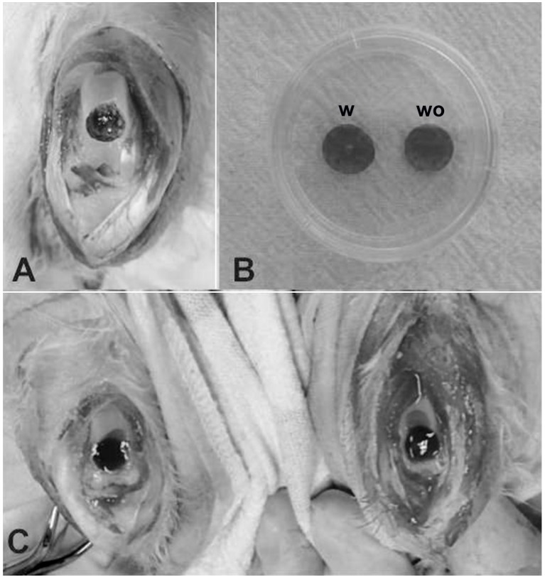
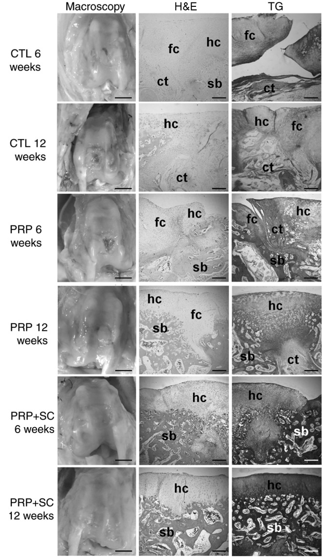
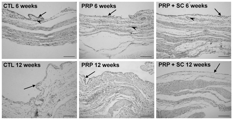
Stem cells in platelet-rich plasma (PRP) scaffolds may be a promising treatment for cartilage repair. Human dental pulp stem cell (hDPSC) subpopulations have been identified to have substantial angiogenic, neurogenic and regenerative potential when compared with other stem cell sources. The present study evaluated the potential of hDPSCs in a PRP scaffold to regenerate full-thickness cartilage defects in rabbits. Full-thickness articular cartilage defects were created in the patellar groove of the femur of 30 rabbits allocated into three experimental groups: Those with an untreated critical defect (CTL), those treated with PRP (PRP) and those treated with stem cells in a PRP scaffold (PRP+SC). The patellar grooves of the femurs from the experimental groups were evaluated macroscopically and histologically at 6 and 12 weeks post-surgery. The synovial membranes were also collected and evaluated for histopathological analysis. The synovial lining cell layer was enlarged in the CTL group compared with the PRP group at 6 weeks (P=0.037) but not with the PRP+SC group. All groups exhibited low-grade synovitis at 6 weeks and no synovitis at 12 weeks. Notably, macroscopic grades for the area of articular cartilage repair for the PRP+SC group were significantly improved compared with those in the CTL (P=0.001) and PRP (P=0.049) groups at 12 weeks. Furthermore, histological scores (modified O'Driscoll scoring system) of the patellar groove articular cartilage in the PRP+SC and PRP groups, in which the articular cartilage was primarily hyaline-like, were significantly higher compared with those in the CTL group at 12 weeks (P=0.002 and P=0.007, respectively). The present results support the therapeutic use of hDPSCs for the treatment of full-thickness articular cartilage defects.
Keywords: cartilage; dental pulp; knee; platelet-rich plasma; stem cells.
Figures
Similar articles
-
Therapeutic Effects of the Addition of Platelet-Rich Plasma to Bioimplants and Early Rehabilitation Exercise on Articular Cartilage Repair.Am J Sports Med. 2018 Jul;46(9):2232-2241. doi: 10.1177/0363546518780955. Epub 2018 Jun 21. Am J Sports Med. 2018. PMID: 29927631
-
Synovial membrane-derived mesenchymal stem cells supported by platelet-rich plasma can repair osteochondral defects in a rabbit model.Arthroscopy. 2013 Jun;29(6):1034-46. doi: 10.1016/j.arthro.2013.02.026. Arthroscopy. 2013. PMID: 23726109
-
Comparison of the Effects of Osteochondral Autograft Transplantation With Platelet-Rich Plasma or Platelet-Rich Fibrin on Osteochondral Defects in a Rabbit Model.Am J Sports Med. 2017 Dec;45(14):3280-3288. doi: 10.1177/0363546517721188. Epub 2017 Aug 30. Am J Sports Med. 2017. PMID: 28853913
-
Repair of full-thickness articular cartilage defects using injectable type II collagen gel embedded with cultured chondrocytes in a rabbit model.J Orthop Sci. 2008 May;13(3):225-32. doi: 10.1007/s00776-008-1220-z. Epub 2008 Jun 6. J Orthop Sci. 2008. PMID: 18528656
-
Leukocyte and Platelet Rich Plasma (L-PRP) Versus Leukocyte and Platelet Rich Fibrin (L-PRF) For Articular Cartilage Repair of the Knee: A Comparative Evaluation in an Animal Model.Iran Red Crescent Med J. 2015 Oct 28;17(10):e19594. doi: 10.5812/ircmj.19594. eCollection 2015 Oct. Iran Red Crescent Med J. 2015. PMID: 26568857 Free PMC article.
Cited by
-
An Update on Mesenchymal Stem Cell-Centered Therapies in Temporomandibular Joint Osteoarthritis.Stem Cells Int. 2021 Apr 1;2021:6619527. doi: 10.1155/2021/6619527. eCollection 2021. Stem Cells Int. 2021. PMID: 33868408 Free PMC article. Review.
-
Cultivation of Cryopreserved Human Dental Pulp Stem Cells-A New Approach to Maintaining Dental Pulp Tissue.Int J Mol Sci. 2022 Sep 29;23(19):11485. doi: 10.3390/ijms231911485. Int J Mol Sci. 2022. PMID: 36232787 Free PMC article.
-
Platelet-Rich Plasma Therapy in the Treatment of Diseases Associated with Orthopedic Injuries.Tissue Eng Part B Rev. 2020 Dec;26(6):571-585. doi: 10.1089/ten.TEB.2019.0292. Epub 2020 Nov 3. Tissue Eng Part B Rev. 2020. PMID: 32380937 Free PMC article. Review.
-
The Role of Platelet-Rich Plasma on the Chondrogenic and Osteogenic Differentiation of Human Amniotic-Fluid-Derived Stem Cells.Int J Environ Res Public Health. 2022 Nov 27;19(23):15786. doi: 10.3390/ijerph192315786. Int J Environ Res Public Health. 2022. PMID: 36497861 Free PMC article.
-
Multidifferentiation potential of dental-derived stem cells.World J Stem Cells. 2021 May 26;13(5):342-365. doi: 10.4252/wjsc.v13.i5.342. World J Stem Cells. 2021. PMID: 34136070 Free PMC article. Review.
References
-
- Dominici M, Le Blanc K, Mueller I, Slaper-Cortenbach I, Marini F, Krause D, Deans R, Keating A, Prockop Dj, Horwitz E. Minimal criteria for defining multipotent mesenchymal stromal cells. The international society for cellular therapy position statement. Cytotherapy. 2006;8:315–317. doi: 10.1080/14653240600855905. - DOI - PubMed
-
- Knutsen G, Drogset JO, Engebretsen L, Grøntvedt T, Isaksen V, Ludvigsen TC, Roberts S, Solheim E, Strand T, Johansen O. A randomized trial comparing autologous chondrocyte implantation with microfracture. Findings at five years. J Bone Joint Surg Am. 2007;89:2105–2112. doi: 10.2106/JBJS.G.00003. - DOI - PubMed
-
- Chen WH, Lo WC, Hsu WC, Wei HJ, Liu HY, Lee CH, Tina Chen SY, Shieh YH, Williams DF, Deng WP. Synergistic anabolic actions of hyaluronic acid and platelet-rich plasma on cartilage regeneration in osteoarthritis therapy. Biomaterials. 2014;35:9599–9607. doi: 10.1016/j.biomaterials.2014.07.058. - DOI - PubMed
LinkOut - more resources
Full Text Sources
Research Materials