

Significance of PD-L1 clones and C-MET expression in hepatocellular carcinoma
- PMID: 31186768
- PMCID: PMC6507339
- DOI: 10.3892/ol.2019.10222
Significance of PD-L1 clones and C-MET expression in hepatocellular carcinoma
Abstract
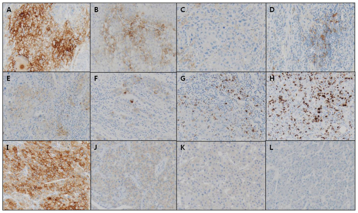
Programmed cell death ligand 1 (PD-L1) is an essential immune checkpoint protein implicated in immune evasion by malignant tumors. Overexpression of programmed cell death protein 1 (PD-1) and its ligand PD-L1 is associated with poor prognosis in various types of cancer. Recently, multiple advances have occurred in the area of cancer immunotherapy. Inhibiting the ligation of PD-1 by PD-L1 has been the major focus of anti-tumor immunotherapy. In diagnostic pathology, it has become crucial to detect PD-L1+ tumor cases using a validated immunohistochemistry (IHC) approach. Preliminary data demonstrate that C-MET promotes survival of some (e.g., renal) cancer types through regulation of PD-L1. However, C-MET expression, and its association with PD-L1, has not been well-characterized in the context of hepatocellular carcinoma (HCC), and no anti-HCC immunotherapy is currently available in Korea. Therefore, it is crucial to investigate the expression of C-MET and PD-L1, and their association with clinicopathologic factors, to facilitate the development of targeted treatments for HCC. PD-L1 expression was examined in tumor cells (TC) and immune cells (IC) of 70 patient-derived HCC specimens using IHC. Two anti-PD-L1 monoclonal antibodies (MAbs), SP263 and SP142, were utilized. Additionally, TC C-MET expression was assessed. Correlations between PD-L1 expression (as identified by both MAbs), C-MET expression and clinicopathologic factors were assessed. More PD-L1+ cases were identified via SP263 than via SP142 when assessing both TC and IC; in the former group, SP236 identified 14/70 positive cases, while SP142 identified only 2/70. In the latter group, SP236 identified 49/70 positive cases, while SP142 identified 30/70. Both MAbs demonstrated a higher frequency of PD-L1 expression by IC than TC. The Edmondson-Steiner grade statistically correlated with a higher frequency of SP236-detected TC PD-L1 expression. C-MET was significantly associated with advanced tumor size and was positively correlated with SP263-detected PD-L1 expression in TC. These results suggest that C-MET may serve a role in regulating PD-L1 expression in HCC. Furthermore, while SP263 generally exhibited a higher sensitivity for PD-L1 detection, concordance in PD-L1+ case detection between the two different MAbs was generally good. These background data may be helpful in the development of targeted anti-HCC immunotherapy focused on PD-L1 or C-MET, and in evaluating selection criteria for target populations best suited to such treatments.
Keywords: C-MET; hepatocellular carcinoma; immunohistochemistry; immunotherapy; programmed cell death ligand 1.
Figures

Similar articles
-
Comparison of PD-L1 detection assays and corresponding significance in evaluation of diffuse large B-cell lymphoma.Cancer Med. 2019 Jul;8(8):3831-3845. doi: 10.1002/cam4.2316. Epub 2019 May 31. Cancer Med. 2019. PMID: 31150165 Free PMC article.
-
Assessment of programmed cell death ligand-1 expression with multiple immunohistochemistry antibody clones in non-small cell lung cancer.J Thorac Dis. 2018 Feb;10(2):816-824. doi: 10.21037/jtd.2018.01.124. J Thorac Dis. 2018. PMID: 29607153 Free PMC article.
-
Multicentric analytical comparability study of programmed death-ligand 1 expression on tumor-infiltrating immune cells and tumor cells in urothelial bladder cancer using four clinically developed immunohistochemistry assays.Virchows Arch. 2019 Nov;475(5):599-608. doi: 10.1007/s00428-019-02610-z. Epub 2019 Jul 2. Virchows Arch. 2019. PMID: 31267201 Free PMC article.
-
Programmed Death-Ligand 1 Immunohistochemistry Testing: A Review of Analytical Assays and Clinical Implementation in Non-Small-Cell Lung Cancer.J Clin Oncol. 2017 Dec 1;35(34):3867-3876. doi: 10.1200/JCO.2017.74.7642. Epub 2017 Oct 20. J Clin Oncol. 2017. PMID: 29053400 Review.
-
Programmed cell death-ligand 1 assessment in urothelial carcinoma: prospect and limitation.J Pathol Transl Med. 2021 May;55(3):163-170. doi: 10.4132/jptm.2021.02.22. Epub 2021 Apr 7. J Pathol Transl Med. 2021. PMID: 33823566 Free PMC article. Review.
Cited by
-
Combination of molecularly targeted therapies and immune checkpoint inhibitors in the new era of unresectable hepatocellular carcinoma treatment.Ther Adv Med Oncol. 2021 May 24;13:17588359211018026. doi: 10.1177/17588359211018026. eCollection 2021. Ther Adv Med Oncol. 2021. PMID: 34104226 Free PMC article. Review.
-
Identifying hub genes of papillary thyroid carcinoma in the TCGA and GEO database using bioinformatics analysis.PeerJ. 2020 Jul 9;8:e9120. doi: 10.7717/peerj.9120. eCollection 2020. PeerJ. 2020. PMID: 32714651 Free PMC article.
-
c-MET and the immunological landscape of cancer: novel therapeutic strategies for enhanced anti-tumor immunity.Front Immunol. 2024 Nov 27;15:1498391. doi: 10.3389/fimmu.2024.1498391. eCollection 2024. Front Immunol. 2024. PMID: 39664377 Free PMC article. Review.
-
Expression of Three Clones of PD-L1 in Lung Cancer: A Single-center Experience.In Vivo. 2023 Jan-Feb;37(1):233-241. doi: 10.21873/invivo.13072. In Vivo. 2023. PMID: 36593005 Free PMC article.
-
c-Met up-regulates the expression of PD-L1 through MAPK/NF-κBp65 pathway.J Mol Med (Berl). 2022 Apr;100(4):585-598. doi: 10.1007/s00109-022-02179-2. Epub 2022 Feb 5. J Mol Med (Berl). 2022. PMID: 35122106
References
LinkOut - more resources
Full Text Sources
Research Materials
Miscellaneous