. 2019 Jul;7(7):e00772.
doi: 10.1002/mgg3.772. Epub 2019 Jun 11.

TET2 missense variants in human neoplasia. A proposal of structural and functional classification

Affiliations

TET2 missense variants in human neoplasia. A proposal of structural and functional classification

Elena Bussaglia et al. Mol Genet Genomic Med. 2019 Jul.

Abstract

Background: The human TET2 gene plays a pivotal role in the epigenetic regulation of normal and malignant hematopoiesis. Somatic TET2 mutations have been repeatedly identified in age-related clonal hematopoiesis and in myeloid neoplasms ranging from acute myeloid leukemia (AML) to myeloproliferative neoplasms. However, there have been no attempts to systematically explore the structural and functional consequences of the hundreds of TET2 missense variants reported to date.

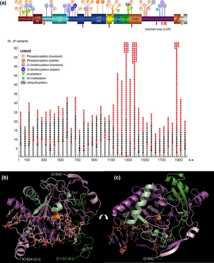
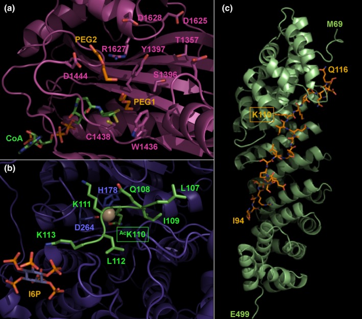
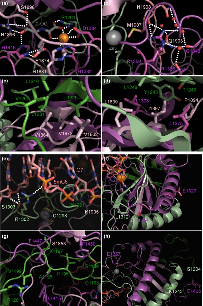
Methods: We have sequenced the TET2 gene in 189 Spanish AML patients using Sanger sequencing and NGS protocols. Next, we performed a thorough bioinformatics analysis of TET2 protein and of the expected impact of all reported TET2 missense variants on protein structure and function, exploiting available structure-and-function information as well as 3D structure prediction tools.

Results: We have identified 38 TET2 allelic variants in the studied patients, including two frequent SNPs: p.G355D (10 cases) and p.I1762V (28 cases). Four of the detected mutations are reported here for the first time: c.122C>T (p.P41L), c.4535C>G (p.A1512G), c.4760A>G (p.D1587G), and c.5087A>T (p.Y1696F). We predict a complex multidomain architecture for the noncatalytic regions of TET2, and in particular the presence of well-conserved α+β globular domains immediately preceding and following the actual catalytic unit. Further, we provide a rigorous interpretation of over 430 missense SNVs that affect the TET2 catalytic domain, and we hypothesize explanations for ~700 additional variants found within the regulatory regions of the protein. Finally, we propose a systematic classification of all missense mutants and SNPs reported to date into three major categories (severe, moderate, and mild), based on their predicted structural and functional impact.

Conclusions: The proposed classification of missense TET2 variants would help to assess their clinical impact on human neoplasia and may guide future structure-and-function investigations of TET family members.

Keywords: 5-methylcytosine; TET2; classification of mutations; epigenetic regulation; neoplasia.


Conflict of interest statement

None of the authors declare any conflict of interest.

Figures

Figure 1
Figure 1
Domain organization and distribution of missense variants in human TET2. (a) The domain architecture of the TET2 protein is schematically represented on top of the figure. Posttranslational modification (PTM) sites were taken from public databases, or predicted, either by similarity with the mouse enzyme (Bauer et al., 2015) or using software given in Methods. Notice that the long polypeptide stretch preceding the actual catalytic unit is predicted to contain several putative, relatively well‐conserved globular modules, separated by more variable PTM‐rich linkers. Single‐nucleotide variations reported in the human TET2 gene are given below the scheme, with the following code: missense mutations are represented by triangles, colored red if identified in hematopoietic and lymphoid neoplasms or black, if reported in solid tumors. Validated SNPs are given as circles, which are colored red and black if they have been in addition reported in cancer patients, or left empty otherwise. (b and c) Two approximately perpendicular views of the three‐dimensional structure of TET2 catalytic unit. Major structural domains are shown as ribbons, colored green (Cys‐N module), light green (Cys‐C), magenta and light pink (N‐ and C‐terminal halves of the DSBH domain, respectively). Zn2+ and Fe2+ ions are represented as gray and orange spheres, respectively; the side chains of their coordinating residues are also shown. The bound DNA oligonucleotide is shown with all its non‐hydrogen atoms, color‐coded (carbon, light pink; nitrogen, blue; oxygen, red; and phosphor, orange). Note the complex interdomain contacts, not only between the N‐ and C‐terminal halves of the catalytic domain, but also between the Zn2+‐binding domains with each other and with the DSHB module. In panel c, note the long insertion between residues Thr1463 and Gly1842 (C‐terminal end of N‐terminal half and N‐terminal start of the C‐terminal half, respectively). DSBH, domain, termed double‐stranded β‐helix
Figure 2
Figure 2
Relevance of the non‐catalytic regions of TET2 protein for enzyme function. Focus is on experimentally verified or predicted roles of residues within the Gly93‐Gln116 TET2 stretch. (a) Close‐up of human p300 catalytic region, highlighting pockets that could accommodate the substrate lysine residue (Lys100; ref. [Zhang et al., 2017]) in TET2, as well as surrounding residues. p300 secondary structure elements are colored pink, and the CoA cofactor is shown with all its nonhydrogen atoms to the bottom‐left, color‐coded (carbon, green; oxygen, red; nitrogen, blue; phosphor, orange and sulfur, yellow). Well‐ordered PEG molecules bound close to the active site in the reported crystal structure (PDB code 4PZR, [Maksimoska, Segura‐Peña, Cole, & Marmorstein, 2014]) are also shown color‐coded (carbon, orange; oxygen, red). Molecule PEG1 overlays with the incoming lysine residue to be acetylated, while PEG2 was proposed to mimic a substrate residue with a small side chain, ideally glycine, flanking the substrate lysine. In TET2, however, Lys110 is flanked by residues with bulkier side chains, and the path of the TET2 peptide in the actual p300·TET2 complex might differ from that with canonical substrates, and/or it would require some rearrangements of residues surrounding the p300 active site for binding in a productive conformation. On the other hand, the shallow negatively charged pocket on p300 termed “P2 site” (top right in this panel) might accommodate Lys113; this interaction is critical for effective substrate binding (Liu et al., 2008). (b) 3D model of TET2 peptide Leu107‐Lys113 with acetylated Lys110 bound to HDAC1. The model has been generated according to the crystal structure of human HDAC1 bound to a substrate mimic based on the sequence of the histone H4 tail (5ICN, [Watson et al., 2016]). HDAC1 secondary structure elements are colored purple. Only side chains of HDAC1 residues that coordinate the catalytic Zn2+ ion are shown. TET2 peptide is green, with all side chains shown color‐coded. Note that the side chain of Lys113 is in the position to engage in electrostatic interactions with the bound inositol hexaphosphate (I6P) molecule, a physiologically relevant activator of HDACs (Watson et al., 2016). (c) 3D model of the Gly93‐Gln116 polypeptide of human TET2 bound to the nuclear importer, importin‐α (IMP‐α). The model has been developed based on the structure of human IMP‐α bound to CPB80 (Dias, Wilson, Rojas, Ambrosio, & Cerione, 2009). The IMP‐α helices are colored lime green, and the TET2 fragment in shown with all is non‐hydrogen atoms, color‐coded (carbon, orange; oxygen, red and nitrogen, blue). Note that the TET2 polypeptide runs in an extended, approximately antiparallel direction to the IMP‐α superhelix
Figure 3
Figure 3
Representative examples of mutations affecting the structure and function of human TET2 catalytic unit. (a) Close‐up of TET2 catalytic site. Shown in the picture are only cofactors Fe2+ (orange sphere) and 2‐OG (color‐coded: carbon, pink; nitrogen, red; oxygen, blue), as well as the side chains of residues directly involved in their coordination (color‐coded as the 2‐OG cofactor, but with carbon atoms green). Note that several TET2 missense mutations affect these residues (e.g., p.Arg1261Cys/His/Leu/Gly, p.His1881Arg/Glu/Asn; class Ia mutants). (b and c), representative examples of proposed type Ib variants. (b) Mutations likely to disrupt the overall 3D structure of the catalytic unit. Shown is a close‐up around residue Arg1359. Note the intricate network of strong H‐bonds centered on its side chain, which directly donates H‐bonds to the carbonyl oxygen from Met1907 (C‐terminal half of the DSBH domain), but is also connected through water molecules to the carbonyls of Gln1348 and Arg1366 (N‐terminal half). Note also that the residue immediately preceding Arg1359 is a Zn2+ ligand. Therefore, mutants p.Arg1359Cys/His/Pro would most likely result in the collapse of the whole 3D structure. (c) Representative example of a mutation introducing a polar or charged residue in the densely packed hydrophobic core. Replacement of Val1864 by a glutamate, as in p.Val1864Glu, would result in major clashes of the mutant Glu1864 carboxylate with the side chains of aliphatic residues shown in the picture and/or with main chain carbonyl oxygen atoms of these and/or other residues. (d) Close‐up around residue Ile1897. Only side chains of residues that make at least one vdW contact with one of the atoms of the Ile1897 side chain are shown, color‐coded according to the domain they belong to. Note that replacement of the aliphatic side chain by a shorter, polar serine, although disfavored, would not be expected to cause major structural rearrangements, as the mutant Ser1897 side chain would not clash with any of the surrounding core residues (class IIa mutation). Note in addition the somehow polar environment created by the nearby Thr1249 side chain. (e) Close‐up of the substrate DNA‐binding site; the side chains of some of the residues mutated in cancer patients are shown with all of their nonhydrogen atoms. Replacement of single residues, such as in mutants p.Arg1302Gly or p.Lys1905Glu, is likely to affect the rate of DNA oxidation, but would not be expected to completely abolish processing of the 5mC residue (class IIc variants). (f) Close‐up showing exposed, well‐conserved residues that are mutated in some cancer patients (e.g., p.Leu1312Val, p.Asp1314Gly, p.Glu1320Ala). Their relative proximity to the bound DNA oligonucleotide suggests that they might form a binding site for a TET2 cofactor such as IDAX/CXXC4, which recruits TET2 to DNA (Ko et al., 2013) (type IId mutants). (g) Close‐up around Ser1189‐Arg1201 TET2 stretch. Only a few side chains of interacting residues are shown for simplicity. Some TET2 missense variants that affect residues within this sequence are likely to be fully tolerated without any important rearrangements of TET2 protein structure (e.g., p.Ile1195Val, p.Val1199Ile; class IIIa). (h) Close‐up showing not conserved, exposed TET2 residues. Missense variants of these residues that introduce physicochemically related residues, commonly found in TET2 from other species (e.g., p.Ser1204Cys, p.Lys1243Arg, p.Glu1405Gln) are unlikely to have any impact on TET2 function (class III variants). DSBH, domain, termed double‐stranded β‐helix

Similar articles

Cited by

References

    1. Abdel‐Wahab, O. , Mullally, A. , Hedvat, C. , Garcia‐Manero, G. , Patel, J. , Wadleigh, M. , … Levine, R. L. (2009). Genetic characterization of TET1, TET2, and TET3 alterations in myeloid malignancies. Blood, 114(1), 144–147. 10.1182/blood-2009-03-210039 - DOI - PMC - PubMed
    1. Abegunde, S. O. , Buckstein, R. , Wells, R. A. , & Rauh, M. J. (2018). An inflammatory environment containing TNFα favors Tet2‐mutant clonal hematopoiesis. Experimental Hematology, 59, 60–65. 10.1016/j.exphem.2017.11.002 - DOI - PubMed
    1. Adzhubei, I. A. , Schmidt, S. , Peshkin, L. , Ramensky, V. E. , Gerasimova, A. , Bork, P. , … Sunyaev, S. R. (2010). A method and server for predicting damaging missense mutations. Nature Methods, 7(4), 248–249. 10.1038/nmeth0410-248 - DOI - PMC - PubMed
    1. Arends, C. M. , Galan‐Sousa, J. , Hoyer, K. , Chan, W. , Jäger, M. , Yoshida, K. , … Damm, F. (2018). Hematopoietic lineage distribution and evolutionary dynamics of clonal hematopoiesis. Leukemia, 32(9), 1908–1919. 10.1038/s41375-018-0047-7 - DOI - PubMed
    1. Aslanyan, M. G. , Kroeze, L. I. , Langemeijer, S. M. C. , Koorenhof‐Scheele, T. N. , Massop, M. , van Hoogen, P. , … Jansen, J. H. (2014). Clinical and biological impact of TET2 mutations and expression in younger adult AML patients treated within the EORTC/GIMEMA AML‐12 clinical trial. Annals of Hematology, 93(8), 1401–1412. 10.1007/s00277-014-2055-7 - DOI - PubMed

Publication types

LinkOut - more resources