Review
. 2019 Jun;8(2):10.1128/ecosalplus.ESP-0003-2019.
doi: 10.1128/ecosalplus.ESP-0003-2019.

Plasmid Localization and Partition in Enterobacteriaceae

Affiliations
Review

Plasmid Localization and Partition in Enterobacteriaceae

Jean-Yves Bouet et al. EcoSal Plus. 2019 Jun.

Abstract

Plasmids are ubiquitous in the microbial world and have been identified in almost all species of bacteria that have been examined. Their localization inside the bacterial cell has been examined for about two decades; typically, they are not randomly distributed, and their positioning depends on copy number and their mode of segregation. Low-copy-number plasmids promote their own stable inheritance in their bacterial hosts by encoding active partition systems, which ensure that copies are positioned in both halves of a dividing cell. High-copy plasmids rely on passive diffusion of some copies, but many remain clustered together in the nucleoid-free regions of the cell. Here we review plasmid localization and partition (Par) systems, with particular emphasis on plasmids from Enterobacteriaceae and on recent results describing the in vivo localization properties and molecular mechanisms of each system. Partition systems also cause plasmid incompatibility such that distinct plasmids (with different replicons) with the same Par system cannot be stably maintained in the same cells. We discuss how partition-mediated incompatibility is a consequence of the partition mechanism.


Figures

Figure 1
Figure 1
Genetic organization of plasmid partition loci. Genes encoding the NTPases and the CBPs are depicted by red and blue arrows, respectively, and the centromere sites are displayed by green boxes. The type I (parABS), type II (parMRC), and type III (tubRZC) partition loci are distinguished by their NTPase signatures, Walker-A (light red), actin-like (dark red), and tubulin-like (orange red), respectively. The CBPs harbor either an HTH2 (light blue) or an RHH2 (dark blue) DNA binding motif. The partition system of R388 of yet-undetermined partition type (gray) encodes only a CBP (purple arrow) with an undetermined (nd) DNA binding motif. For type III, note that the order of the NTPase and CBP is inverse from that of types I and II. All plasmids diagrammed are found in Enterobacteriaceae except pTAR, pSM19035, and pBtoxis, which are included since their properties are discussed in this review. The historical names of genes and centromeres are indicated within the arrows and below the boxes, respectively (reviewed in reference 14). Note that in the body of the review we have simplified this nomenclature for type I systems and use ParA, ParB, and parS (with subscripts indicating the plasmid) for the ATPase, CBP, and centromere, respectively. The schematic representation is drawn at the indicated scale.
Figure 2
Figure 2
Mini-F plasmids, partition proteins, and nucleoid localization. (A) Colocalization of mini-F plasmids and ParBF clusters in a growing E. coli cell. (a) The mini-F plasmid pJYB273 is visualized with the ParB pMT1-mTurquoise2/parSpMT1 labeling system. (b) ParBF-mVenus fusion protein is expressed from its endogenous locus on pJYB273. (c) The overlay displays the phase-contrast along with the two fluorescent channels. The dashed yellow lines represent the contour length of the cell. (B) Dual localization of ParAF and the nucleoid. The E. coli strain DLT3057 expresses the HU-mCherry fusion that labels the nucleoid and carries a mini-F with the parAF-mVenus allele (pJYB243). The growing cell was observed in phase contrast (a) and in the yellow (b) and red (c) channels to image ParAF and the nucleoid, respectively. The overlay (d) displays the combination of all three channels, showing that ParAF localized over the nucleoid. C ParAF oscillates from pole to pole. An E. coli cell carrying the mini-F parAF-mVenus (pJYB243) was imaged every 20 s for 15 min. A selection of images (times in seconds) were displayed showing that ParAF-mVenus proteins oscillate in a collective and coordinated fashion. Scale bars: 1 μm.
Figure 3
Figure 3
The organization and composition of centromeres are highly diverse. Direct and inverted repeats are depicted by oriented arrows with the same color, indicating conserved motifs. For parSF, the black inverted arrows represent the 16-bp inverted repeat ParB binding sites within the 43-bp direct repeats (green arrows). The centromere of P1 is composed of two 6-bp box B (blue) and four 7-bp box A (red) motifs present on both sides of an integration host factor (IHF) binding site (gray rectangle). The RK2 centromere (OB3) consists of one inverted repeat of 13 bp. For pB171, the parC1 and parC2 regions of par2 are composed of 17 (2 clusters) and 18 (3 clusters) repeats, respectively, of a 6-bp motif. The parS site of par1 comprises only two identical 10-bp motifs (orange arrows) in direct orientation separated by a 31-bp direct repeat; the blue arrows overlapping the −35 and −10 promoter sequences correspond to the beginning of parC1 from the par2 locus involved in the cross-regulation between the two Par systems of pB171. pSM19035 carries three parS loci composed of contiguous repeats of 7 bp in direct or inverse orientations (only parS1 and parS2 are depicted). For parSTP228, the two centromere regions, parH (left) and OF (right), delineated by a vertical dashed line, are composed of 12 and 8 degenerated repeats (4 bp) separated by AT-rich spacers (4 bp), respectively. The pTAR centromere contains 13 repeats of 9 bp, each separated by 8 bp, encompassing the −35 and −10 promoter boxes. The centromere of plasmid R1 comprises two arrays, spaced by 39 bp, composed of five direct repeats of 11 bp. The pBtoxis centromere, tubC, comprises two arrays of three and four 12-bp motifs, separated by 54 bp. For parSR388, the two arrays spaced by 43 bp are each composed of five direct repeats of 9 bp separated by 2 bp; the putative −35 and −10 promoter sequences are deduced from the sequence. Note that (i) the scale for the large sopC centromeres is different from all others (separated by the horizontal gray dashed line), and (ii) only 9 out of 13 repeats of the pTAR parS are drawn. The parS centromeres are depicted in the same order with the same color code (colored vertical lines on the left) as the partition loci to which they belong (Fig. 1).
Figure 4
Figure 4
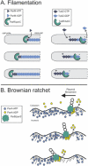
Mechanisms of partition NTPase-driven plasmid movement. (A) Filamentation. (Left) Filament growth and catastrophe. Plasmid R1, the paradigm for type II plasmid partition, uses ATP-dependent polymerization of the actin-like ParM ATPase to push plasmids towards the poles. Plasmids (via their ParR/parC partition complexes) are inserted at the growing end of the filaments, which are polar, by associating with the barbed end of a ParM molecule. Filaments are capped by partition complexes and ATP subunits, while individual monomers within the filaments hydrolyze ATP to ADP. The other “pointed” end of the filament is proposed to be capped by association with an antiparallel ParM filament (not shown), which itself associates with another plasmid via ParR/parC complexes for bidirectional plasmid movement (111). Loss of the cap results in “catastrophe,” or rapid filament disassembly (not shown). (Right) Treadmilling. In type III partition systems such as that of pBtoxis, the tubulin-like TubZ GTPase polymerizes by addition of TubZ-GTP to the plus end and depolymerizes by loss of TubZ-GDP from the minus end, a behavior known as treadmilling. The plasmid (via its TubR/tubC partition complex) tracks with the minus end, so it is pulled from midcell to the cell pole. (B) Brownian ratchet partition systems rely on the ATP-dependent nonspecific DNA binding activity of the partition ATPase (ParA), which binds to the bacterial nucleoid. The plasmid (via the ParB/parS partition complex) attaches to ParA on the nucleoid and then stimulates ParA release from DNA by ATP hydrolysis or conformational change. Because ParA rebinding to the nucleoid is slow (40), a void of ParA is created on the bacterial chromosome, which serves as a barrier to motion so that the ParB/parS/plasmid complex moves towards the remaining ParA on the nucleoid. Further details and variations of this mechanism are described in the main text.
Figure 5
Figure 5
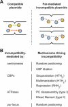
Partition-mediated incompatibility phenotypes. (A) Schematic representation of the incompatibility at the onset of cell division. Two different replicons, represented with orange and blue colors, are fully compatible if their partition systems (red and green circles) are distinct (left) or are incompatible if their partition systems cross-react (gray circles) (right). In the latter case, sibling plasmids would frequently be inherited in the same daughter cell, leading to mutual exclusion. (B) Mechanisms driving incompatibility phenotypes. Each partition component leads to mutual exclusion of plasmids sharing parts of the partition locus (see main text for details).

Similar articles

Cited by

References

    1. Onogi T, Miki T, Hiraga S. 2002. Behavior of sister copies of mini-F plasmid after synchronized plasmid replication in Escherichia coli cells. J Bacteriol 184:3142–3145. 10.1128/JB.184.11.3142-3145.2002. [PubMed] - DOI - PMC - PubMed
    1. Wang X, Montero Llopis P, Rudner DZ. 2013. Organization and segregation of bacterial chromosomes. Nat Rev Genet 14:191–203. 10.1038/nrg3375. [PubMed] - DOI - PMC - PubMed
    1. Badrinarayanan A, Le TBK, Laub MT. 2015. Bacterial chromosome organization and segregation. Annu Rev Cell Dev Biol 31:171–199. 10.1146/annurev-cellbio-100814-125211. [PubMed] - DOI - PMC - PubMed
    1. Jacob F, Brenner S, Cuzin F. 1963. On the regulation of DNA replication in bacteria. Cold Spring Harb Symp Quant Biol 228:329–348. 10.1101/SQB.1963.028.01.048. - DOI
    1. Green EW, Schaechter M. 1972. The mode of segregation of the bacterial cell membrane. Proc Natl Acad Sci U S A 69:2312–2316. 10.1073/pnas.69.8.2312. [PubMed] - DOI - PMC - PubMed

Publication types

Substances

LinkOut - more resources