Review
. 2019 Jun 12;9(3):1155-1212.
doi: 10.1002/cphy.c180037.

Tissue Engineering of the Microvasculature

Affiliations
Review

Tissue Engineering of the Microvasculature

Joe Tien. Compr Physiol. .

Abstract

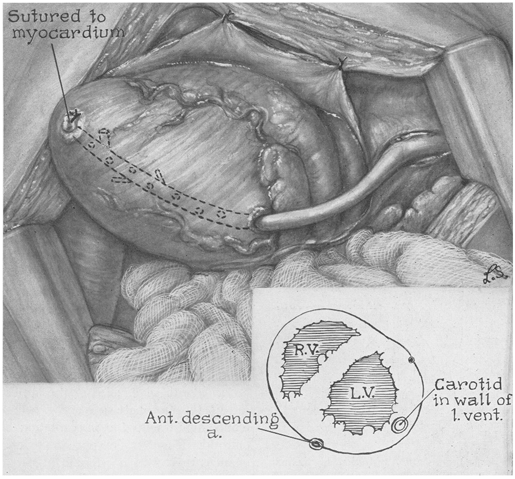
The ability to generate new microvessels in desired numbers and at desired locations has been a long-sought goal in vascular medicine, engineering, and biology. Historically, the need to revascularize ischemic tissues nonsurgically (so-called therapeutic vascularization) served as the main driving force for the development of new methods of vascular growth. More recently, vascularization of engineered tissues and the generation of vascularized microphysiological systems have provided additional targets for these methods, and have required adaptation of therapeutic vascularization to biomaterial scaffolds and to microscale devices. Three complementary strategies have been investigated to engineer microvasculature: angiogenesis (the sprouting of existing vessels), vasculogenesis (the coalescence of adult or progenitor cells into vessels), and microfluidics (the vascularization of scaffolds that possess the open geometry of microvascular networks). Over the past several decades, vascularization techniques have grown tremendously in sophistication, from the crude implantation of arteries into myocardial tunnels by Vineberg in the 1940s, to the current use of micropatterning techniques to control the exact shape and placement of vessels within a scaffold. This review provides a broad historical view of methods to engineer the microvasculature, and offers a common framework for organizing and analyzing the numerous studies in this area of tissue engineering and regenerative medicine. © 2019 American Physiological Society. Compr Physiol 9:1155-1212, 2019.


Figures

Figure 1.
Figure 1.
Vascularization strategies based on (A) angiogenesis into a graft, (B) vasculogenesis within a graft, and (C) direct seeding and perfusion of a microfluidic scaffold. GF, growth factor; ECM, extracellular matrix. Adapted with permission from (584).
Figure 2.
Figure 2.
Quantitative metrics of microvascular physiology. (A) Calculation of endothelial hydraulic conductivity relies on measurement of filtration speed as a function of vascular pressure. Reprinted with permission from (313). (B) Calculation of solute permeability relies on measurement of solute accumulation over time. Reproduced with permission from (227).
Figure 3.
Figure 3.
Binding of VEGFs to their receptors, the VEGFRs and neuropilins. Reproduced with permission from (247).
Figure 4.
Figure 4.
Diagram of the Vineberg procedure for vascularizing ischemic myocardium, using a carotid artery implant. RV, right ventricle; LV, left ventricle. Reproduced with permission from (479).
Figure 5.
Figure 5.
Dose-dependent collateral growth two months after implantation of FGF2-loaded heparin/alginate beads around occluded coronary arteries in the ischemic pig heart. bFGF, basic fibroblast growth factor (FGF2). Reproduced with permission from (350).
Figure 6.
Figure 6.
Density of vascular ingrowth into polyHEMA implants after one month. Reproduced with permission from (361).
Figure 7.
Figure 7.
Cellularity of fibrovascular ingrowth into porous PTFE chambers in the presence of various growth factors after ten days. Reproduced with permission from (541).
Figure 8.
Figure 8.
Number of viable hepatocytes in PLGA sponge implants as a function of time. Hepatocytes were added directly to scaffolds before implantation; scaffolds were not pre-vascularized. Reproduced with permission from (388).
Figure 9.
Figure 9.
Strategy for vascularization of grafts from a surgically constructed arteriovenous (AV) loop. Angiogenesis from the loop invades into overlying tissue. A, artery; V, vein. Reproduced with permission from (137).
Figure 10.
Figure 10.
Generation of centimeter-scale tissue with an arteriovenous (AV) loop in a polycarbonate chamber. (A) AV loop overlaid on a polycarbonate base. (B) Addition of rat cardiomyocytes and Matrigel around the AV loop. (C) Explant of pedicled myocardium that formed by four weeks. Scale bar refers to 1 cm. Reproduced with permission from (394).
Figure 11.
Figure 11.
VEGF-driven angiogenesis into collagen gels in microfluidic devices. Treatment of devices with poly-D-lysine (PDL) enabled ECs to sprout from a monolayer towards a VEGF source. Arrows denote the direction of VEGF transport. Scale bars refer to 100 μm. EGM2mv, endothelial cell growth media. Reproduced with permission from (92).
Figure 12.
Figure 12.
Derivation and characterization of EPCs. (A) Spindle-shaped EPCs that were derived from one-week-old cultures of CD34-enriched peripheral blood-derived mononuclear cells. Reproduced with permission from (23). (B) Flow cytometry of EPCs and monocytes. EPCs express blood cell markers, including CD45 and the monocyte activation marker CD11c. Adapted with permission from (459).
Figure 13.
Figure 13.
Derivation and characterization of EOCs. (Left) Colony of cord blood-derived EOCs that emerged after mononuclear cells were cultured for nine days. (Right) Flow cytometry of EOCs. EOCs express EC markers, such as CD31 and CD144 (VE-cadherin), but do not express markers of blood cells, such as CD45 and the macrophage/monocyte marker CD14. Adapted with permission from (232).
Figure 14.
Figure 14.
Incorporation of bone marrow-derived, lacZ-expressing EPCs at sites of ischemia. Sections were co-stained by X-gal (blue) and for lectin (A) and CD31 (B). Scale bar refers to 25 μm. Reproduced with permission from (22).
Figure 15.
Figure 15.
Vascular mimicry in uveal melanoma. Red blood cells (top, arrowheads) fill vessel-like structures that do not appear to be lined by ECs. Reproduced with permission from (358).
Figure 16.
Figure 16.
Fine structure of vessels that formed from normal and Bcl2-expressing HUVECs in collagen-fibronectin gels one month after implantation. (A) Implants of normal HUVECs. (B, C) Implants of Bcl2-expressing HUVECs. The lumens are perfused with red blood cells (RBC); asterisks denote mural cells. Reproduced with permission from (494).
Figure 17.
Figure 17.
Organization of human microvascular ECs into blood and lymphatic vessels in a dermal graft after two weeks. Immunostains are shown for CD31, blood vessel marker laminin (Lam1,2), and lymphatic markers LYVE-1 and podoplanin (hPDN). Scale bars refer to 100 μm. Adapted with permission from (360).
Figure 18.
Figure 18.
Transformation of endothelial aggregates into perfused vessels. (A) Random self-organized HUVEC networks and patterned HUVEC cords, before and after implantation. (B) Two-week-old implants after perfusion with species-specific lectins. Vessels in implants consist of ECs that are derived from graft (human, red) and host (mouse, green). Scale bars refer to 250 μm (A, upper left), 500 μm (A, upper middle and upper right), 100 μm (A, lower), and 150 μm (B). Reproduced with permission from (38).
Figure 19.
Figure 19.
Increase in perfused vascular density over time in subcutaneous polymer implants that contained adipose-derived microvascular fragments (black circles) or that did not (clear circles). (Left) Vascular density near the surface of implants. (Right) Vascular density at the center of implants. Adapted with permission from (316).
Figure 20.
Figure 20.
Vascularized myocardial constructs that contained human cardiomyocytes, HUVECs, and mouse fibroblasts in a PLA/PLGA sponge after two weeks. (Left) Immunostain for human CD31 (red) and von Willebrand factor (green). (Right) Immunostain for human CD31 (brown). Reproduced with permission from (332).
Figure 21.
Figure 21.
Perfused vascular networks in microscale fibrin gels within microfluidic devices. Fibroblasts, in the same or separate gel, were included to promote vasculogenesis. In (B), the resulting networks were perfused with a solution of fluorescent dextran. Reproduced with permission from (277, 395).
Figure 22.
Figure 22.
Branching vascular-like pattern that was etched into a silicon wafer. Such patterns could be seeded with ECs to generate patterned cultures. Feature widths were on the order of ~10 μm. Adapted with permission from (249).
Figure 23.
Figure 23.
Vascularization of a microfluidic type I collagen scaffold that was formed by combining a micropatterned and planar gel. Both en face and reconstructed 3D views of HUVEC-seeded structures are shown. Scale bar refers to 100 μm. Reproduced with permission from (677).
Figure 24.
Figure 24.
Vascularization of a microfluidic type I collagen scaffold that was formed around a removable needle. (Top) Unseeded scaffold. (Bottom) Scaffold that was seeded with HUVECs. Insets show cross-sectional views. Scale bar refers to 100 μm. Reproduced with permission from (90).
Figure 25.
Figure 25.
Vascularization of a microfluidic fibrin scaffold that was formed around a sacrificial 3D-printed network of sugar-based fibers. Channels were seeded with HUVECs (red), and the scaffold bulk contained 10T1/2 mouse fibroblasts (green). Scale bar refers to 1 mm. Reproduced with permission from (382).
Figure 26.
Figure 26.
Vascularization of a microfluidic polyethylene glycol gel that was patterned by photodegradation. (Left) Visualization of interconnected channels by perfusion with fluorescent dextran. (Right) 3D confocal reconstruction of a vascularized channel that was stained for ZO-1 (green). Reproduced with permission from (196).
Figure 27.
Figure 27.
Perfusion-decellularization of whole hearts preserves the vascular architecture. (A) Corrosion casts of native and decellularized hearts. Scale bars refer to 1 mm (top) and 250 μm (bottom). (B) Transplanted decellularized heart before and after re-establishment of blood flow. The recipient animal was heparinized to minimize thrombosis. Reproduced with permission from (424).
Figure 28.
Figure 28.
Physical mechanisms for stable vascularization of microfluidic scaffolds. Maintenance of vascular adhesion can be viewed as (A) a balance of outward (stabilizing) and inward (destabilizing) stresses, including pressures, contractile stress, and adhesion stress, or as (B) a balance of adhesion (stabilizing) and stored elastic (destabilizing) energies. Adapted with permission from (643).

References

    1. Aicher A, Brenner W, Zuhayra M, Badorff C, Massoudi S, Assmus B, Eckey T, Henze E, Zeiher AM, Dimmeler S. Assessment of the tissue distribution of transplanted human endothelial progenitor cells by radioactive labeling. Circulation 107: 2134–2139, 2003. - PubMed
    1. Aicher A, Heeschen C, Mildner-Rihm C, Urbich C, Ihling C, Technau-Ihling K, Zeiher AM, Dimmeler S. Essential role of endothelial nitric oxide synthase for mobilization of stem and progenitor cells. Nat Med 9: 1370–1376, 2003. - PubMed
    1. Aicher A, Rentsch M, Sasaki K, Ellwart JW, Fändrich F, Siebert R, Cooke JP, Dimmeler S, Heeschen C. Nonbone marrow-derived circulating progenitor cells contribute to postnatal neovascularization following tissue ischemia. Circ Res 100: 581–589, 2007. - PubMed
    1. Akahori T, Kobayashi A, Komaki M, Hattori H, Nakahama K-I, Ichinose S, Abe M, Takeda S, Morita I. Implantation of capillary structure engineered by optical lithography improves hind limb ischemia in mice. Tissue Eng A 16: 953–959, 2010. - PubMed
    1. Akita S, Tamai N, Myoui A, Nishikawa M, Kaito T, Takaoka K, Yoshikawa H. Capillary vessel network integration by inserting a vascular pedicle enhances bone formation in tissue-engineered bone using interconnected porous hydroxyapatite ceramics. Tissue Eng 10: 789–795, 2004. - PubMed

FURTHER READING

    1. Briquez PS, Clegg LE, Martino MM, Mac Gabhann F, Hubbell JA. Design principles for therapeutic angiogenic materials. Nat Rev Mater 1: 1–15, 2016.
    1. Cooke JP, Losordo DW. Modulating the vascular response to limb ischemia: angiogenic and cell therapies. Circ Res 116: 1561–1578, 2015. - PMC - PubMed
    1. Lähteenvuo J, Ylä-Herttuala S. Advances and challenges in cardiovascular gene therapy. Hum Gene Ther 28: 1024–1032, 2017. - PubMed
    1. Medina RJ, Barber CL, Sabatier F, Dignat-George F, Melero-Martin JM, Khosrotehrani K, Ohneda O, Randi AM, Chan JKY, Yamaguchi T, Van Hinsbergh VWM, Yoder MC, Stitt AW. Endothelial progenitors: a consensus statement on nomenclature. Stem Cells Transl Med 6: 1316–1320, 2017. - PMC - PubMed
    1. Park KM, Gerecht S. Harnessing developmental processes for vascular engineering and regeneration. Development 141: 2760–2769, 2014. - PMC - PubMed

CROSS-REFERENCES

    1. Cardiovascular Physiology: Overview of the microcirculation (legacy)

    1. Cardiovascular Physiology: Physiology and pathobiology of microvascular endothelium (legacy)

    1. Cardiovascular Physiology: Angiogenesis

    1. Cardiovascular Physiology: Development of a vascular network

    1. Cardiovascular Physiology: Lymphatics

Publication types

MeSH terms

Substances

LinkOut - more resources