

Clinical importance of the occipital artery in vascular lesions: A review of the literature
- PMID: 31188082
- PMCID: PMC6728704
- DOI: 10.1177/1971400919857245
Clinical importance of the occipital artery in vascular lesions: A review of the literature
Abstract
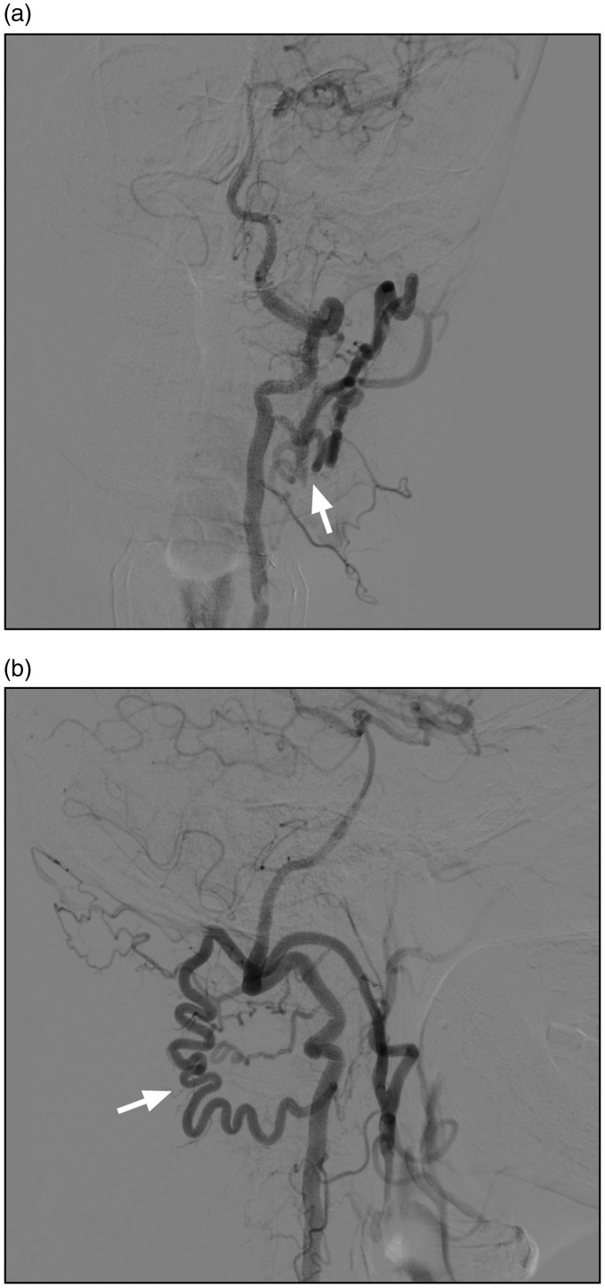
The occipital artery (OA) is a critical artery in vascular lesions. However, a comprehensive review of the importance of the OA is currently lacking. In this study, we used the PubMed database to perform a review of the literature on the OA to increase our understanding of its role in vascular lesions. We also provided our typical cases to illustrate the importance of the OA. The OA has several variations. For example, it may arise from the internal carotid artery or anastomose with the vertebral artery. Therefore, the OA may provide a crucial collateral vascular supply source and should be preserved in these cases. The OA is a good donor artery. Consequently, it is used in extra- to intracranial bypasses for moyamoya disease (MMD) or aneurysms. The OA can be involved in dural arteriovenous fistula (DAVF) and is a feasible artery for the embolisation of DAVF. True aneurysms and pseudoaneurysms can occur in the OA; surgical resection and embolisation are the effective treatment approaches. Direct high-flow AVF can occur in the OA; embolisation treatment is a good option in such cases. The OA can also be involved in MMD and brain arteriovenous malformation (AVM) by forming transdural collaterals. For a patient in the prone position, if occipital and suboccipital craniotomies are performed, the OA can also be used for intraoperative angiography. In brief, the OA is a very important artery in vascular lesions.
Keywords: Occipital artery; clinical importance; review; vascular lesion.
Figures


References
-
- Oner Z, Oner S, Kahraman AS. The right vertebral artery originating from the right occipital artery and the absence of the transverse foramen: a rare anatomical variation. Surg Radiol Anat 2017; 39: 1397–1400. - PubMed
-
- Ates O, Ahmed AS, Niemann D, et al. The occipital artery for posterior circulation bypass: microsurgical anatomy. Neurosurg Focus 2008; 24: E9. - PubMed
-
- Roski RA, Spetzler RF, Hopkins LN. Occipital artery to posterior inferior cerebellar artery bypass for vertebrobasilar ischemia. Neurosurgery 1982; 10: 44–49. - PubMed
-
- Alvernia JE, Fraser K, Lanzino G. The occipital artery: a microanatomical study. Neurosurgery 2006; 58: ONS114–22. discussion ONS114-22. - PubMed
-
- Lasjaunias P, Theron J, Moret J. The occipital artery. Anatomy – normal arteriographic aspects – embryological significance. Neuroradiology 1978; 15: 31–37. - PubMed
Publication types
MeSH terms
LinkOut - more resources
Full Text Sources
