. 2019 Jun 12;14(6):e0216776.
doi: 10.1371/journal.pone.0216776. eCollection 2019.

Layer-by-layer ultraviolet assisted extrusion-based (UAE) bioprinting of hydrogel constructs with high aspect ratio for soft tissue engineering applications

Affiliations

Layer-by-layer ultraviolet assisted extrusion-based (UAE) bioprinting of hydrogel constructs with high aspect ratio for soft tissue engineering applications

Pei Zhuang et al. PLoS One. .

Abstract

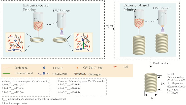
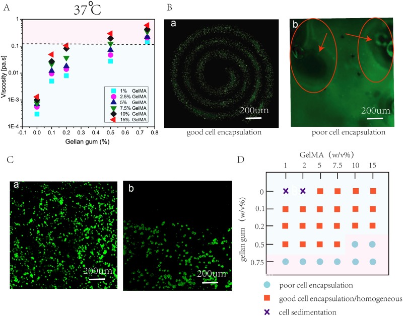
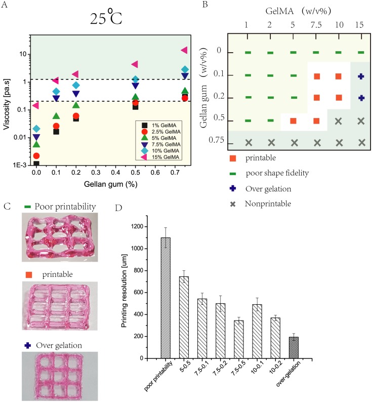
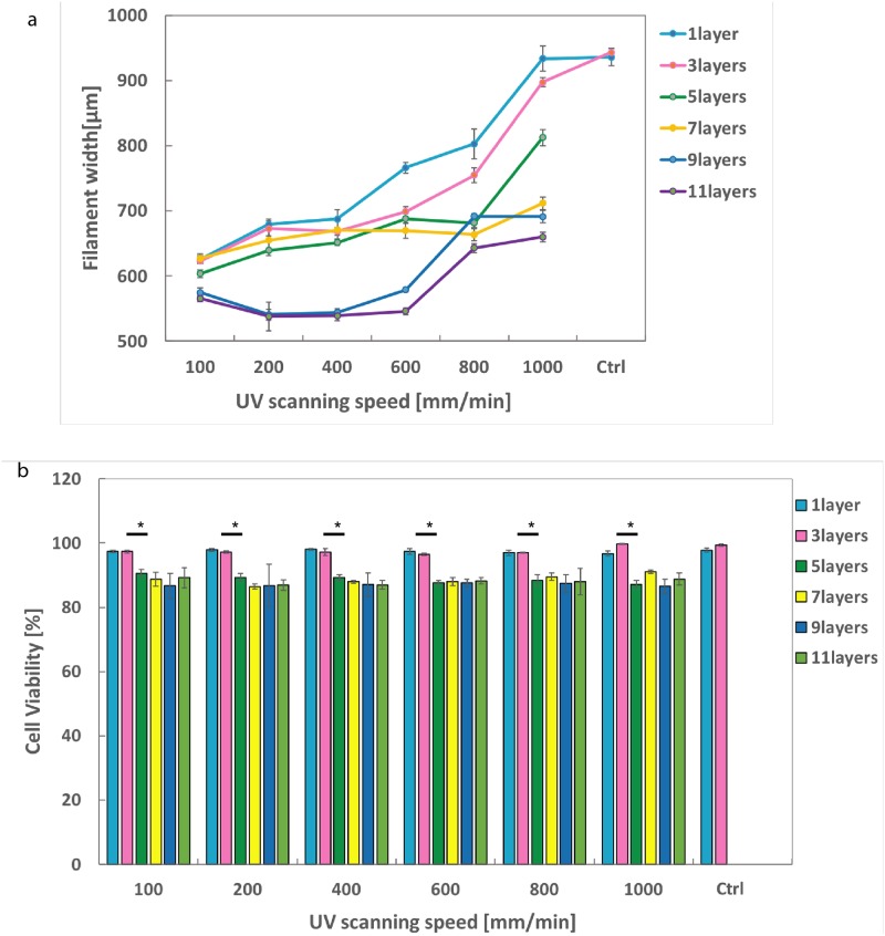
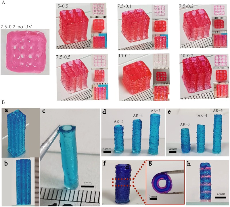
One of the major challenges in the field of soft tissue engineering using bioprinting is fabricating complex tissue constructs with desired structure integrity and mechanical property. To accomplish such requirements, most of the reported works incorporated reinforcement materials such as poly(ϵ-caprolactone) (PCL) polymer within the 3D bioprinted constructs. Although this approach has made some progress in constructing soft tissue-engineered scaffolds, the mechanical compliance mismatch and long degradation period are not ideal for soft tissue engineering. Herein, we present a facile bioprinting strategy that combines the rapid extrusion-based bioprinting technique with an in-built ultraviolet (UV) curing system to facilitate the layer-by-layer UV curing of bioprinted photo-curable GelMA-based hydrogels to achieve soft yet stable cell-laden constructs with high aspect ratio for soft tissue engineering. GelMA is supplemented with a viscosity enhancer (gellan gum) to improve the bio-ink printability and shape fidelity while maintaining the biocompatibility before crosslinking via a layer-by-layer UV curing process. This approach could eventually fabricate soft tissue constructs with high aspect ratio (length to diameter) of ≥ 5. The effects of UV source on printing resolution and cell viability were also studied. As a proof-of-concept, small building units (3D lattice and tubular constructs) with high aspect ratio are fabricated. Furthermore, we have also demonstrated the ability to perform multi-material printing of tissue constructs with high aspect ratio along both the longitudinal and transverse directions for potential applications in tissue engineering of soft tissues. This layer-by-layer ultraviolet assisted extrusion-based (UAE) Bioprinting may provide a novel strategy to develop soft tissue constructs with desirable structure integrity.


Conflict of interest statement

The authors have declared that no competing interests exist.

Figures

Fig 1
Fig 1. Schematic drawing of layer-by-layer UV-assisted bioprinting strategy.
The gellam gun in the bio-ink serves as a viscosity enhancer to improve the bio-ink printability (via formation of ionic bonds between GelMA chain and gellan gum) during the extrusion printing process prior to further UV crosslinking (to form chemical bond between adjacent GelMA chains) of each individual printed layer. This layer-by-layer UV-assisted bioprinting strategy is repeated to eventually achieve fabrication of complex 3D structures with high aspect ratio.
Fig 2
Fig 2. Bio-ink formulation involves characterization of rheological properties, ease of cell encapsulation and influence of cell sedimentation within bio-inks.
A) Viscosity of 30 different GelMA-GG bio-inks at a constant shear rate of 100s−1 at 37°C indicate higher bio-ink viscosity with increasing polymer concentrations. B) Representative images to highlight the influence of bio-ink viscosity on cell encapsulation; a low viscous bio-ink facilitates good cell encapsulation (in spiral pattern with 7.5-0.2% w/v GelMA-GG) whereas a highly viscous bio-inks results in poor cell encapsulation. C) Representative images to highlight the influence of bio-ink viscosity and density on cell sedimentation; a low polymer concentration (low viscosity and density) and without gellan gum leads to cell sedimentation whereas a high polymer concentration (high viscosity and density) results in a homogeneous cell distribution with negligible cell sedimentation. D) An overview of the different GelMA-GG bio-inks in terms of cell encapsulation and cell distribution.
Fig 3
Fig 3. The bioprinting phase involves characterization of rheological properties, determination of suitable UV scanning speed and selection of suitable bio-inks.
A) Rheological properties of 30 different GelMA-GG bio-inks at a constant shear rate of 100s−1 at 25°C indicated higher bio-ink viscosity with increasing polymer concentrations. B) An overview of the different GelMA-GG bio-inks in terms of printability and cell encapsulation. C) Representative images of printed constructs to distinguish among the three different classifications; (Top) poor printability, (Middle) good printability, (Bottom) over-gelation. D) Influence of bio-ink on printing resolution, a more viscous bio-ink results in higher printing resolution due to significantly less spreading of the shear-thinning bio-inks upon contact with the substrate surface.
Fig 4
Fig 4
a) The effects of UV scanning speed (100, 200, 400, 600, 800, 1000 mm/min) on filaments width. b) The effects of UV scanning speed (100, 200, 400, 600, 800, 1000 mm/min) on cell viability. Constructs with no UV curing were used as control group. *—p < 0.05, **—p < 0.001.
Fig 5
Fig 5
A) Left: Printed grid construct with no layer-by-layer UV curing using 7.5-0.2 group. Right: Printed grid pattern (W× L× H = 9mm × 9mm × 10mm) with the 6 selected GelMA-GG bio-inks. B) a. Printed grid construct (W× L × H = 9mm × 9mm × 30mm). b. Side view of the printed construct (W × L × H = 9mm × 9mm × 30mm. c-e. Tubular structures printed with GelMA-GG bio-ink (7.5-0.2) with different AR which is bioprintable and cell permissive. f-h. Multiple materials deposition with the layer-by-layer UV curing strategy.
Fig 6
Fig 6. Bio-ink properties (mechanical stiffness and microstructure).
A) Cyclic compression test. B) Compressive modulus of GelMA-GG bio-inks with different concentrations. C) FE-SEM imaging of GelMA-GG with varied concentrations; scale bar = 200 nm. D) Pore size distribution of GelMA-GG hydrogel bio-inks.
Fig 7
Fig 7. Live/dead staining of C2C12 in manual-cast cell-laden bio-inks with varied concentrations on Days 0, 7 and 14, with pink arrows showing cell elongation and spreading.
Fig 8
Fig 8. C2C12 cell viability and proliferation study of cell printing on Day 1,4 and 7; scale bar is 500 μm.
*—p < 0.05, **—p < 0.001.

References

    1. Chua CK, Yeong WY. Bioprinting: principles and applications. vol. 1 World Scientific Publishing Co Inc; 2014.
    1. Chua CK, Leong KF. 3D Printing and Additive Manufacturing: Principles and Applications (with Companion Media Pack) of Rapid Prototyping Fifth Edition World Scientific Publishing Company; 2017.
    1. Ng WL, Wang S, Yeong WY, Naing MW. Skin bioprinting: impending reality or fantasy? Trends in Biotechnology. 2016;34(9):689–699. 10.1016/j.tibtech.2016.04.006 - DOI - PubMed
    1. Zhuang P, Sun AX, An J, Chua CK, Chew SY. 3D neural tissue models: From spheroids to bioprinting. Biomaterials. 2018;154:113–133. 10.1016/j.biomaterials.2017.10.002 - DOI - PubMed
    1. Ng WL, Qi JTZ, Yeong WY, Naing MW. Proof-of-concept: 3D bioprinting of pigmented human skin constructs. Biofabrication. 2018;10(2):025005 10.1088/1758-5090/aa9e1e - DOI - PubMed

Publication types