

Functional connectivity of the raphe nucleus as a predictor of the response to selective serotonin reuptake inhibitors in obsessive-compulsive disorder
- PMID: 31189178
- PMCID: PMC6898154
- DOI: 10.1038/s41386-019-0436-2
Functional connectivity of the raphe nucleus as a predictor of the response to selective serotonin reuptake inhibitors in obsessive-compulsive disorder
Abstract
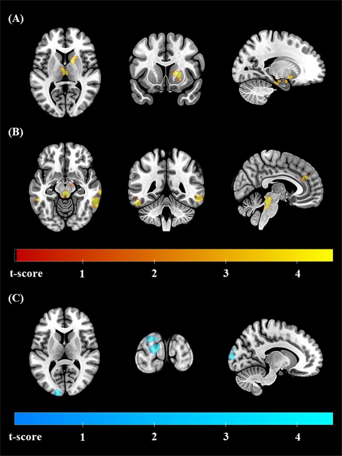
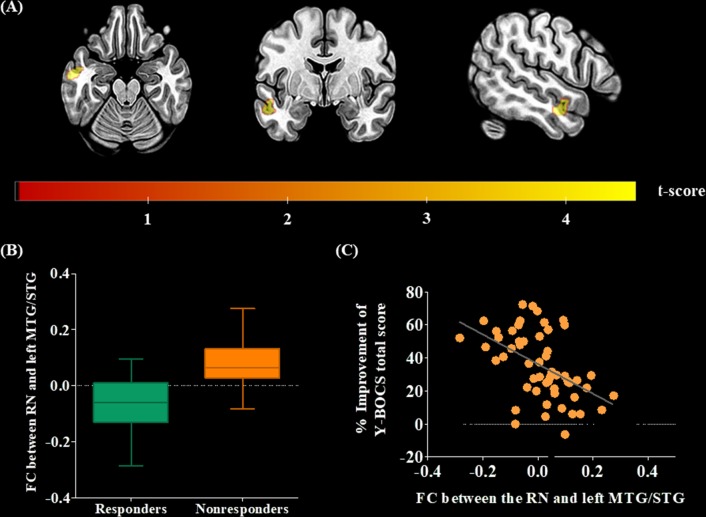
Selective serotonin reuptake inhibitors (SSRIs) are first-line pharmacological agents for treating obsessive-compulsive disorder (OCD). However, because nearly half of patients show insufficient SSRI responses, serotonergic dysfunction in heterogeneous OCD patients should be investigated for precision medicine. We aimed to determine whether functional connectivity (FC) of the raphe nucleus (RN), the major source of most serotonergic neurons, was altered in OCD patients and could predict the SSRI response. A total of 102 medication-free OCD patients and 101 matched healthy control (HC) subjects participated in resting-state functional magnetic resonance imaging. Among them, 54 OCD patients were treated with SSRIs for 16 weeks, resulting in 26 responders and 28 nonresponders. Seed-based whole brain FC with the RN as a seed region was compared between the OCD and HC groups, as well as between SSRI responders and nonresponders. FC cluster values showing significant group differences were used to investigate factors correlated with symptomatic severity before treatment and predictive of SSRI response. Compared to HCs, OCD patients exhibited significantly larger FC between the RN and temporal cortices including the middle temporal gyrus (MTG), paracingulate gyrus, amygdala, hippocampus, putamen, thalamus, and brain stem. Greater RN-left MTG FC was positively correlated with OC symptom severity at baseline. In addition, larger FC of the RN-left MTG was also found in SSRI nonresponders compared to responders, which was a significant predictor of SSRI response after 16 weeks. The FC of RN may reflect the neurobiological underpinning of OCD and could aid future precision medicine as a differential brain-based biomarker.
Figures
Similar articles
-
Abnormal intrinsic brain activity of the sensory-motor area as a predictor of the response to selective serotonin reuptake inhibitors in treatment-naïve obsessive-compulsive disorder.J Affect Disord. 2025 Nov 1;388:119457. doi: 10.1016/j.jad.2025.119457. Epub 2025 May 24. J Affect Disord. 2025. PMID: 40419153
-
The effects of pharmacological treatment on functional brain connectome in obsessive-compulsive disorder.Biol Psychiatry. 2014 Apr 15;75(8):606-14. doi: 10.1016/j.biopsych.2013.09.002. Epub 2013 Oct 4. Biol Psychiatry. 2014. PMID: 24099506
-
The effects of selective serotonin reuptake inhibitors on brain functional networks during goal-directed planning in obsessive-compulsive disorder.Sci Rep. 2020 Nov 26;10(1):20619. doi: 10.1038/s41598-020-77814-4. Sci Rep. 2020. PMID: 33244182 Free PMC article.
-
Structural and functional brain imaging after treatment with selective-serotonin reuptake-inhibitors in obsessive-compulsive disorder: A mini review.J Affect Disord. 2024 Jan 15;345:141-148. doi: 10.1016/j.jad.2023.10.034. Epub 2023 Oct 10. J Affect Disord. 2024. PMID: 37820957 Review.
-
Pharmacological management of treatment-resistant obsessive-compulsive disorder.CNS Drugs. 2011 Jul;25(7):585-96. doi: 10.2165/11587860-000000000-00000. CNS Drugs. 2011. PMID: 21699270 Review.
Cited by
-
Network-based functional connectivity predicts response to exposure therapy in unmedicated adults with obsessive-compulsive disorder.Neuropsychopharmacology. 2021 Apr;46(5):1035-1044. doi: 10.1038/s41386-020-00929-9. Epub 2021 Jan 14. Neuropsychopharmacology. 2021. PMID: 33446895 Free PMC article.
-
Compulsive-like Behaviors in Amyloid-β 1-42-Induced Alzheimer's Disease in Mice Are Associated With Hippocampo-cortical Neural Circuit Dysfunction.Biol Psychiatry Glob Open Sci. 2023 Mar 7;3(4):773-784. doi: 10.1016/j.bpsgos.2023.02.009. eCollection 2023 Oct. Biol Psychiatry Glob Open Sci. 2023. PMID: 37881551 Free PMC article.
-
Triple-Network Dysconnectivity in Patients With First-Episode Psychosis and Individuals at Clinical High Risk for Psychosis.Psychiatry Investig. 2022 Dec;19(12):1037-1045. doi: 10.30773/pi.2022.0091. Epub 2022 Dec 22. Psychiatry Investig. 2022. PMID: 36588438 Free PMC article.
-
Eye movement as a biomarker of impaired organizational strategies during visual memory encoding in obsessive-compulsive disorder.Sci Rep. 2021 Sep 15;11(1):18402. doi: 10.1038/s41598-021-97885-1. Sci Rep. 2021. PMID: 34526587 Free PMC article.
-
Potential therapeutic mechanism of deep brain stimulation of the nucleus accumbens in obsessive-compulsive disorder.Front Cell Neurosci. 2023 Jan 4;16:1057887. doi: 10.3389/fncel.2022.1057887. eCollection 2022. Front Cell Neurosci. 2023. PMID: 36687525 Free PMC article.
References
-
- Lochner C, Stein DJ. Heterogeneity of obsessive-compulsive disorder: a literature review. Harv Rev Psychiatry. 2003;11:113–32. - PubMed
-
- Stein DJ, Koen N, Fineberg N, Fontenelle LF, Matsunaga H, Osser D, et al. A 2012 evidence-based algorithm for the pharmacotherapy for obsessive-compulsive disorder. Curr Psychiatry Rep. 2012;14:211–9. - PubMed
-
- Koran LM, Hanna GL, Hollander E, Nestadt G, Simpson HB, American Psychiatric Association. Practice guideline for the treatment of patients with obsessive-compulsive disorder. Am J Psychiatry. 2007;164:5–53. - PubMed
-
- Kim E, Howes OD, Park JW, Kim SN, Shin SA, Kim BH, et al. Altered serotonin transporter binding potential in patients with obsessive-compulsive disorder under escitalopram treatment: [11C]DASB PET study. Psychol Med. 2016;46:357–66. - PubMed
-
- Hasselbalch SG, Hansen ES, Jakobsen TB, Pinborg LH, Lonborg JH, Bolwig TG. Reduced midbrain-pons serotonin transporter binding in patients with obsessive-compulsive disorder. Acta Psychiatr Scand. 2007;115:388–94. - PubMed
Publication types
MeSH terms
Substances
LinkOut - more resources
Full Text Sources
Medical
