. 2019 Jun 26;476(12):1725-1740.
doi: 10.1042/BCJ20180714.

AMP-activated protein kinase complexes containing the β2 regulatory subunit are up-regulated during and contribute to adipogenesis

Affiliations

AMP-activated protein kinase complexes containing the β2 regulatory subunit are up-regulated during and contribute to adipogenesis

Omar J Katwan et al. Biochem J. .

Abstract

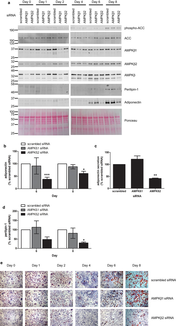
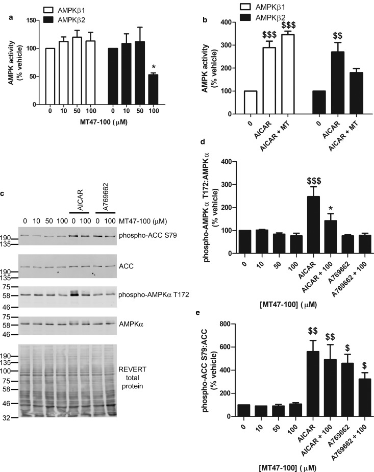
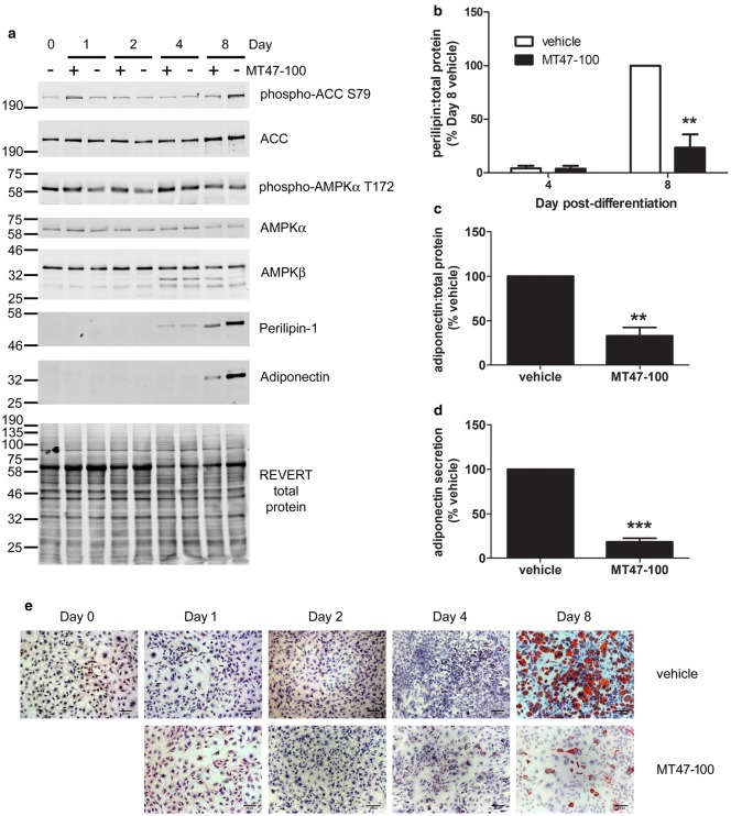
AMP-activated protein kinase (AMPK) is a heterotrimer of α-catalytic and β- and γ-regulatory subunits that acts to regulate cellular and whole-body nutrient metabolism. The key role of AMPK in sensing energy status has led to significant interest in AMPK as a therapeutic target for dysfunctional metabolism in type 2 diabetes, insulin resistance and obesity. Despite the actions of AMPK in the liver and skeletal muscle being extensively studied, the role of AMPK in adipose tissue and adipocytes remains less well characterised. Small molecules that selectively influence AMPK heterotrimers containing specific AMPKβ subunit isoforms have been developed, including MT47-100, which selectively inhibits complexes containing AMPKβ2. AMPKβ1 and AMPKβ2 are the principal AMPKβ subunit isoforms in rodent liver and skeletal muscle, respectively, yet the contribution of specific AMPKβ isoforms to adipose tissue function, however, remains largely unknown. This study therefore sought to determine the contribution of AMPKβ subunit isoforms to adipocyte biology, focussing on adipogenesis. AMPKβ2 was the principal AMPKβ isoform in 3T3-L1 adipocytes, isolated rodent adipocytes and human subcutaneous adipose tissue, as assessed by the contribution to total cellular AMPK activity. Down-regulation of AMPKβ2 with siRNA inhibited lipid accumulation, cellular adiponectin levels and adiponectin secretion during 3T3-L1 adipogenesis, whereas down-regulation of AMPKβ1 had no effect. Incubation of 3T3-L1 cells with MT47-100 selectively inhibited AMPK complexes containing AMPKβ2 whilst simultaneously inhibiting cellular lipid accumulation as well as cellular levels and secretion of adiponectin. Taken together, these data indicate that increased expression of AMPKβ2 is an important feature of efficient adipogenesis.

Keywords: AMPK; adipocytes; adipogenesis.


Conflict of interest statement

The Authors declare that there are no competing interests associated with the manuscript.

Figures

Figure 1.
Figure 1.. AMPKβ2 levels increase during 3T3-L1 cell adipogenesis.
(ac) 3T3-L1 preadipocytes were differentiated into adipocytes over 12 days and lysates prepared. Representative immunoblots are shown a) with the molecular masses (in kDa) indicated. Quantification of (b) AMPKα1:AMPKα2 or (c) AMPKβ2:AMPKβ1, assessed using individual isoform-specific antibodies. (dh) 3T3-L1 preadipocytes were differentiated into adipocytes and (d) RNA or (eh) lysates prepared at the indicated times during adipogenesis. (d) mRNA levels were assessed by qPCR and data shown represents Prkab2 (AMPKβ2) mRNA expression relative to Prkab1 (AMPKβ1). (eh) Lysate proteins were resolved by SDS–PAGE and immunoblotted with the antibodies indicated. (e) Representative immunoblots are shown with the molecular masses (in kDa) indicated. Quantification of AMPKβ2:AMPKβ1 assessed using (f) individual isoform-specific antibodies or (g) antibodies recognising both AMPKβ isoforms and (h) LKB1:AMPKα levels over the duration of adipogenesis. Data is representative of three independent experiments, * P < 0.05, ** P < 0.01, *** P < 0.001 relative to preadipocyte levels (c) two-tailed t-test, (d and fh) one-way ANOVA.
Figure 2.
Figure 2.. Isoform-specific AMPK activity in 3T3-L1 preadipocytes and adipocytes.
AMPK was immunoprecipitated in lysates (100 µg) from 3T3-L1 preadipocytes or adipocytes using (a) antibodies specific to AMPKα1 or AMPKα2 or (b) antibodies specific to AMPKβ1 or AMPKβ2 and AMPK activity assayed. The values are expressed as % total AMPK activity (AMPKα1 + AMPKα2 or AMPKβ1 + AMPKβ2, respectively). Results shown are from three (AMPKα isoforms in preadipocytes), four (AMPKα isoforms in adipocytes), five (AMPKβ isoforms in preadipocytes) or six (AMPKβ isoforms in adipocytes) independent experiments. *** P < 0.001 relative to % activity in preadipocytes (unpaired t-test).
Figure 3.
Figure 3.. Isoform-specific AMPK activity in rodent adipocytes and human subcutaneous adipose tissue.
AMPK was assayed in AMPK isoform-specific immunoprecipitates of lysates (100 µg) prepared from (a and b) rodent adipocytes isolated from mesenteric or epididymal adipose tissue or (c) human subcutaneous adipose tissue. Results shown are from five (mouse and rat epididymal adipocytes), four (mouse mesenteric adipose tissue), three (rat mesenteric adipocytes) or 10 (human subcutaneous adipose tissue) individual rodents or volunteers.
Figure 4.
Figure 4.. Down-regulation of AMPKβ2 suppresses adiponectin levels and lipid accumulation in 3T3-L1 adipocytes.
3T3-L1 preadipocytes were incubated with siRNA targeted to AMPKβ1, AMPKβ2 or scrambled siRNA for 48 h prior to differentiation into adipocytes. (a, b and d) Lysates were prepared after the indicated durations after initiation of differentiation and resolved by SDS–PAGE and immunoblotting with the indicated antibodies. (a) Representative immunoblots are shown, repeated on two to five further occasions. Densitometric analysis of (b) adiponectin or (d) perilipin-1 levels (days 6 and 8), normalised to Ponceau staining. (c) Conditioned media was collected from 3T3-L1 adipocytes at day 8 post-differentiation and adiponectin measured by ELISA in three independent experiments. (e) Cells were fixed and stained with oil red O at the times indicated during differentiation. Representative images from three independent experiments are shown, scale bar represents 20 µm. * P < 0.05, ** P < 0.01, *** P < 0.001 relative to scrambled siRNA (one-way ANOVA).
Figure 5.
Figure 5.. MT47-100 inhibits AMPK activity of complexes containing AMPKβ2 and suppresses AICAR-stimulated AMPKα T172 phosphorylation in 3T3-L1 preadipocytes.
3T3-L1 preadipocytes were incubated in the presence or absence of the indicated concentrations of MT47-100 for (a) 1 h or (be) 30 min prior to incubation in the presence or absence of AICAR (2 mmol/l) or A769662 (100 µmol/l) for 1 h and cell lysates prepared. (a and b) AMPKβ1 or AMPKβ2 were immunoprecipitated from lysates and AMPK activity subsequently assessed in immunoprecipitates. Data shown are from (a) five or (b) three independent experiments. (ce) Lysate proteins were resolved by SDS–PAGE and immunoblotted with the indicated antibodies. (c) Representative immunoblots are shown, repeated on two further occasions. Densitometric analysis of (d) phospho-AMPKα or (e) phospho-ACC relative to AMPKα and ACC, respectively. *P < 0.05 relative to absence of MT47-100, $P < 0.05, $$P < 0.01, $$$P < 0.001 relative to absence of AICAR or A769662 (one-way ANOVA).
Figure 6.
Figure 6.. MT47-100 suppresses adiponectin secretion and lipid accumulation during adipogenesis.
3T3-L1 preadipocytes were differentiated in the presence or absence of MT47-100 (100 µmol/l), with media changed on days 3 and 6, when MT47-100 was re-introduced to the culture medium. (ac) Cell lysates were prepared at the times indicated and proteins resolved by SDS–PAGE and immunoblotting with the antibodies indicated. (a) Representative immunoblots, repeated with similar results on two further occasions are shown. Densitometric analysis of (b) perilipin-1 or (c) adiponectin levels relative to REVERT total protein stain at 8 days post-differentiation from three independent experiments. (d) Conditioned media were collected from 3T3-L1 adipocytes differentiated in the absence or presence of MT47-100 (100 µmol/l) for 8 days and adiponectin measured by ELISA in three independent experiments. (e) Cells were fixed and stained with oil red O at the times indicated during differentiation. Representative images from three independent experiments are shown, scale bar represents 20 µm. ** P < 0.01, *** P < 0.001 relative to absence of MT47-100 (b) 2-way ANOVA (c and d) unpaired t-test.
Figure 7.
Figure 7.. MT47-100 inhibits cellular PPARγ and C/EBPα levels during differentiation of 3T3-L1 cells.
(a) 3T3-L1 preadipocytes in six-well plates were differentiated in the presence of troglitazone (1 µmol/l) and the absence or presence of MT47-100 (100 µmol/l), trypsinised and counted using a Nexcelom Cellometer Auto 1000 on the days indicated after commencement of differentiation. Data shown represent the mean ± SEM cells/well from three independent experiments. *** P < 0.001 relative to day 0 (two-way ANOVA). (bd) 3T3-L1 preadipocytes were differentiated in the absence or presence of MT47-100 (100 µmol/l) as well as troglitazone (1 µmol/l) and cell lysates were prepared 3 days post-differentiation and equal volumes resolved by SDS–PAGE and immunoblotting with the antibodies indicated. (b) Representative immunoblots, repeated with similar results on two further occasions are shown. (c and d) Densitometric analysis of protein levels relative to total lysate protein, assessed with REVERT total protein stain (Li-Cor Biosciences). Data shown represents the mean ± SEM relative to vehicle control. * P < 0.05, ** P < 0.01 relative to absence of MT47-100 (two-way ANOVA).

Similar articles

Cited by

References

    1. Carling D. (2017) AMPK signalling in health and disease. Curr. Opin. Cell Biol. 45, 31–37 10.1016/j.ceb.2017.01.005 - DOI - PubMed
    1. Salt I.P. and Hardie D.G. (2017) AMP-activated protein kinase: an ubiquitous signaling pathway with key roles in the cardiovascular system. Circ. Res. 120, 1825–1841 10.1161/CIRCRESAHA.117.309633 - DOI - PMC - PubMed
    1. Day E.A., Ford R.J. and Steinberg G.R. (2017) AMPK as a therapeutic target for treating metabolic diseases. Trends Endocrinol. Metab. 28, 545–560 10.1016/j.tem.2017.05.004 - DOI - PubMed
    1. Bijland S., Mancini S.J. and Salt I.P. (2013) Role of AMP-activated protein kinase in adipose tissue metabolism and inflammation. Clin. Sci. 124, 491–507 10.1042/CS20120536 - DOI - PubMed
    1. Thornton C., Snowden M.A. and Carling D. (1998) Identification of a novel AMP-activated protein kinase beta subunit isoform that is highly expressed in skeletal muscle. J. Biol. Chem. 273, 12443–12450 10.1074/jbc.273.20.12443 - DOI - PubMed

Publication types