. 2019 Jun 17;8(6):bio044974.
doi: 10.1242/bio.044974.

Ubiquitin-mediated proteasome degradation regulates optic fissure fusion

Affiliations

Ubiquitin-mediated proteasome degradation regulates optic fissure fusion

Warlen Pereira Piedade et al. Biol Open. .

Abstract

Optic fissure fusion is a critical event during retinal development. Failure of fusion leads to coloboma, a potentially blinding congenital disorder. Pax2a is an essential regulator of optic fissure fusion and the target of numerous morphogenetic pathways. In our current study, we examined the negative regulator of pax2a expression, Nz2, and the mechanism modulating Nlz2 activity during optic fissure fusion. Upregulation of Nlz2 in zebrafish embryos resulted in downregulation of pax2a expression and fissure fusion failure. Conversely, upregulation of pax2a expression also led to fissure fusion failure suggesting Pax2 levels require modulation to ensure proper fusion. Interestingly, we discovered Nlz2 is a target of the E3 ubiquitin ligase Siah. We show that zebrafish siah1 expression is regulated by Hedgehog signaling and that Siah1 can directly target Nlz2 for proteasomal degradation, in turn regulating the levels of pax2a mRNA. Finally, we show that both activation and inhibition of Siah activity leads to failure of optic fissure fusion dependent on ubiquitin-mediated proteasomal degradation of Nlz2. In conclusion, we outline a novel, proteasome-mediated degradation regulatory pathway involved in optic fissure fusion.

Keywords: Coloboma; Nlz2; Optic fissure; Pax2; Proteasome; Retina; SIAH.


Figures

Fig. 1.
Fig. 1.
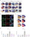
Nlz2 regulates optic fissure closure by inhibiting pax2a gene expression. (A) WISH for pax2a in 24 hpf embryos injected with varying amounts of Nlz2 mRNA. OF-associated pax2a signal is decreased in response to increasing Nlz2 mRNA. (B) qPCR results at 24 hpf for pax2a expression±s.d. *P<0.05 compared to uninjected, $P<0.05 compared to Nlz2 50 pg. (C) 72 hpf Tg[rx3:GFP] (green) embryos injected with Pax2a, Nlz2 or Pax2+Nlz2 mRNA stained for laminin (red) and DAPI (blue). Arrowheads indicate persistence of laminin signal. Scale bar: 50 µm. (D) Proportion of embryos with failure of fusion (1) or completed fusion (0)±s.d. Region of analysis is outlined by dashed lines. *P<0.05 compared to uninjected, one-way ANOVA; P<0.0001. Dashed boxes outline region of inset.
Fig. 2.
Fig. 2.
Siah targets Nlz2 for proteasomal degradation. (A) Conservation of Nlz2 ‘degron’ motif sequence. (B) Western blot analysis of nlz2-FLAG protein stability in response to siah1-myc, siah1ΔR-myc or siah1-myc+MG132. Actin was used as a loading control. * indicates potential ubiquitination products. Nlz2-FLAG band intensity quantification is shown. (C) Co-immunoprecipitation of nlz2-FLAG co-transfected with HA-ubiquitin, siah1-myc or siah1ΔR-myc probed for FLAG (green), MYC (red) and HA (B/W). * indicates potential ubiquitination. (D) Upper panels: endogenous Siah activity reporter assay expression in eyes of 24 hpf GFP-NxN or GFP-VSP mRNA-injected embryos, ±12.5 µM MG132. mCherry mRNA was co-injected for normalization. Scale bar: 50 µm. Lower panel: quantification of normalized GFP fluorescence intensity in the eye±s.d. *P<0.05 compared to GFP-NxN, #P<0.05 compared to GFP-VSP, one-way ANOVA; P<0.0001.
Fig. 3.
Fig. 3.
siah1, siah2l and nlz2 gene expression during eye morphogenesis. (A) WISH for siah1, siah2l and nlz2 between 24 and 48 hpf. (B) Two-color fluorescent WISH (FWISH) for nlz2, siah1, siah2 and pax2a. Arrowheads indicate co-localization. (C) siah1 WISH after treatment with RA (AGN194310), BMP (DMH1), hedgehog (cyclopamie) inhibitors, hedgehog agonist (purmorphamine) or in smo^hi1640Tg embryos at 24 hpf. (D) qPCR results for siah1 and pax2a expression±s.d. *P<0.05 compared to respective controls.
Fig. 4.
Fig. 4.
Siah activity indirectly regulates pax2a expression. (A) WISH for pax2a after injection of Siah1, Siah1ΔR, Siah2l or Siah2lΔR±MG132 at 24 hpf. (B) qPCR results for pax2a expression at 24 hpf. *P<0.05 compared to uninjected. (C) WISH for pax2a gene expression upon co-injection of Siah1+Nlz2 or Nlz2nxn. (D) qPCR results for pax2a expression at 24 hpf. *P<0.05 compared to uninjected, @P<0.05 compared to Nlz2, #P<0.05 compared to Siah1, $P<0.05 compared to Siah1+Nlz2. (E) Tg[rx3:GFP] (green) embryos injected with siah1, siah2l, siah1ΔR or siah2lΔR±100 pg Nlz2 mRNA stained for laminin (red) and DAPI (blue) at 72 hpf. Arrowheads indicate persistence of laminin. Scale bar: 50 µm. (F) Proportion of embryos with failure of fusion (1) or completed fusion (0). Region of analysis is outlined by dashed lines. *P<0.05 compared to uninjected, #P<0.05 compared to Siah1, $P<0.05 compared to Siah1+Nlz2. One-way ANOVA; P<0.0001. Dashed boxes indicate region of inset.

References

    1. Babcock H. E., Dutta S., Alur R. P., Brocker C., Vasiliou V., Vitale S., Abu-Asab M. and Brooks B. P. (2014). aldh7a1 regulates eye and limb development in zebrafish. PLoS ONE 9, e101782 10.1371/journal.pone.0101782 - DOI - PMC - PubMed
    1. Bernstein C. S., Anderson M. T., Gohel C., Slater K., Gross J. M. and Agarwala S. (2018). The cellular bases of choroid fissure formation and closure. Dev. Biol. 440, 137-151. 10.1016/j.ydbio.2018.05.010 - DOI - PMC - PubMed
    1. Bhattacharjee S., Renganaath K., Mehrotra R. and Mehrotra S. (2013). Combinatorial control of gene expression. Biomed. Res. Int. 2013, 407263 10.1155/2013/407263 - DOI - PMC - PubMed
    1. Bogdan S., Senkel S., Esser F., Ryffel G. U. and Pogge V. Strandmann E. (2001). Misexpression of Xsiah-2 induces a small eye phenotype in Xenopus. Mech. Dev. 103, 61-69. 10.1016/S0925-4773(01)00332-X - DOI - PubMed
    1. Brown J. D., Dutta S., Bharti K., Bonner R. F., Munson P. J., Dawid I. B., Akhtar A. L., Onojafe I. F., Alur R. P., Gross J. M. et al. (2009). Expression profiling during ocular development identifies 2 Nlz genes with a critical role in optic fissure closure. Proc. Natl. Acad. Sci. USA 106, 1462-1467. 10.1073/pnas.0812017106 - DOI - PMC - PubMed