. 2019 Jul 10;39(7):BSR20190946.
doi: 10.1042/BSR20190946. Print 2019 Jul 31.

DEPDC1 promotes cell proliferation and suppresses sensitivity to chemotherapy in human hepatocellular carcinoma

Affiliations

DEPDC1 promotes cell proliferation and suppresses sensitivity to chemotherapy in human hepatocellular carcinoma

Chao Zhou et al. Biosci Rep. .

Retraction in

Abstract

Background: Hepatocellular carcinoma (HCC) is one of the major causes of tumor-related morbidity and mortality worldwide. Accumulating evidence has revealed that aberrant expression of crucial cancer-related genes contributes to hepatocellular carcinogenesis. This study aimed to characterize the biological role of DEP domain containing 1 (DEPDC1), a novel cancer-related gene, in HCC and illuminate the potential molecular mechanisms involved.

Materials and methods: Quantitative real-time PCR (qRT-PCR), Western blotting and immunohistochemical (IHC) staining were used to characterize the expression patterns of DEPDC1 in tumorous tissues and adjacent normal tissues. Kaplan-Meier survival analysis was launched to evaluate the relationship between DEPDC1 expression and overall survival. CCK8 assay, colony formation and flow cytometry were performed to investigate the effects of DEPDC1 on HCC cell viability, clonogenic capability and cell apoptosis. Murine xenograft models were established to determine the effect of DEPDC1 on tumor growth in vivo SP600125, a JNK specific inhibitor, was applied to carriy out mechanistic studies.

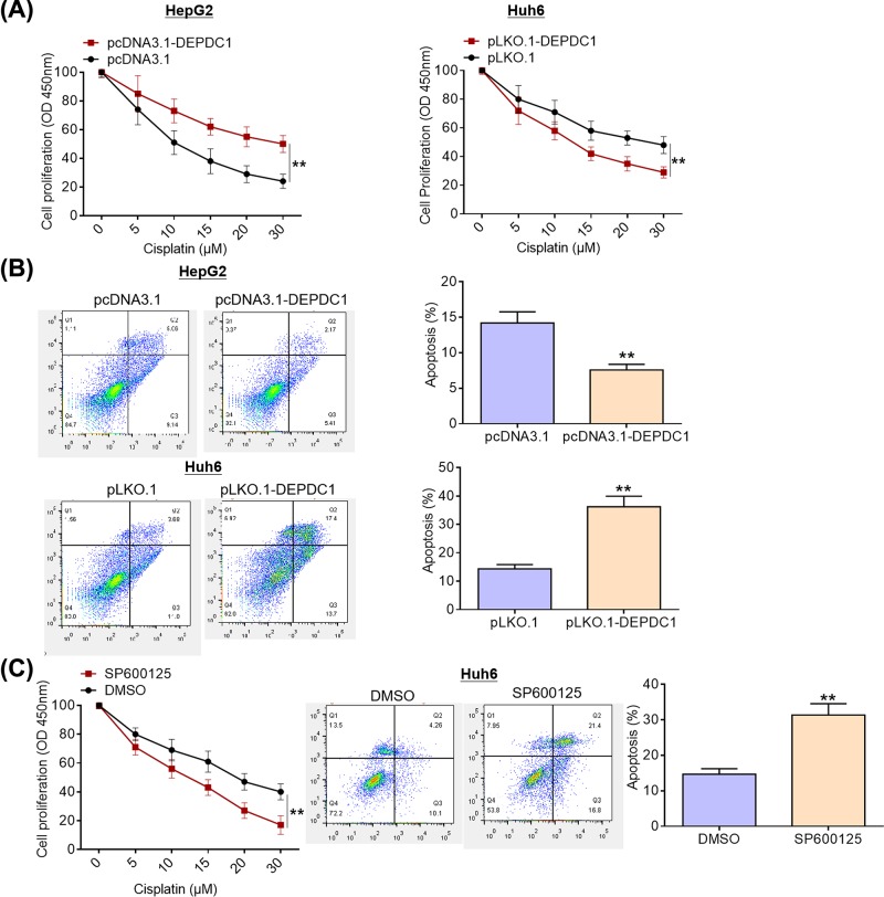
Results: DEPDC1 was significantly up-regulated in HCC tissues compared with para-cancerous tissues. Besides, patients with high DEPDC1 expression experienced a significantly shorter overall survival. Functional investigations demonstrated that DEPDC1 overexpression facilitated HCC cell proliferation and suppressed cell apoptosis, whereas DEPDC1 depletion inhibited cell proliferation and promoted cell apoptosis. Furthermore, DEPDC1 ablation suppressed tumorigenecity of HCC cells in murine xenograft models. Mechanistic studies uncovered that JNK signaling pathway mediated the promoting effects of DEPDC1 on HCC cell viability and chemotherapy resistance.

Conclusion: Collectively, our data may provide some evidence for DEPDC1 as a candidate therapeutic target for HCC.

Keywords: DEPDC1; JNK signaling pathway; cell proliferation; chemotherapy resistance; hepatocellular carcinoma.


Conflict of interest statement

The authors declare that there are no competing interests associated with the manuscript.

Figures

Figure 1
Figure 1. Increased expression of DEPDC1 predicts poor prognosis of HCC patients
(A) DEPDC1 mRNA expression levels in 60 pairs of HCC tissues and adjacent non-cancerous tissues were examined by qRT-PCR analysis. (B) DEPDC1 protein expression levels in HCC tissues and matched normal tissues were detected by Western blotting analysis. (C) DEPDC1 protein expression in tumorous tissues and corresponding para-cancerous tissues were visualized by IHC. (D) HCC tissues were classified into high DEPDC1 expression group and low DEPDC1 expression group based on its mRNA expression levels. (E) DEPDC1 mRNA expression levels in normal human liver L02 cells and four HCC cell lines (HepG2, SK-Hep1, Huh7 and Huh6) were determined by qRT-PCR analysis; **P < 0.01; DEPDC1, DEP domain containing 1; HCC, hepatocellular carcinoma; qRT-PCR, quantitative real-time polymerase chain reaction; IHC, immunohistochemistry.
Figure 2
Figure 2. DEPDC1 facilitates HCC cell proliferation and survival
(A) Transfection efficiency was evaluated by Western blotting assays after transfection with DEPDC1 expression vector or shRNA-DEPDC1. (B) Cell viability was measured by CCK8 assays after transfection with DEPDC1 expression vector or shRNA-DEPDC1. (C) Clonogenic ability was detected by colony formation assays after transfection with DEPDC1 expression vector or shRNA-DEPDC1. (D) Cell apoptosis was evaluated by flow cytometry after transfection with DEPDC1 expression vector or shRNA-DEPDC1; **P < 0.01; DEPDC1, DEP domain containing 1; HCC, hepatocellular carcinoma; shRNA, short hairpin RNA.
Figure 3
Figure 3. DEPDC1 ablation suppresses tumorigenicity of HCC cells in murine xenograft models
(A) shRNA-DEPDC1 treated Huh6 cells were subcutaneously injected into the flanks of the mice (n = 5). Tumor volume was measured by slide caliper every 3 days; the mice were killed and tumors were weighed at day 21 post-implantation. (B) Ki67 protein expression in the collected tumors were visualized by IHC staining. (C) Apoptotic cells in the harvested tumors were determined by TUNEL assays; **P < 0.01; DEPDC1, DEP domain containing 1; HCC, hepatocellular carcinoma; shRNA, short hairpin RNA; TUNEL, terminal deoxynucleotidyl mediated nick end labeling.
Figure 4
Figure 4. DEPDC1 promotes activation of JNK signaling pathway in HCC cells
(A) JNK and p-JNK protein expression levels were examined by Western blotting in DEPDC1 overexpression group and DEPDC1 depletion group, respectively. (B) JNK and p-JNK protein expression levels were determined by Western blotting after usage of JNK specific inhibitor SP600125 in DEPDC1 overexpression group. (C) FCM analysis was performed to evaluate the effect of JNK specific inhibitor SP600125 on apoptosis of HepG2 cells overexpressing DEPDC1; **P<0.01; DEPDC1, DEP domain containing 1; JNK, c-Jun N-terminal kinase; HCC, hepatocellular carcinoma; p-JNK, phosphorylated c-Jun N-terminal kinase.
Figure 5
Figure 5. DEPDC1 depletion enhances sensitivity of HCC cells to chemotherapy through JNK signaling pathway
(A) Cell viability was detected by CCK8 assay after treatment with different concentrations of cisplatin for 12 h. (B) Cell apoptosis was evaluated using flow cytometry after treatment with 10 μM cisplatin for 12 h. (C) Cell viability from DMSO group and SP600125 group was determined by CCK8 assays after treatment with various concentration of cisplatin for 12 h, respectively; cell apoptosis from DMSO and SP600125 group was examined by flow cytometry after treatment with 10 μM cisplatin for 12 h; **P < 0.01; DEPDC1, DEP domain containing 1; HCC, hepatocellular carcinoma; JNK, c-Jun N-terminal kinase.

References

    1. Giorgio M.D. and Fagiuoli S. (2007) Management of hepatocellular carcinoma. Dig. Dis. 25, 279–281 10.1159/000103902 - DOI - PubMed
    1. Suzuki K., Hayashi N. and Yamada Y. (2010) Expression of the c-met protooncogene in human hepatocellular carcinoma. Hepatology 20, 1231–1236 10.1002/hep.1840200520 - DOI - PubMed
    1. Fernández-Alvarez A., Llorenteizquierdo C. and Mayoral R. (2012) Evaluation of epigenetic modulation of cyclooxygenase-2 as a prognostic marker for hepatocellular carcinoma. Oncogenesis 1, e23. 10.1038/oncsis.2012.23 - DOI - PMC - PubMed
    1. Siegel R.L., Miller K.D. and Jemal A. (2017) Cancer statistics, 2017. CA Cancer J. Clin. 67, 7–30 10.3322/caac.21387 - DOI - PubMed
    1. Pan Q.Z., Pan K. and Weng D.S. (2015) Annexin A3 promotes tumorigenesis and resistance to chemotherapy in hepatocellular carcinoma. Mol. Carcinog. 54, 598–607 10.1002/mc.22126 - DOI - PubMed

Publication types

MeSH terms