. 2019 Jun 12;10(6):458.
doi: 10.1038/s41419-019-1672-4.

Ets-1 deficiency alleviates nonalcoholic steatohepatitis via weakening TGF-β1 signaling-mediated hepatocyte apoptosis

Affiliations

Ets-1 deficiency alleviates nonalcoholic steatohepatitis via weakening TGF-β1 signaling-mediated hepatocyte apoptosis

Dechen Liu et al. Cell Death Dis. .

Abstract

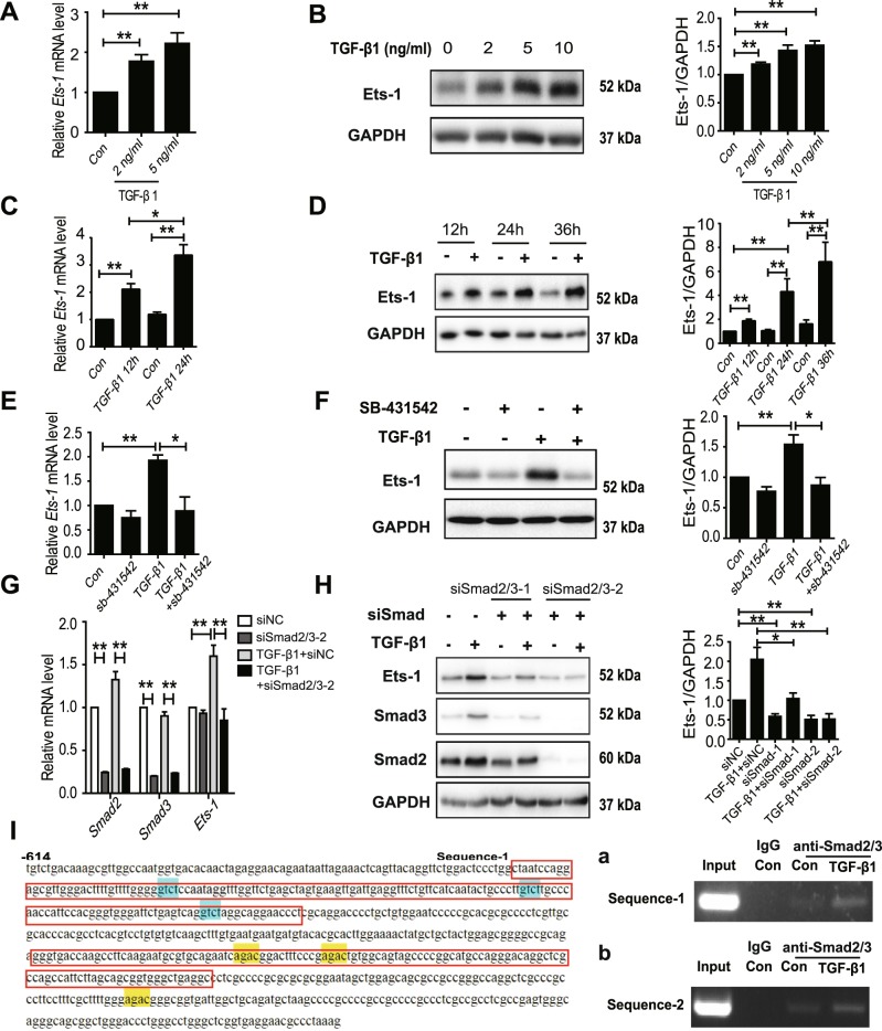
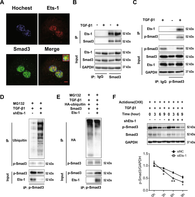
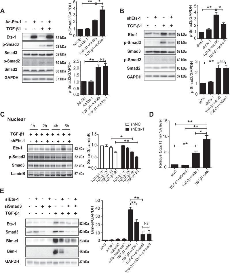
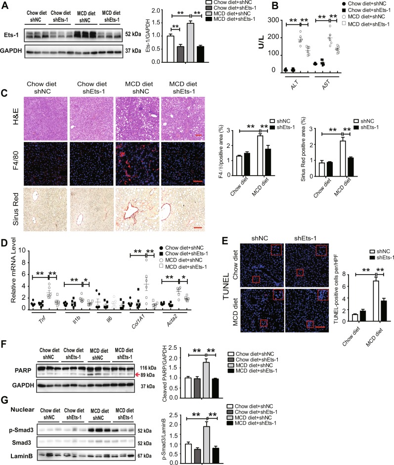
Hepatocyte apoptosis is a hallmark of nonalcoholic steatohepatitis (NASH) and contributes to liver injury, fibrosis, and inflammation. However, the molecular mechanisms underlying excessive hepatocyte apoptosis in NASH remain largely unknown. This study aimed to explore whether and how the v-ets avian erythroblastosis virus E26 oncogene homolog 1 (Ets-1) is involved in diet-induced hepatocyte apoptosis in mice. The study found that the expression level of hepatic Ets-1 was elevated in a NASH mouse model as a result of the activation of transforming growth factor beta1 (TGF-β1) signaling. In the presence of TGF-β1, phosphorylated mothers against decapentaplegic homolog 2/3 (p-Smad2/3) translocated to the binding sites of the Ets-1 promoter to upregulate the expression of Ets-1 in primary hepatocytes. In addition, Ets-1 bound directly to phosphorylated Smad3 (p-Smad3), thereby preventing the ubiquitination and proteasomal degradation of p-Smad3 and enhancing the activity of TGF-β1/Smad3 signaling. Consequently, elevated Ets-1 stimulated TGF-β1-induced hepatocyte apoptosis. However, Ets-1 knockdown alleviated diet-induced hepatocyte apoptosis and NASH with reduced liver injury, inflammation, and fibrosis. Taken together, Ets-1 had an adverse impact on hepatocyte survival under TGF-β1 treatment and accelerated the development of NASH in mice.


Conflict of interest statement

The authors declare that they have no conflict of interest.

Figures

Fig. 1
Fig. 1. The expression of Ets-1 was associated with the progression of NASH.
a, b Expression of Ets-1 was examined in biopsies from normal controls (n = 5) and patients with NASH (n = 12) (GSE24807). a Some genes that changed significantly are listed. CXCL10, CXC motif chemokine ligand 10; SH3BP5, SH3 domain–binding protein 5; CYP2R1, cytochrome P450 2R1; FABP1, fatty acid-binding protein 1; TGFB1, transforming growth factor beta1. b The correlation between ETS-1 and TGFB1 was determined by using the linear regression test (P < 0.01). ce WT mice were fed with chow diet (n = 5) and MCD diet (n = 5) for 8 weeks and sacrificed. Hepatic mRNA levels of Tgfb1 (c) and Ets-1 (d) were examined using qRT-PCR. e Lysates of liver tissues were used for immunoblotting. Quantitative data represent mean ± standard error of the mean (SEM). *P < 0.05 and **P < 0.01
Fig. 2
Fig. 2. The expression of Ets-1 was directly regulated by TGF-β1-Smad2/3.
ad Primary hepatocytes from WT mice were (a, b) treated with different concentrations of TGF-β1 for 24 h or (c, d) harvested at different time points under the treatment of 10 ng/mL TGF-β1. a, c The relative mRNA level of Ets-1 was measured using qPCR. b, d Immunoblots of Ets-1 are shown. e, f WT primary hepatocytes were pretreated with SB-431542 (10 μM) for 12 h. Then, 10 ng/mL TGF-β1 was added to the cells for 24 h. e The relative expression of Ets-1 was measured using qPCR. f Immunoblots of Ets-1. g, h siRNAs of the negative control (siNC) and Smad2/3 (siSmad2/3-1 and siSmad2/3-2) were added to primary hepatocytes for 36 h, before incubation with 10 ng/mL TGF-β1 for 12 h (g) or 24 h (h). g The expression of Smad2, Smad3, and Ets-1 were examined. h Immunoblotting for Smad3, Smad2, and Ets-1 was performed. i The promotor of Ets-1 between –614 bp and –3 bp is shown. The locations of Sequence-1 and Sequence-2 are marked by a red box. The sequences of (C/AGAC) were Smad2/3-specific DNA-binding elements. a, b The ChIP assay of Ets-1 promoter used primary hepatocytes treated with TGF-β1 (10 ng/mL) for 6 h. An anti-Smad2/3 polyclonal antibody was used for precipitation. PCR analysis of the input and immunoprecipitation with IgG and an anti-Smad2/3 antibody were performed. Quantitative data are expressed as mean ± SEM (at least three independent experiments). *P < 0.05 and **P < 0.01
Fig. 3
Fig. 3. Ets-1 diminished the ubiquitination and proteasomal degradation of p-Smad3.
a Primary hepatocytes were stained with Hoechst in blue, anti-Ets-1 antibody in red and anti-Smad3 antibody in green, followed by assessment using confocal microscopy. Scale bar: 5 μm. b, c Hepatocytes were treated with TGF-β1 (10 ng/mL) for 6 h. b Cell lysates were immunoprecipitated with an anti-Smad3 antibody, followed by immunoblotting with an anti-Smad3 or anti-Ets-1 antibody. c Total lysates of cells were subjected to co-IP analysis with an anti-phosphorylated Smad3 (p-Smad3) antibody and immunoblot of Ets-1 or p-Smad3. d, e Hepatocytes were transfected with shEts-1 adenovirus (d) or co-transfected with HA-ubiquitin, Smad3 and Ets-1 plasmids (e) for 24 h and then treated with TGF-β1 for another 2 h. Finally, MG132 (25 μM) was added for 4 h. The lysates were used for immunoprecipitation with p-Smad3, and immunoblot was examined using p-Smad3, anti-ubiquitin (d) or anti-HA (e) antibody. f Hepatocytes were infected with shNC and the shEts-1 adenovirus for 24 h and treated with TGF-β1 (10 ng/mL) for 2 h. Cycloheximide (CHX, 50 ng/mL) was added, and the cells were harvested for 3, 6 and 9 h. Immunoblotting for p-Smad3 and Smad3 were performed. Quantitative data represent mean ± SEM. *P < 0.05 and **P < 0.01
Fig. 4
Fig. 4. Ets-1 enhanced the activity of TGF-β1/Smad3 signaling.
a, b Primary hepatocytes treated with Gfp and Ets-1 (a) or shNC and shEts-1 (b) recombinant adenovirus for 36 h. The cells were incubated with TGF-β1 (10 ng/mL) for 6 h, and the lysates were used for immunoblot analysis. (c) TGF-β1 (10 ng/mL) was added to hepatocytes for 1, 2, 4 and 6 h. The nuclear fraction was subjected to immunoblotting. d, e Primary hepatocytes were treated with siNC, siSmad3 or siEts-1 for 36 h and incubated with TGF-β1 (10 ng/mL) for 24 h. d mRNA level of Bcl2l11 is shown. e Total lysates were used for immunoblotting of Ets-1, Smad3 and Bim. Quantitative data are presented as mean ± SEM (at least three independent experiments). NS (negative significance); *P < 0.05 and **P < 0.01
Fig. 5
Fig. 5. Ets-1 accelerated hepatocyte apoptosis induced by TGF-β1/Smad3 signaling.
a Hepatocytes were treated with TGF-β1 (10 ng/mL) for 12, 24 and 36 h and then used for immunoblot analysis. b, c siRNAs of NC, Smad3 (b) or Ets-1 (c) were added to hepatocytes for 36 h and stimulated with TGF-β1 (10 ng/mL) for 24 h. Total lysates were subjected to immunoblotting of cleaved-caspase3, Smad3 (b) or Ets-1 (c). d Primary hepatocytes were treated with shNC and shEts-1 recombinant adenovirus for 36 h. Then, the cells were incubated with TGF-β1 (10 ng/mL) for 24 h. The cells were stained with TUNEL. Scale bar: 100 μm. Representative images are on the left, and quantitative analysis is on the right. e Hepatocytes were transfected with shNC or shEts-1 and Gfp or Smad3 recombinant adenovirus for 36 h. Then, the cells were stimulated with TGF-β1 (2 ng/mL) for 24 h. Lysates were used for immunoblotting. f Hepatocytes were transfected with Gfp and Ets-1 recombinant adenovirus for 24 h, and then TGF-β1 (2 ng/mL) was added for 36 h. Total lysates of cells were used for immunoblotting. Data are presented as mean ± SEM (at least three independent experiments). *P < 0.05 and **P < 0.01
Fig. 6
Fig. 6. Ets-1 was elevated in the liver tissues of NASH mice.
WT mice were injected with AAV8-shNC virus and AAV8-shEts-1 virus through the tail vein for 2 weeks and divided into four groups: chow diet+shNC (n = 6), chow diet+shEts-1 (n = 6), MCD diet+shNC (n = 6), and MCD diet+shEts-1 (n = 6). The mice were fed with a chow diet or an MCD diet for 8 weeks and sacrificed. a The lysates of liver tissues were used for immunoblotting of Ets-1. b ALT and AST levels in serum, U/L: enzyme activity per liter of liquid. c Liver sections were stained with H&E, Sirius Red, and F4/80. Scale bar: 100 μm. The left panel is a representative image, and the right panel is the quantification of Sirius Red–positive and F4/80-positive areas. The results were quantified using the Image J software. d Hepatic mRNA levels of inflammation-related proteins (Tnf, Il1b and Il6) and fibrosis-related proteins (Col1A1 and Acta2) were quantified using qRT-PCR. e Liver specimens were stained with TUNEL. A representative image is shown on the left, with the corresponding statistical result on the right. f, g Total lysates (f) or nuclear lysates (g) of liver tissues were used for immunoblotting. The left panels are representative images, and the right panels are corresponding statistical results. The red arrow is referring to cleaved PARP. Quantitative data represent mean ± SEM. *P < 0.05; **P < 0.01
Fig. 7
Fig. 7
Working model: Ets-1 maintained nuclear p-Smad3 and enhanced hepatocyte apoptosis induced by TGF-β1, thereby promoting NASH progression

References

    1. Younossi Z, et al. Global burden of NAFLD and NASH: trends, predictions, risk factors and prevention. Nat. Rev. Gastroenterol. Hepatol. 2017;15:11. doi: 10.1038/nrgastro.2017.109. - DOI - PubMed
    1. Zhang X, She Z, Li H. Time to step-up the fight against NAFLD. Hepatology. 2018;67:2068. doi: 10.1002/hep.29845. - DOI - PubMed
    1. Diehl AM, Day C. Cause, pathogenesis, and treatment of nonalcoholic steatohepatitis. N Engl. J. Med. 2017;377:2063. doi: 10.1056/NEJMra1503519. - DOI - PubMed
    1. Machado MV, Diehl AM. Pathogenesis of nonalcoholic steatohepatitis. Gastroenterology. 2016;150:1769. doi: 10.1053/j.gastro.2016.02.066. - DOI - PMC - PubMed
    1. Sunny NE, Bril F, Cusi K. Mitochondrial adaptation in nonalcoholic fatty liver disease: novel mechanisms and treatment strategies. Trends Endocrinol. Metab. 2017;28:250. doi: 10.1016/j.tem.2016.11.006. - DOI - PubMed

Publication types

MeSH terms